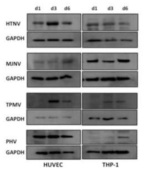
Fig 3. Western blot analysis of N protein expression.
HUVEC or THP-1 cells were infected with HTNV, MJNV, TPMV or PHV (MOI of 1.0). Cells were lysed at 1, 3, 6 days postinfection as indicated. Total protein levels were determined, and an equivalent amount of whole-cell lysate was separated by 10% SDS-polyacrylamide gel electrophoresis. Proteins were detected by Western blotting using anti-nucleocapsid monoclonal mouse or polyclonal rat antibody, followed by HRP-conjugated species-specific secondary antibodies. Anti-GAPDH monoclonal antibody was used as a loading control.

