

Abstract
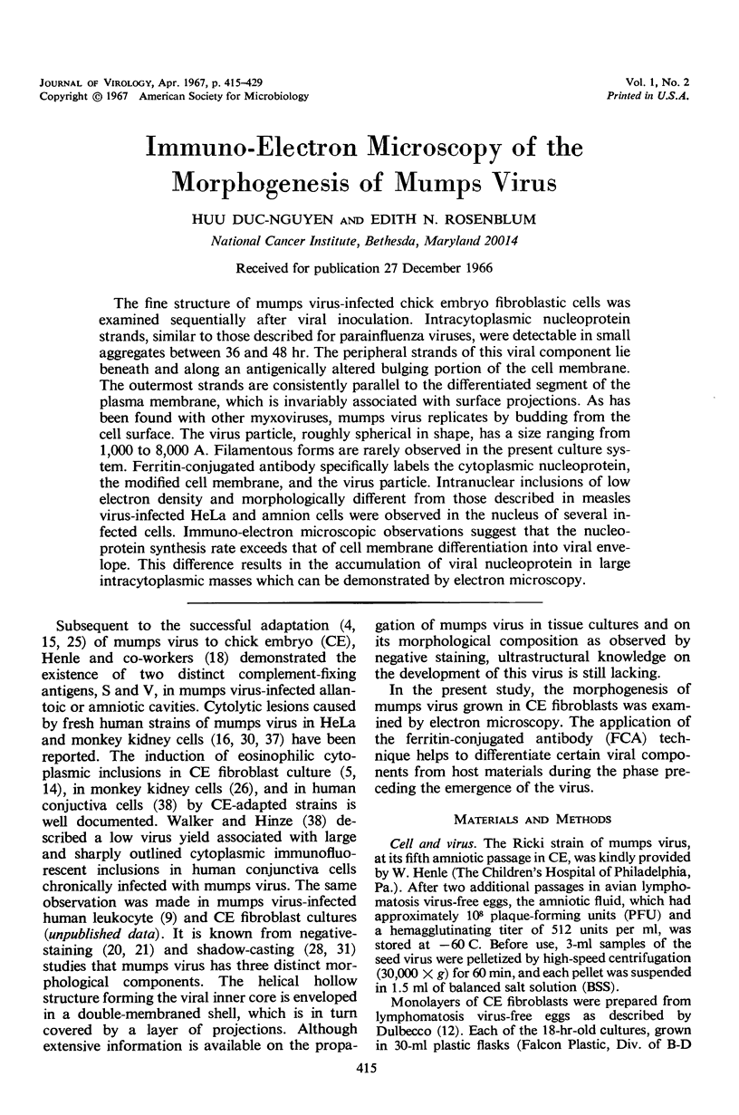
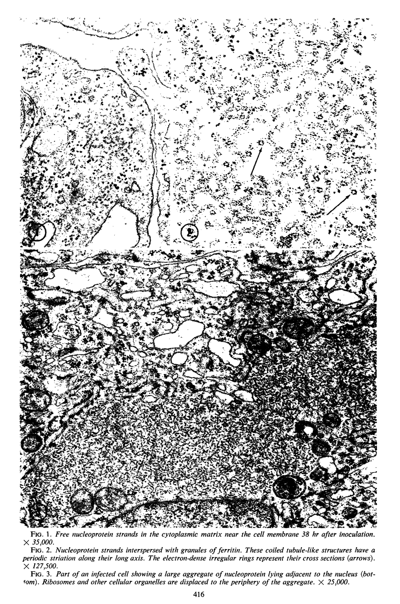
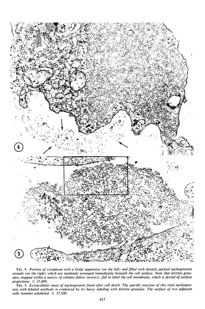
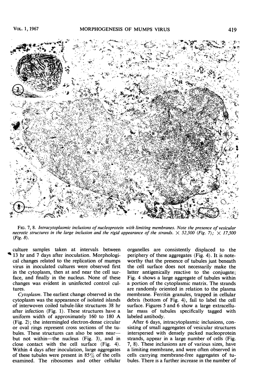
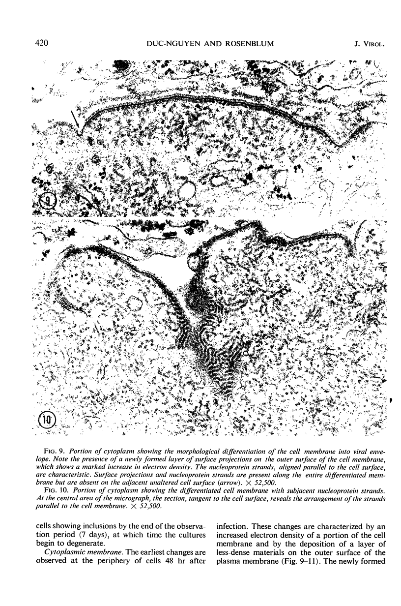
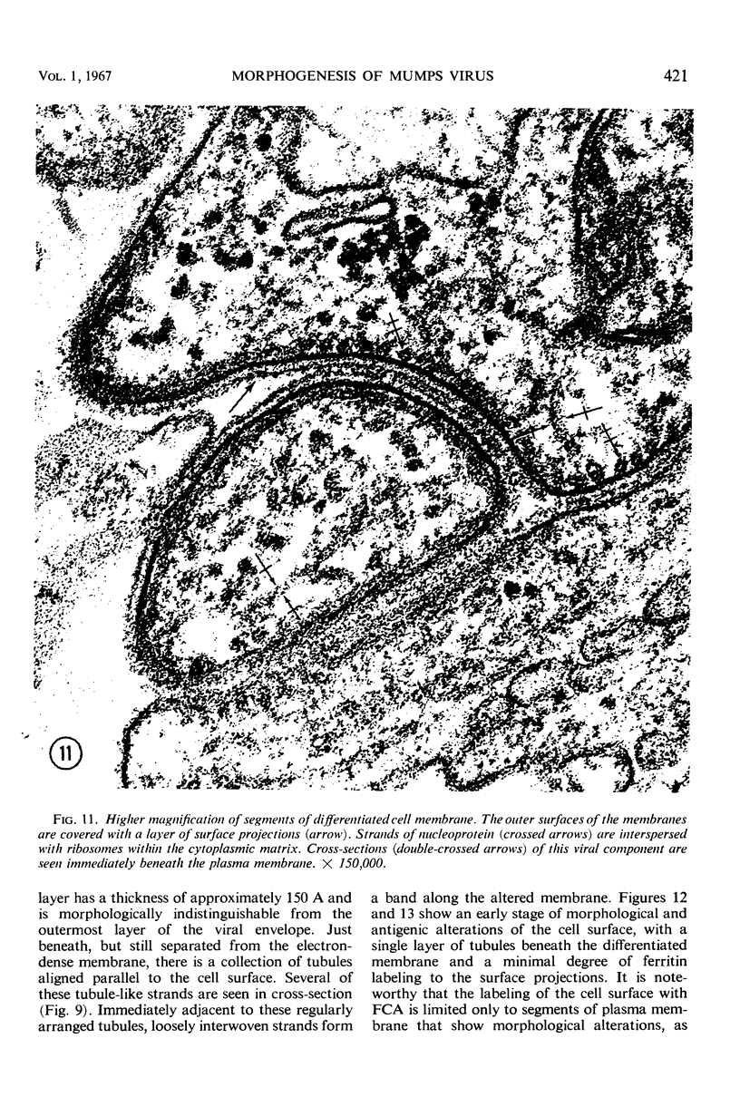
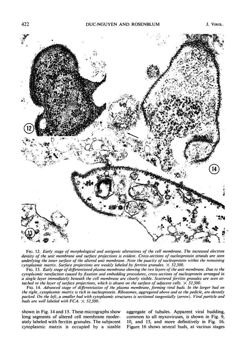
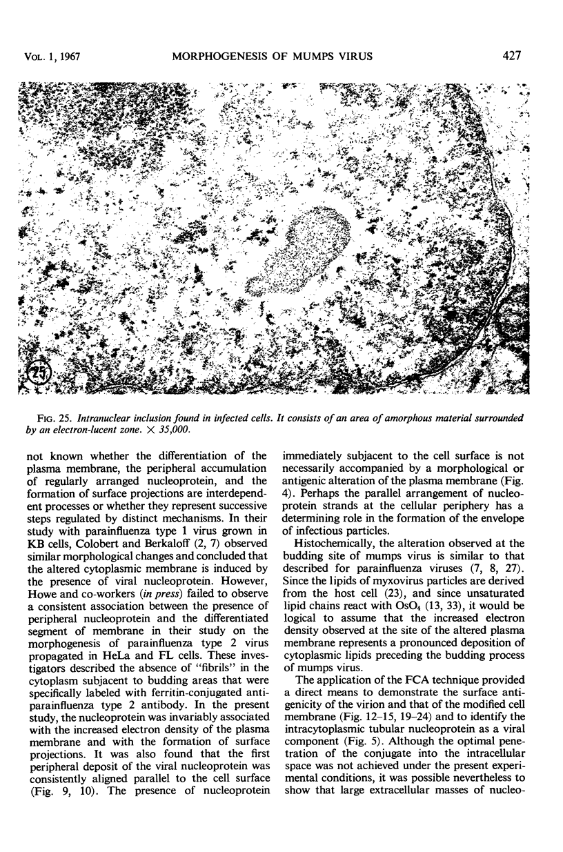
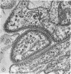
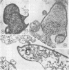
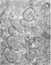
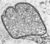
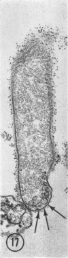
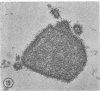
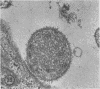
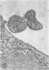
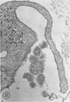
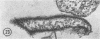
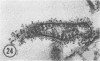
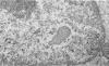
The fine structure of mumps virus-infected chick embryo fibroblastic cells was examined sequentially after viral inoculation. Intracytoplasmic nucleoprotein strands, similar to those described for parainfluenza viruses, were detectable in small aggregates between 36 and 48 hr. The peripheral strands of this viral component lie beneath and along an antigenically altered bulging portion of the cell membrane. The outermost strands are consistently parallel to the differentiated segment of the plasma membrane, which is invariably associated with surface projections. As has been found with other myxoviruses, mumps virus replicates by budding from the cell surface. The virus particle, roughly spherical in shape, has a size ranging from 1,000 to 8,000 A. Filamentous forms are rarely observed in the present culture system. Ferritin-conjugated antibody specifically labels the cytoplasmic nucleoprotein, the modified cell membrane, and the virus particle. Intranuclear inclusions of low electron density and morphologically different from those described in measles virus-infected HeLa and amnion cells were observed in the nucleus of several infected cells. Immuno-electron microscopic observations suggest that the nucleoprotein synthesis rate exceeds that of cell membrane differentiation into viral envelope. This difference results in the accumulation of viral nucleoprotein in large intracytoplasmic masses which can be demonstrated by electron microscopy.
Full text
PDF
Images in this article

Selected References
These references are in PubMed. This may not be the complete list of references from this article.
- BAKER R. F., GORDON I., RAPP F. Electron-dense crystallites in nuclei of human amnion cells infected with measles virus. Nature. 1960 Mar 12;185:790–791. doi: 10.1038/185790a0. [DOI] [PubMed] [Google Scholar]
- BRANDT C. D. Inclusion body formation with Newcastle disease and mumps viruses in cultures of chick embryo cells. Virology. 1958 Apr;5(2):177–191. doi: 10.1016/0042-6822(58)90017-5. [DOI] [PubMed] [Google Scholar]
- CASPAR D. L., DULBECCO R., KLUG A., LWOFF A., STOKER M. G., TOURNIER P., WILDY P. Proposals. Cold Spring Harb Symp Quant Biol. 1962;27:49–50. doi: 10.1101/sqb.1962.027.001.007. [DOI] [PubMed] [Google Scholar]
- COLOBERT L., BERKALOFF A. LIB'ERATION DU VIRUS SENDAUI PAR DES CELLULES PORTEUSES D'UNE INFECTION CHRONIQUE. Ann Inst Pasteur (Paris) 1964 Apr;106:581–587. [PubMed] [Google Scholar]
- Compans R. W., Holmes K. V., Dales S., Choppin P. W. An electron microscopic study of moderate and virulent virus-cell interactions of the parainfluenza virus SV5. Virology. 1966 Nov;30(3):411–426. doi: 10.1016/0042-6822(66)90119-x. [DOI] [PubMed] [Google Scholar]
- Duc-Nguyen H., Henle W. Replication of mumps virus in human leukocyte cultures. J Bacteriol. 1966 Jul;92(1):258–265. doi: 10.1128/jb.92.1.258-265.1966. [DOI] [PMC free article] [PubMed] [Google Scholar]
- Dulbecco R. Production of Plaques in Monolayer Tissue Cultures by Single Particles of an Animal Virus. Proc Natl Acad Sci U S A. 1952 Aug;38(8):747–752. doi: 10.1073/pnas.38.8.747. [DOI] [PMC free article] [PubMed] [Google Scholar]
- FINEAN J. B. Electron microscope and x-ray diffraction studies of a saturated synthetic phospholipide. J Biophys Biochem Cytol. 1959 Aug;6(1):123–124. doi: 10.1083/jcb.6.1.123. [DOI] [PMC free article] [PubMed] [Google Scholar]
- GRESSER I., ENDERS J. F. Cytopathogenicity of mumps virus in cultures of chick embryo and human amnion cells. Proc Soc Exp Biol Med. 1961 Aug-Sep;107:804–807. doi: 10.3181/00379727-107-26761. [DOI] [PubMed] [Google Scholar]
- HENLE G., DEINHARDT F., GIRARDI A. Cytolytic effects of mumps virus in tissue cultures of epithelial cells. Proc Soc Exp Biol Med. 1954 Nov;87(2):386–393. doi: 10.3181/00379727-87-21390. [DOI] [PubMed] [Google Scholar]
- HENLE G., HENLE W., BURGOON J. S., BASHE W. J., Jr, STOKES J., Jr Studies on the prevention of mumps. I. The determination of susceptibility. J Immunol. 1951 May;66(5):535–549. [PubMed] [Google Scholar]
- HORNE R. W., WATERSON A. P., WILDY P., FARNHAM A. E. The structure and composition of the myxoviruses. I. Electron microscope studies of the structure of myxovirus particles by negative staining techniques. Virology. 1960 May;11:79–98. doi: 10.1016/0042-6822(60)90056-8. [DOI] [PubMed] [Google Scholar]
- Holmes K. V., Choppin P. W. On the role of the response of the cell membrane in determining virus virulence. Contrasting effects of the parainfluenza virus SV5 in two cell types. J Exp Med. 1966 Sep 1;124(3):501–520. doi: 10.1084/jem.124.3.501. [DOI] [PMC free article] [PubMed] [Google Scholar]
- Huu Duc-Nguyen, Rosenblum E. N., Zeigel R. F. Persistent infection of a rat kidney cell line with Rauscher murine leukemia virus. J Bacteriol. 1966 Oct;92(4):1133–1140. doi: 10.1128/jb.92.4.1133-1140.1966. [DOI] [PMC free article] [PubMed] [Google Scholar]
- KALLMAN F., ADAMS J. M., WILLIAMS R. C., IMAGAWA D. T. Fine structure of cellular inclusions in measles virus infections. J Biophys Biochem Cytol. 1959 Dec;6:379–382. doi: 10.1083/jcb.6.3.379. [DOI] [PMC free article] [PubMed] [Google Scholar]
- KATES M., ALLISON A. C., TYRELL D. A., JAMES A. T. Origin of lipids in influenza virus. Cold Spring Harb Symp Quant Biol. 1962;27:293–301. doi: 10.1101/sqb.1962.027.001.027. [DOI] [PubMed] [Google Scholar]
- Levens J. H., Enders J. F. THE HEMOAGGLUTINATIVE PROPERTIES OF AMNIOTIC FLUID FROM EMBRYONATED EGGS INFECTED WITH MUMPS VIRUS. Science. 1945 Aug 3;102(2640):117–120. doi: 10.1126/science.102.2640.117. [DOI] [PubMed] [Google Scholar]
- MAASS G., MANNWEILER K. [Cytological and biological studies on the behavior of mumps virus in monkey kidney epithelium cultures]. Arch Gesamte Virusforsch. 1960;10:195–207. [PubMed] [Google Scholar]
- NASTUK W. L., PLESCIA O. J., OSSERMAN K. E. Changes in serum complement activity in patients with myasthenia gravis. Proc Soc Exp Biol Med. 1960 Oct;105:177–184. doi: 10.3181/00379727-105-26050. [DOI] [PubMed] [Google Scholar]
- Prose P. H., Balk S. D., Liebhaber H., Krugman S. Studies of a myxovirus recovered from patients with infectious hepatitis. II. Fine structure and electron microscopic demonstration of intracytoplasmic internal component and viral filament formation. J Exp Med. 1965 Dec 1;122(6):1151–1160. doi: 10.1084/jem.122.6.1151. [DOI] [PMC free article] [PubMed] [Google Scholar]
- RAY B. G., SWAIN R. H. An investigation of the mumps virus by electron microscopy. J Pathol Bacteriol. 1954 Jan;67(1):247–252. doi: 10.1002/path.1700670130. [DOI] [PubMed] [Google Scholar]
- RIFKIND R. A., HSU K. C., MORGAN C. IMMUNOCHEMICAL STAINING FOR ELECTRON MICROSCOPY. J Histochem Cytochem. 1964 Feb;12:131–136. doi: 10.1177/12.2.131. [DOI] [PubMed] [Google Scholar]
- RUSSELL P. K., MORGAN H. R. Mumps viral cytolysin. I. Action on human epithelial cells in tissue culture. J Infect Dis. 1959 Jan-Feb;104(1):38–40. doi: 10.1093/infdis/104.1.38. [DOI] [PubMed] [Google Scholar]
- SINGER S. J. Preparation of an electron-dense antibody conjugate. Nature. 1959 May 30;183(4674):1523–1524. doi: 10.1038/1831523a0. [DOI] [PubMed] [Google Scholar]
- STOECKENIUS W. An electron microscope study of myelin figures. J Biophys Biochem Cytol. 1959 May 25;5(3):491–500. doi: 10.1083/jcb.5.3.491. [DOI] [PMC free article] [PubMed] [Google Scholar]
- TAWARA J. T., GOODMAN J. R., IMAGAWA DT ADAMS J. M. Fine structure of cellular inclusions in experimental measles. Virology. 1961 Aug;14:410–416. doi: 10.1016/0042-6822(61)90332-4. [DOI] [PubMed] [Google Scholar]
- TAWARA J. FINE STRUCTURE OF FILAMENTS IN DOG KIDNEY CELL CULTURES INFECTED WITH MEASLES VIRUS. Virology. 1965 Feb;25:322–322. doi: 10.1016/0042-6822(65)90209-6. [DOI] [PubMed] [Google Scholar]
- UTZ J. P., KASEL J. A., CRAMBLETT H. G., SZWED C. F., PARROTT R. H. Clinical and laboratory studies of mumps. I. Laboratory diagnosis by tissue-culture technics. N Engl J Med. 1957 Sep 12;257(11):497–502. doi: 10.1056/NEJM195709122571103. [DOI] [PubMed] [Google Scholar]
- WALKER D. L., HINZE H. C. A carrier state of mumps virus in human conjunctiva cells. I. General characteristics. J Exp Med. 1962 Nov 1;116:739–750. doi: 10.1084/jem.116.5.739. [DOI] [PMC free article] [PubMed] [Google Scholar]