Abstract
Plexiform neurofibromatosis is a relatively common but potentially devastating manifestation of neurofibromatosis type 1 (NF1). It produces very hideous deformity if the face is involved. Surgical management remains the mainstay of therapy, but in the head and neck region it is limited by the infiltrating nature of these tumors, inherent operative morbidity and high rate of regrowth. We present two cases of facial neurofibromatosis managed in our hospital. The first patient presented with overhanging mass of skin folds on the right side of her face, completely obliterating her right eye. The other patient was a young male having a huge, unsightly swelling over the right cheek, resulting in pulled down right eyelids and right pinna. Physical examination revealed the presence of café au lait macules, freckling in the axillary region and multiple neurofibromas over the trunk. Reconstructive surgical procedure in the form of subtotal excision of tumor mass followed by re draping of the facial skin was performed in both cases. There was evidence of regrowth of the tumor on review after 6 months.
Keywords: Facial plexiform neurofibromatosis, regrowth, subtotal excision
INTRODUCTION
Plexiform neurofibromas (PNF) are benign tumors that originate from nerve sheath cells or subcutaneous peripheral nerves, and can involve multiple fascicles.[1] Massive plexiform neurofibromatosis results in functional disability and disfigurement, and usually occur in as much as 30% of the patients with neurofibromatosis type I.[2] Such tumors are generally present at birth, and often progress slowly during early childhood. The condition can cause disfigurement by pulling down of important structures. Because of the large size of the tumors and involvement of multiple fascicles of nerves, the risk of neurological and functional destruction upon tumor resection is high. Surgical interventions are thus commonly postponed to as long as possible.[3]
Most cases require repeat surgery as complete excision is generally not possible due to the infiltrating nature of these tumors.[4] Such neurofibromas infiltrate multiple tissue planes and are thus much more difficult or impossible to resect.[3]
We present two such cases managed in our hospital to share our experience and the lessons learnt.
CASE REPORTS
Case 1
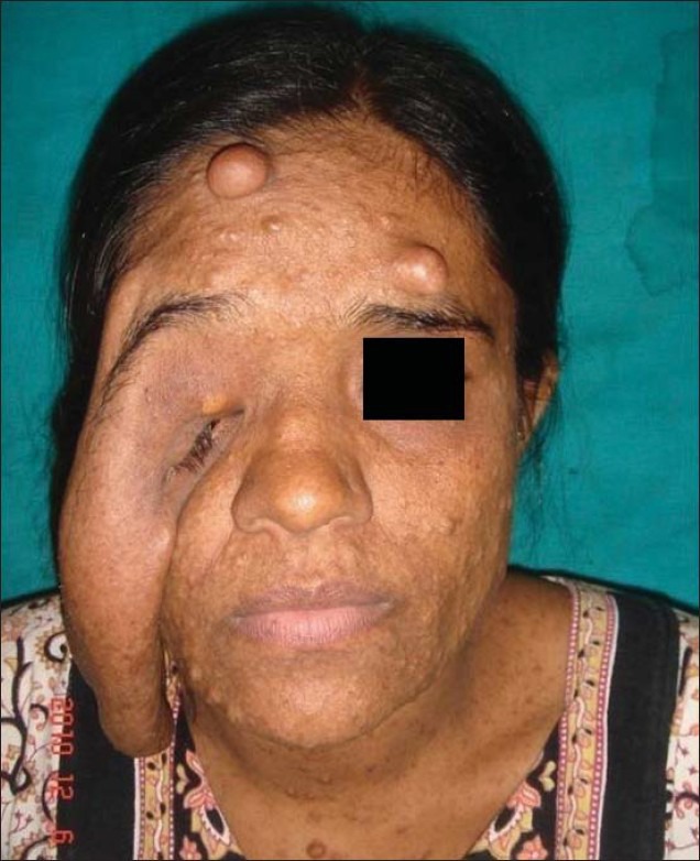
A 29-year-old female patient presented to us with a history of overhanging folds of loose skin affecting the right side of her face and right forearm [Figure 1]. The swelling appeared during childhood and progressed slowly. She complained of occasional pain and itching on the affected part. The patient initially sought treatment elsewhere, about 10 years back, when excision of forearm swelling was carried out, but the swelling recurred. She came to us now with complaints of inability to open her right eye and sought improvement in her facial appearance.
Figure 1.

Pre-operative picture showing facial plexiform neurofibromatosis
On physical examination, the face was disfigured due to overhanging folds of skin affecting the temporal, orbital and cheek areas. She was unable to open her right eye due to the overhanging folds affecting both eyelids, and the eye was pulled inferiorly. Vision acuity of the right eye was however not affected. Café au lait macules (some measuring over 15 mm) as well as several freckles were observed in the axillary, back and chest regions. She had multiple neurofibromas over her forehead, neck and trunk regions. She also had a similar swelling and an old healed scar over the right forearm.
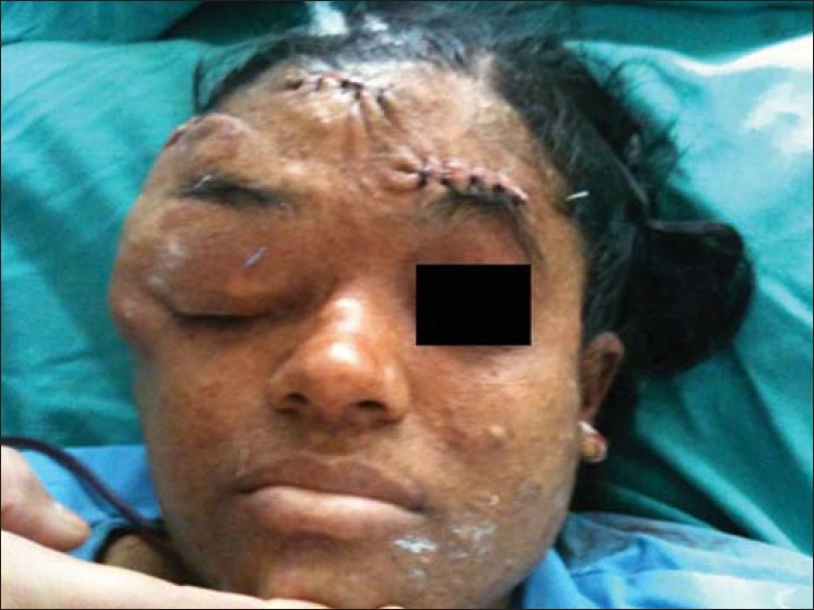
Reconstructive surgical procedure in the form of subtotal excision of tumor and re-draping of the facial skin was performed. Operative findings revealed grossly thickened subcutaneous nerves and neo-vessels in the tumor mass, which was infiltrating deeply, and no clear tissue planes were identifiable. The neo-vessels were very friable and bled profusely during dissection. Complete excision was not possible; hence, subtotal excision and redraping of skin flap was carried out. Anchoring sutures were applied over the lateral canthal region between the skin flap and the lateral orbital margin to prevent pulling down of eyelids [Figure 2]. The post-operative period was uneventful. However, on review after 6 months, there was partial regrowth of the swelling.
Figure 2.

Immediate post operative showing reasonable correction
Case 2
This 18-year-old boy was brought to our out patient department by his parents with a history of progressively increasing swelling over the right side of his face since childhood.
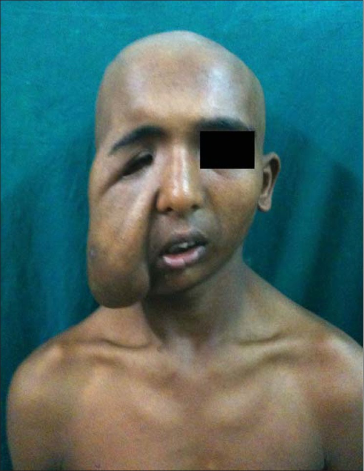
Physical examination revealed a huge soft tissue swelling involving the right mandibular region and cheek, extending up to the nasolabial fold pulling down the right angle of the mouth and right eye, partially obstructing the field of vision in his right eye [Figure 3]. He had few neurofibromas over his trunk, axillary freckles and café au lait macules over his back. Magnetic resonance imaging (MRI) of the face excluded any intracranial extension.
Figure 3.

Facial plexiform neurofibromatosis in a male patient
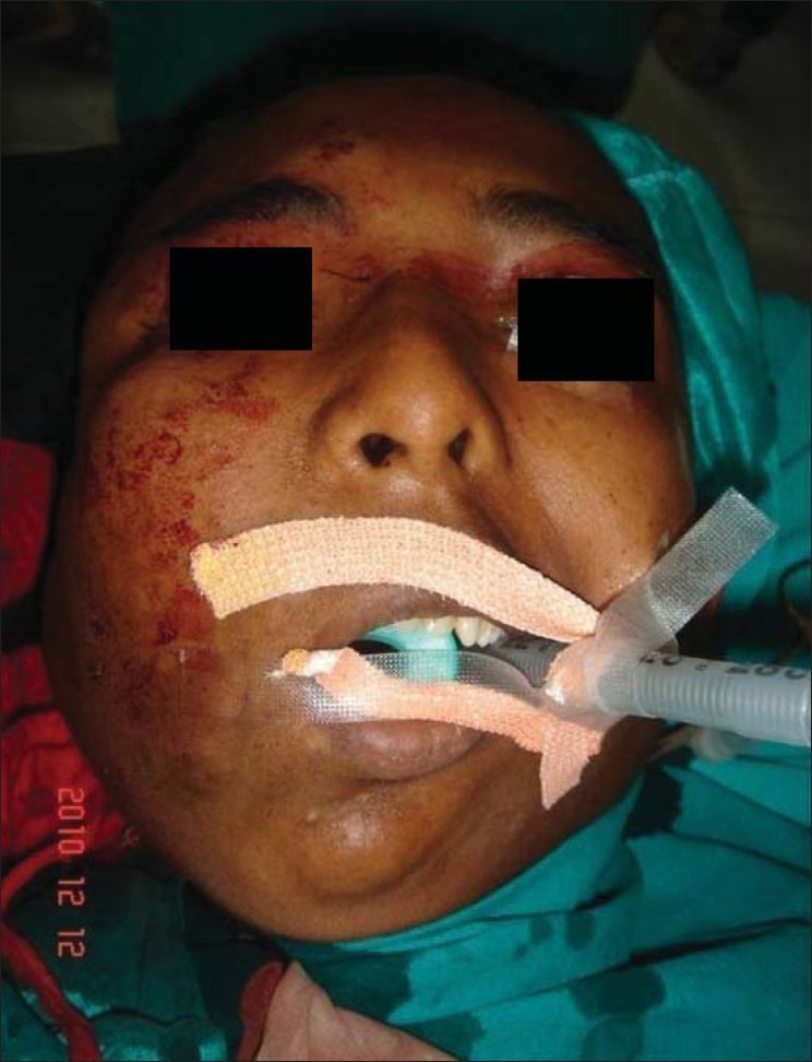
He was taken up for reconstructive surgery and the swelling was approached through pre-auricular incision extending superiorly into the temporal area. Tumescent fluid infiltration was carried out in the tumor mass to reduce the blood loss. The tumor mass was infiltrating deeply and the tissue planes could not be defined. There was gross thickening of facial nerve in the pre-auricular region as well as its branches. Subtotal excision of tumor mass and readjustment of facial flap was carried out with reasonable post-op correction [Figure 4]. However, 6-monthly review in this case as well showed evidence of some regrowth.
Figure 4.

Immediate post operative result
DISCUSSION
PNF is a rare type of generalised neurofibromatosis, which occurs due to overgrowth of neural tissue in the subcutaneous tissue. The diffuse and soft nature of PNF is often compared with “a bag of worms.”[5]
The lesions can occur anywhere along a nerve, and may appear on the face,[5] orbit and globe,[6] and frequently involve the cranial and upper cervical nerves.[7] The condition can be quite disfiguring, as observed in the cases being presented, and hemifacial hypertrophy can occur.[8] Complications include bleeding from trauma, neurological deficits and psychological disturbance because of abnormal anatomy.[5]
Histologically, these are peripheral nerve sheath tumors containing all elements of the peripheral nerve, and are characterized by an increase in endo-neural matrix with separation of nerve fascicles and proliferation of Schwann cells.[9] PNF can turn malignant in 4-5% of cases.[8]
Needle et al,[4] analyzed the largest series of surgically managed PNFs and demonstrated that 54% recurred within a 10-year period, with the greatest risk of recurrence found in lesions involving the head and neck region. We encountered recurrence in both the cases being presented here.
Jeffrey et al,[10] in their series of 39 cases had 10 patients with trigeminal nerve involvement who underwent a total of 11 surgical procedures.
Friedrich et al[3] studied nine cases of PNF having no neurological or functional deficit, and were able to carry out total resection of the tumors and have advocated total resection in small PNF as a preventive strategy for later disfigurement and functional deficits.
Resection and debulking of invasive PNF is however associated with a high rate of recurrence. In one pediatric series, complete resections developed recurrence in 20% and incomplete resections had a recurrence in up to 45% of the cases.[4] One of the limiting factors is vascularity of these lesions and their abnormal propensity to bleed. These tumors bleed profusely during surgery because of the friable nature of the neo-vessels. Adequate blood should be arranged before taking up these cases for resection.
Mukherji[11] compared them with angiomas, and also commented on the friability of the vessels. We also noticed friability of vessels and excessive neo-vascularisation in our cases, and used tumescent fluid infiltration in the second case, which was found to be quite effective in minimising the bleeding.
Surgical management remains the mainstay of treatment for these tumors, but functional disturbances are almost inevitable while resecting tumors involving the head and neck region.[1] No chemotherapeutic agent has yet been identified that reduces the size of these tumors.[10]
CONCLUSION
Plexiform neurofibromatosis remains a devastating manifestation of Von-Recklinghausen's neurofibromatosis. Extensive surgical procedures have to be weighed against functional disturbances that are almost inevitable while resecting invasive neurofibromas in the head and neck region.[12] Diagnosis is not difficult; however, MRI evaluation of tumors involving the head and neck region can help in determining the local infiltration and precise anatomy. We advocate MRI evaluation pre-operatively and during review on follow-up.
We found the tumescent technique to be quite effective in reducing blood loss, helping in easier dissection. We recommend that adequate blood should be arranged before undertaking surgical excision of facial PNF; the tumescent technique should be employed in all cases and, after excising the overhanging folds, skin flap should be re-draped and few anchoring sutures between the inner surface of the flap and the underlying periosteum should be placed to obviate the pulling down of the soft tissues and vital structures post operatively. We found that extensive surgical resections in the head and neck region are risky due to the infiltrating nature of these tumors, which can lead to functional disability and a high rate of regrowth. Hence, surgery should be undertaken only after giving due consideration to the possible psychological benefits by making these patients socially more acceptable in the society. Periodic clinical examination and MRI evaluation are required for about 2 years for timely detection and repeat surgery to achieve further correction.
Footnotes
Source of Support: Nil
Conflict of Interest: Nil
REFERENCES
- 1.Kleinhues P, Cavenee WK. Lyon: IARC Press Lyon; 2000. Tumours of the nervous system. In World Health Classification of Tumours. [Google Scholar]
- 2.Huson SM, Hughes RA. London: Chapman and Hall Medical; 1994. The Neurofibromatosis: A Pathogenetic and Clinical Overview. [Google Scholar]
- 3.Friedrich RE, Schmelzle R, Hartmann M, Fünsterer C, Mautner VF. Resection of small plexiform neurofibromas in neurofibromatosis type 1 children. World J Surg Oncol. 2005;3:3–6. doi: 10.1186/1477-7819-3-6. [DOI] [PMC free article] [PubMed] [Google Scholar]
- 4.Needle MN, Cnaan A, Dattilo J, Chatten J, Phillips PC, Shochat S, et al. Prognostic signs in the surgical management of plexiform neurofibroma: The Children's Hospital of Philadelphia experience, 1974-1994. J Pediatr. 1997;131:678–82. doi: 10.1016/s0022-3476(97)70092-1. [DOI] [PubMed] [Google Scholar]
- 5.Patil K, Mahima VG, Shetty SK, Lahari K. Facial plexiform neurofibroma in a child with neurofibromatosis type I: A case report. J Indian Soc Pedod Prev Dent. 2007;25:30–5. doi: 10.4103/0970-4388.31987. [DOI] [PubMed] [Google Scholar]
- 6.Arute JE, Nwosu Paul JC, ErahPatrick O. Facial Plexiform Neurofibromatosis in a 16- year Old Female from Eastern Nigeria: A Rare Presentation. Int J Health Res. 2008;1:105–10. [Google Scholar]
- 7.Ferrozzi F, Zuccoli G, Bacchini E, Piazza P, Sigorini M, Virdis R. Extracerebral neoplastic manifestations in neurofibromatosis 1: Integrated diagnostic imaging. Radiol Med (Torino) 1998;96:562–9. [PubMed] [Google Scholar]
- 8.Ducatman BS, Scheithauer BW, Piepgras DG, Reiman HM, Istrup DM. Malignant peripheral nerve sheath tumors. Cancer. 1986;57:2006–21. doi: 10.1002/1097-0142(19860515)57:10<2006::aid-cncr2820571022>3.0.co;2-6. [DOI] [PubMed] [Google Scholar]
- 9.Wenig BM. Philadelphia, Pa: WB Saunders Co; 1993. Atlas of Head and Neck Pathology; pp. 156–9. [Google Scholar]
- 10.Wise JB, Cryer JE, Belasco JB, Jacobs I, Elden L. Management of Head and Neck Plexiform Neurofibromas in Pediatric Patients With Neurofibromatosis Type 1. Arch Otolaryngol Head Neck Surg. 2005;131:712–8. doi: 10.1001/archotol.131.8.712. [DOI] [PubMed] [Google Scholar]
- 11.Mukherji MM. Giant neurofibroma of the head and neck. Plast Recon Surg. 1974;53:184–9. doi: 10.1097/00006534-197402000-00010. [DOI] [PubMed] [Google Scholar]
- 12.Wise JB, Patel SG, Shah JP. Management issues in massive pediatric facial plexiform neurofibroma with neurofibromatosis type 1. Head Neck. 2002;24:207–11. doi: 10.1002/hed.10001. [DOI] [PubMed] [Google Scholar]


