Abstract
A 26-year-old man (human immunodeficiency virus-positive and not taking highly active antiretroviral treatment [HAART]) presented to the emergency room with 2 months of malaise, 20 kg weight loss, high spiking fevers, generalized lymph nodes, night sweats, dry cough, and chest pain when swallowing. On physical examination, he had multiple cervical lymphadenopathies. Suspecting a systemic opportunistic infection, a contrasted chest computed tomography (CT) was done, showing an esophageal to mediastinum fistulae. Two days after admission, a fluoroscopic contrasted endoscopy was done that showed two esophageal fistulae from scrofula to esophagus and then, to mediastinum. A bronchoalveolar lavage and a cervical lymphadenopathy biopsy were done, both showing multiple acid-fast bacillae, where cultures grew Mycobacterium tuberculosis.
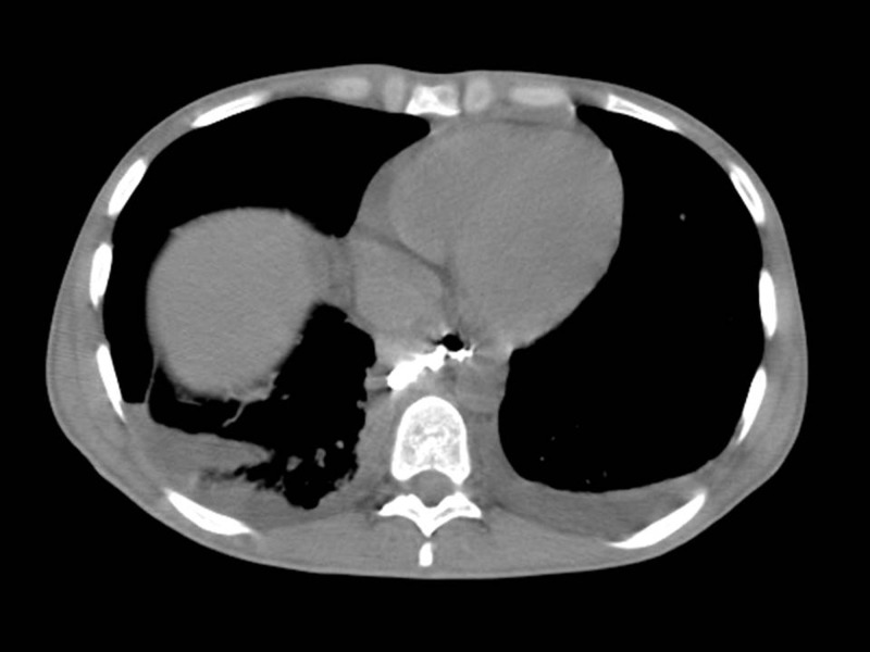
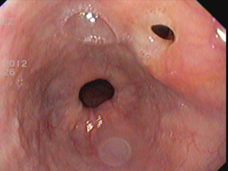
A 26-year-old man from Medellin, Colombia (human immunodeficiency virus [HIV]-positive since February of 2012, last CD4 count = 188 cells/mL, HIV viral load = 6.58 log in May of 2012, highly active antiretroviral treatment [HAART]-naive) presented to the emergency room with 2 months of malaise, 20 kg weight loss, high spiking fevers, generalized lymphadenopathy, night sweats, dry cough, and chest pain when swallowing. On physical examination, he was febrile, his temperature was 39°C, his weight was 48 kg, he had blood pressure of 90/50 mmHg, his pulse was 120 beats/min, he had multiple cervical lymphadenopathies and oral candidiasis, his lungs were clear, a systolic murmur on the apex was heard on cardiac examination, and hepatosplenomegaly was found on abdominal examination. Suspecting a systemic opportunistic infection, a contrasted chest computed tomography (CT) was performed, and it showed an esophageal to mediastinum fistulae (Figure 1). Two days after admission, a fluoroscopic contrasted endoscopy was performed, and it showed two esophageal fistulae from scrofula to esophagus and then, to mediastinum (Figure 2 and Supplemental Video). A bronchoalveolar lavage and a cervical lymphadenopathy biopsy were done, both showing multiple acid-fast bacillae where, subsequently, cultures grew Mycobacterium tuberculosis; then, a gastrostomy was placed, and he was started on four antituberculosis medications. He was discharged 1 month after admission, and at a follow-up visit to the acquired immunodeficiency syndrome (AIDS) outpatient clinic, the patient reported no odynophagia, and a control endoscopy showed no fistulae; then, the gastrostomy was removed, and no sequelae was documented.
Figure 1.
Contrasted chest CT with an esophageal to mediastinum fistulae.
Figure 2.
Endoscopy with esophageal fistulae from esophagus to mediastinum.
Mediastinal fistulae formation secondary to tuberculosis scrofula is an unusual complication, and it is most commonly seen in immunosuppressed or malnourished patients with advanced disease.1,2 The diagnosis is suspected in patients with tuberculosis lymphadenopathy and odynophagia, but invasive diagnostic methods are needed to show it, requiring gastrostomy to feed the patient while the fistulae closes by the antituberculosis treatment.3,4
Supplementary Material
Footnotes
Authors' addresses: Juan Cataño, Infectious Diseases Section, Internal Medicine Department, University of Antioquia Medical School, Fundacion Antioqueña de Infectologia, Medellin, Colombia, E-mail: kataju@hotmail.com. John Cardeño, Clinica Las Vegas, Infectious Diseases, Medellin, Antioquia, Colombia, E-mail: johncardeno@hotmail.com.
References
- 1.Brighenti S, Andersson J. Local immune responses in human tuberculosis: learning from the site of infection. J Infect Dis. 2012;205((Suppl 2)):S316–S324. doi: 10.1093/infdis/jis043. [DOI] [PubMed] [Google Scholar]
- 2.Fontanilla J-M, Barnes A, von Reyn CF. Current diagnosis and management of peripheral tuberculous lymphadenitis. Clin Infect Dis. 2011;53:555–562. doi: 10.1093/cid/cir454. [DOI] [PubMed] [Google Scholar]
- 3.Gupta KB, Manchanda M, Vermas M. Tuberculous oesophagopleural fistula. J Indian Med Assoc. 2011;109:504–505. [PubMed] [Google Scholar]
- 4.Rana SS, Bhasin DK, Singh K. Endoscopic ultrasound features of mediastinal tuberculosis. Endoscopy. 2012;44:106–107. doi: 10.1055/s-0030-1257085. [DOI] [PubMed] [Google Scholar]
Associated Data
This section collects any data citations, data availability statements, or supplementary materials included in this article.


