

Abstract
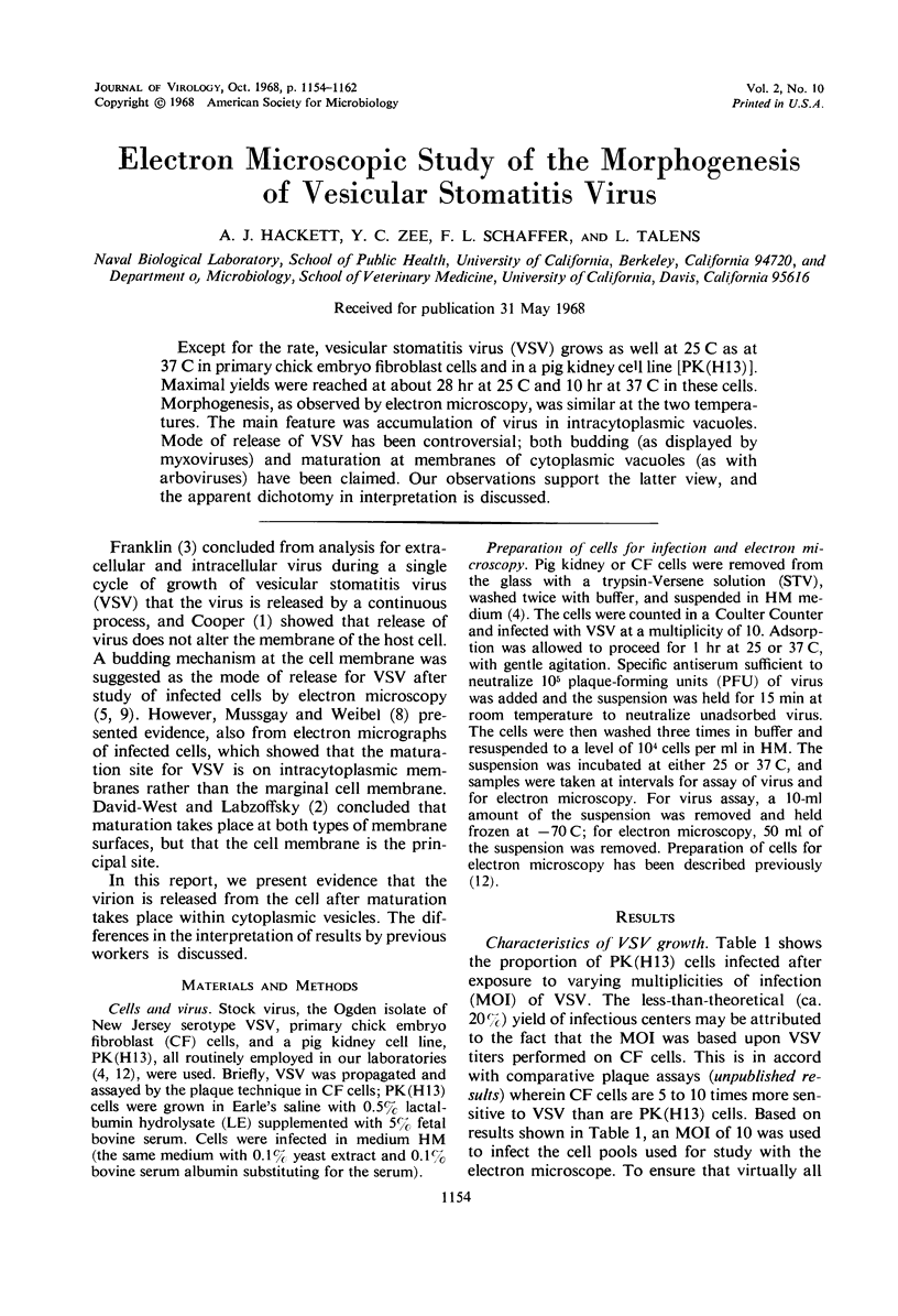
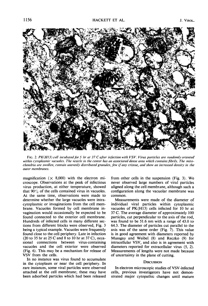
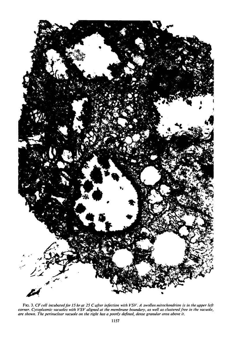
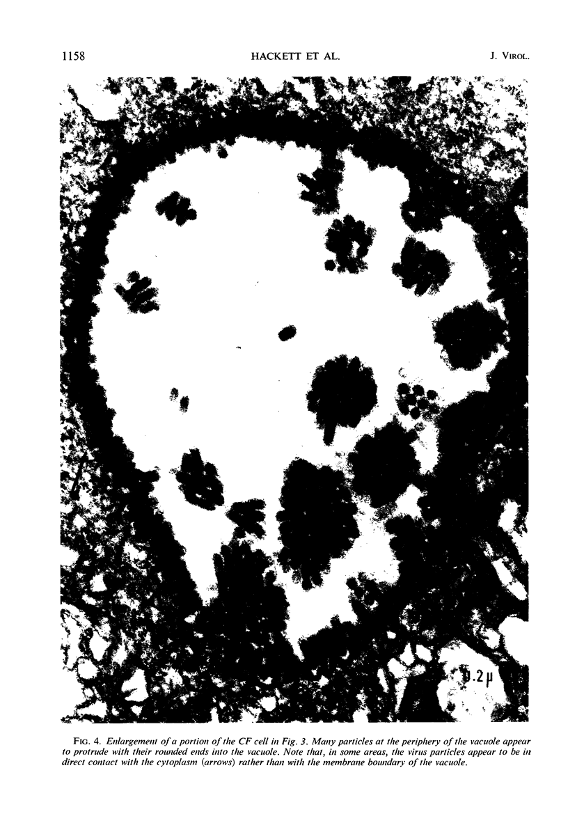
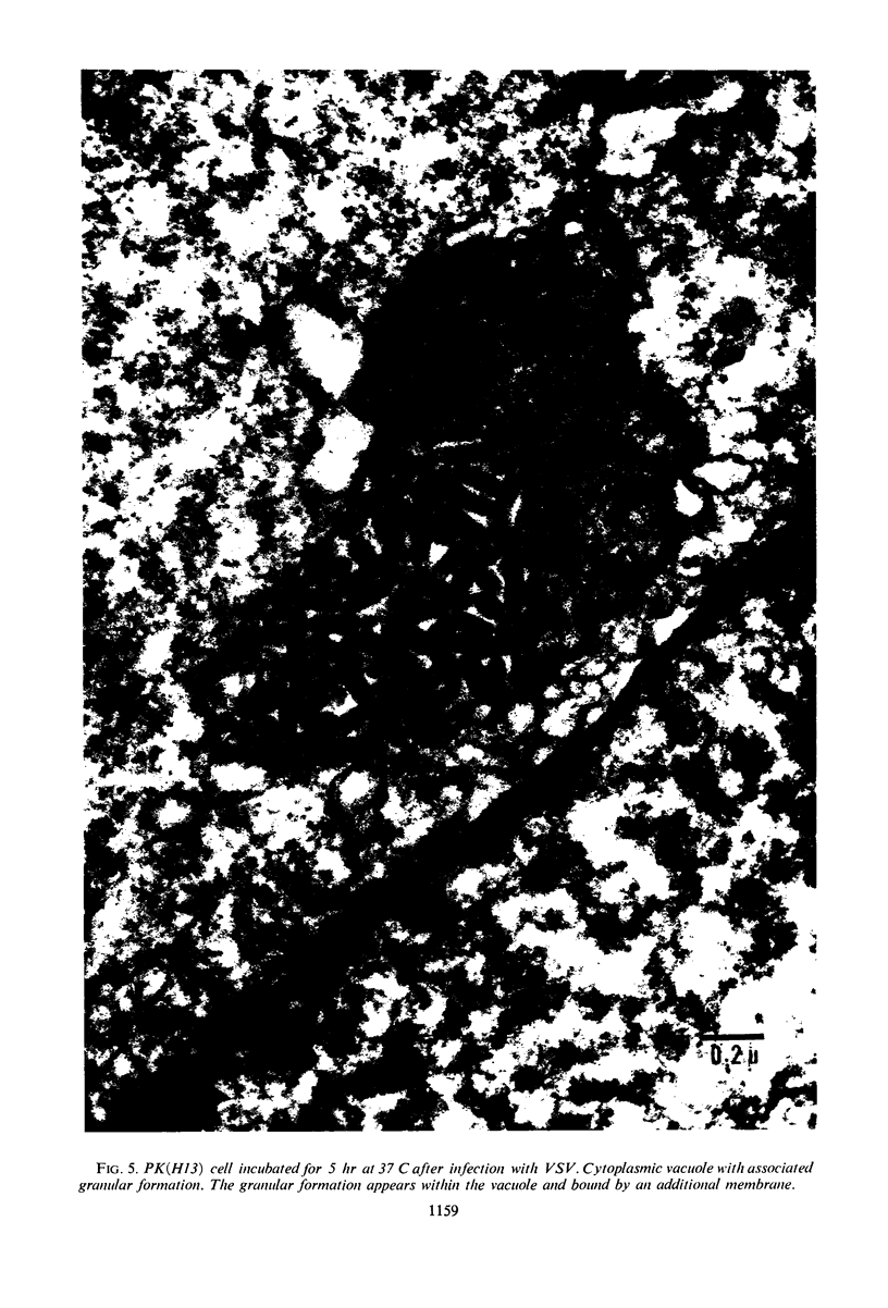
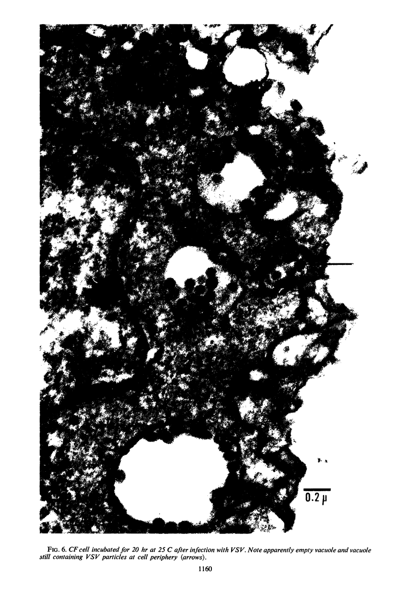
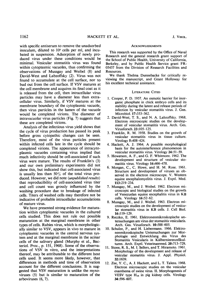
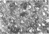
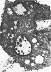
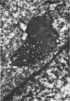
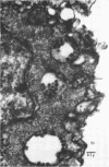
Except for the rate, vesicular stomatitis virus (VSV) grows as well at 25 C as at 37 C in primary chick embryo fibroblast cells and in a pig kidney cell line [PK(H13)]. Maximal yields were reached at about 28 hr at 25 C and 10 hr at 37 C in these cells. Morphogenesis, as observed by electron microscopy, was similar at the two temperatures. The main feature was accumulation of virus in intracytoplasmic vacuoles. Mode of release of VSV has been controversial; both budding (as displayed by myxoviruses) and maturation at membranes of cytoplasmic vacuoles (as with arboviruses) have been claimed. Our observations support the latter view, and the apparent dichotomy in interpretation is discussed.
Full text
PDF
Images in this article
Selected References
These references are in PubMed. This may not be the complete list of references from this article.
- COOPER P. D. An osmotic barrier for inorganic phosphate in chick embryo cells and its stability during the latent and release periods of infection by vesicular stomatitis virus. J Gen Microbiol. 1957 Oct;17(2):353–362. doi: 10.1099/00221287-17-2-353. [DOI] [PubMed] [Google Scholar]
- David-West T. S., Labzoffsky N. A. Electron microscopic studies on the development of vesicular stomatitis virus. Arch Gesamte Virusforsch. 1968;23(1):105–125. doi: 10.1007/BF01242119. [DOI] [PubMed] [Google Scholar]
- FRANKLIN R. M. Studies on the growth of vesicular stomatitis virus in tissue culture. Virology. 1958 Jun;5(3):408–424. doi: 10.1016/0042-6822(58)90036-9. [DOI] [PubMed] [Google Scholar]
- HACKETT A. J. A POSSIBLE MORPHOLOGIC BASIS FOR THE AUTOINTERFERENCE PHENOMENON IN VESICULAR STOMATITIS VIRUS. Virology. 1964 Sep;24:51–59. doi: 10.1016/0042-6822(64)90147-3. [DOI] [PubMed] [Google Scholar]
- HOWATSON A. F., WHITMORE G. F. The development and structure of vesicular stomatitis virus. Virology. 1962 Apr;16:466–478. doi: 10.1016/0042-6822(62)90228-3. [DOI] [PubMed] [Google Scholar]
- MORGAN C., HOWE C., ROSE H. M. Structure and development of viruses as observed in the electron microscope. V. Western equine encephalomyelitis virus. J Exp Med. 1961 Jan 1;113:219–234. doi: 10.1084/jem.113.1.219. [DOI] [PMC free article] [PubMed] [Google Scholar]
- MUSSGAY M., WEIBEL J. Electron microscopic and biological studies on the growth of Venezuelan equine encephalitis virus in KB cells. Virology. 1962 Jan;16:52–62. doi: 10.1016/0042-6822(62)90201-5. [DOI] [PubMed] [Google Scholar]
- MUSSGAY M., WEIBEL J. Electron microscopic studies on the development of vesicular stomatitis virus in KB cells. J Cell Biol. 1963 Jan;16:119–129. doi: 10.1083/jcb.16.1.119. [DOI] [PMC free article] [PubMed] [Google Scholar]
- RECZKO E. [Electron microscopic research on the virus of vesicular stomatitis]. Arch Gesamte Virusforsch. 1961;10:588–605. [PubMed] [Google Scholar]
- Schulze P., Liebermann H. Elektronenmikroskopische Untersuchungen zur Morphologie und Entwicklung des Virus der Stomatitis vesicularis in Kälbernierenzellkulturen. Arch Exp Veterinarmed. 1966 Sep;20(4):713–729. [PubMed] [Google Scholar]
- Zee Y. C., Hackett A. J., Talens L. T. Electron microscopic studies on the vesicular exanthema of swine virus. II. Morphogenesis of VESV type H54 in pig kidney cells. Virology. 1968 Apr;34(4):596–607. doi: 10.1016/0042-6822(68)90081-0. [DOI] [PubMed] [Google Scholar]