

Abstract
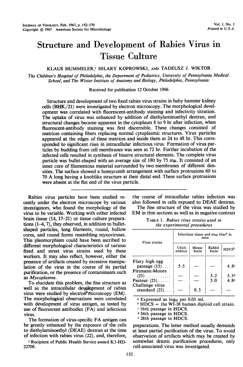
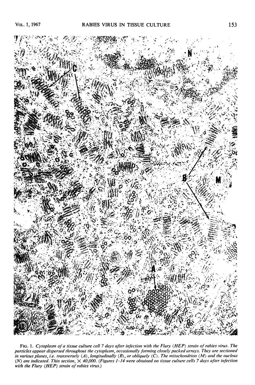
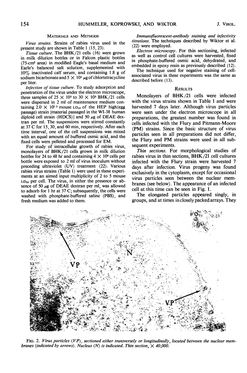
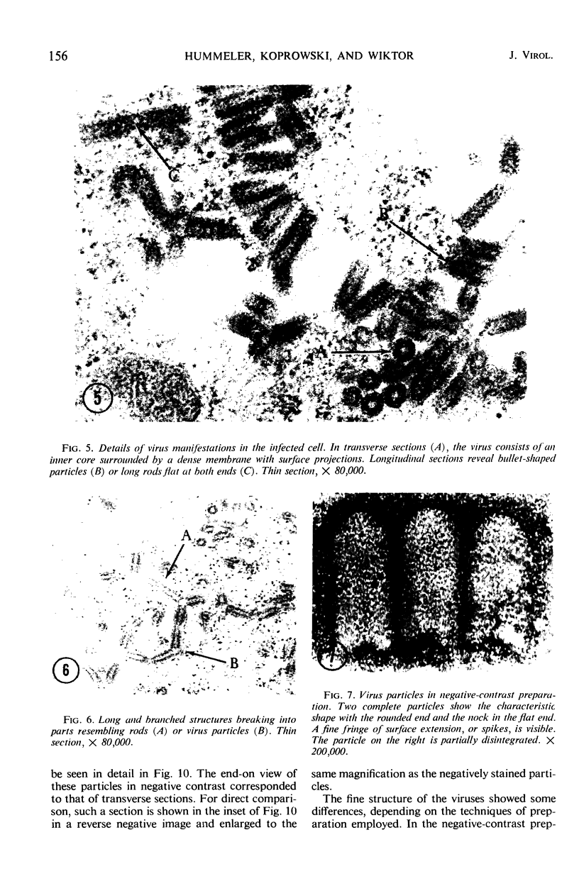
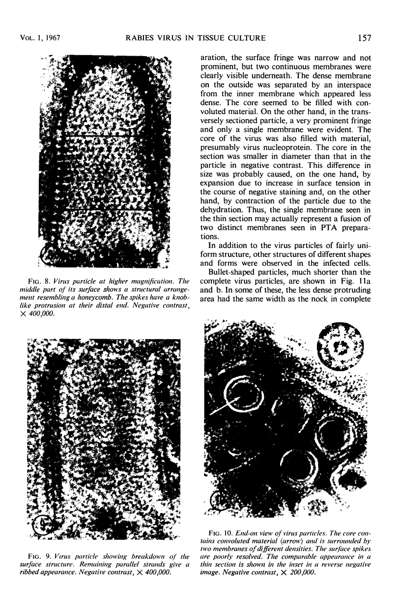
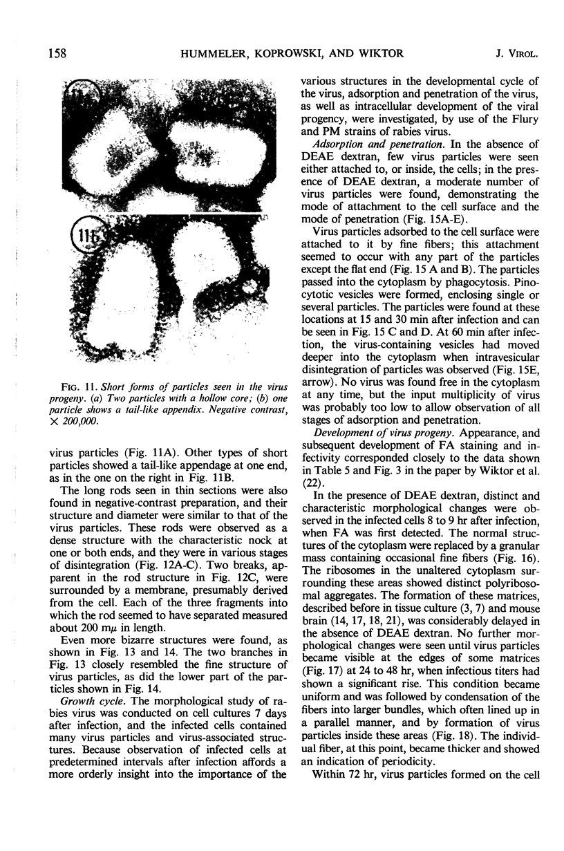
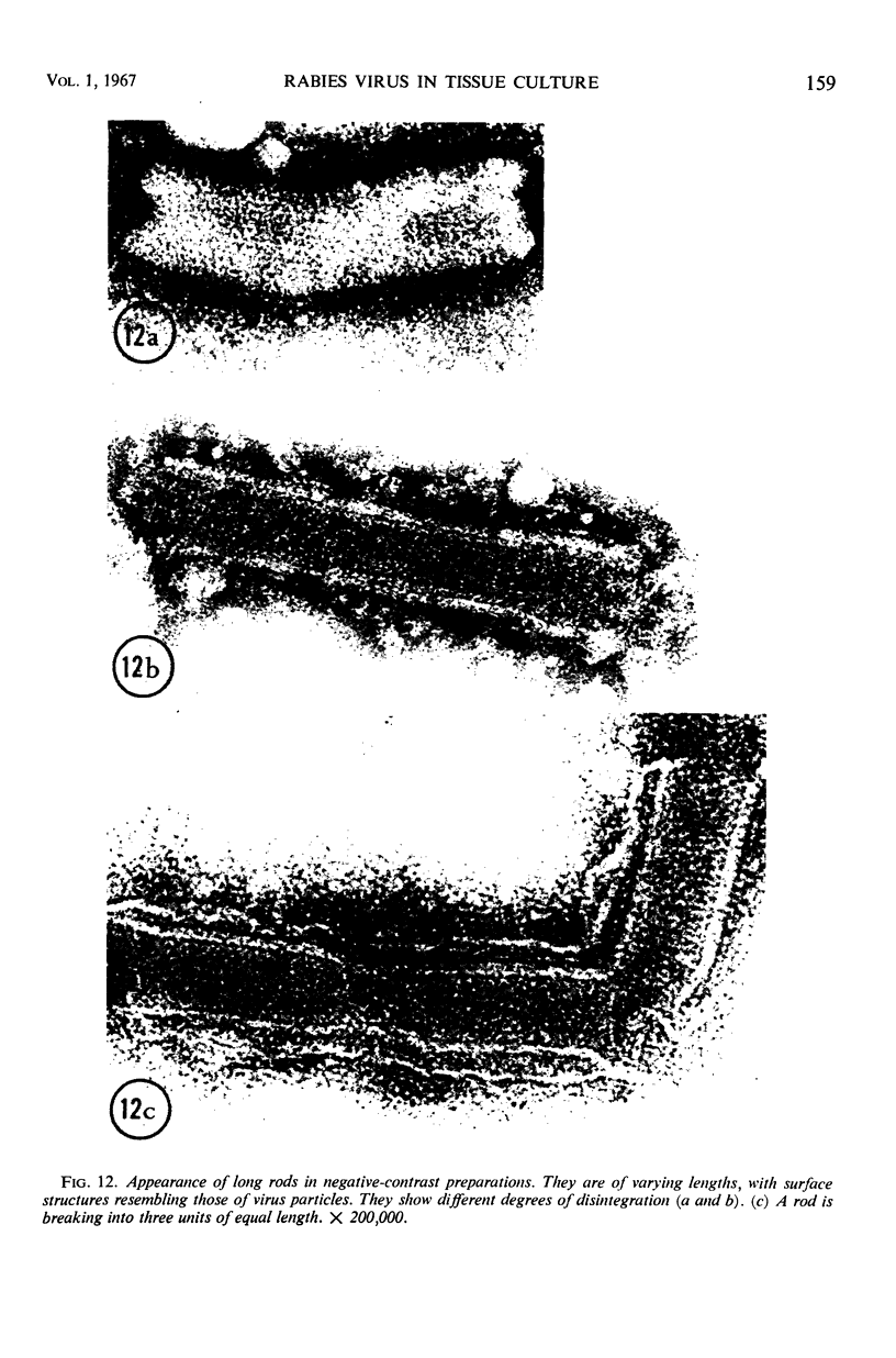
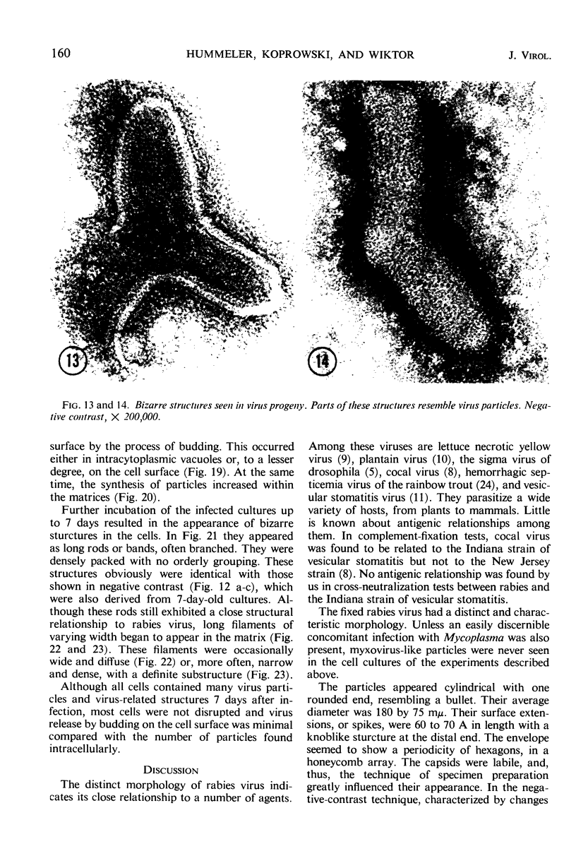
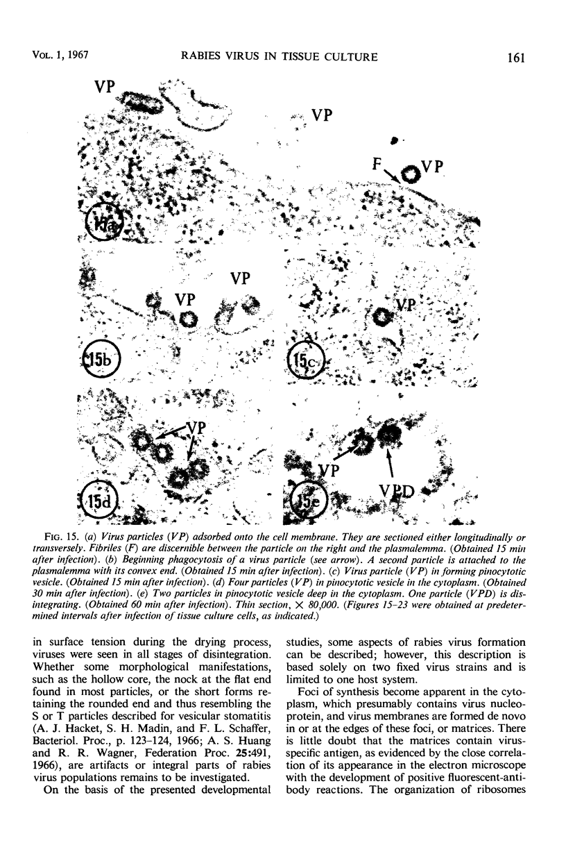
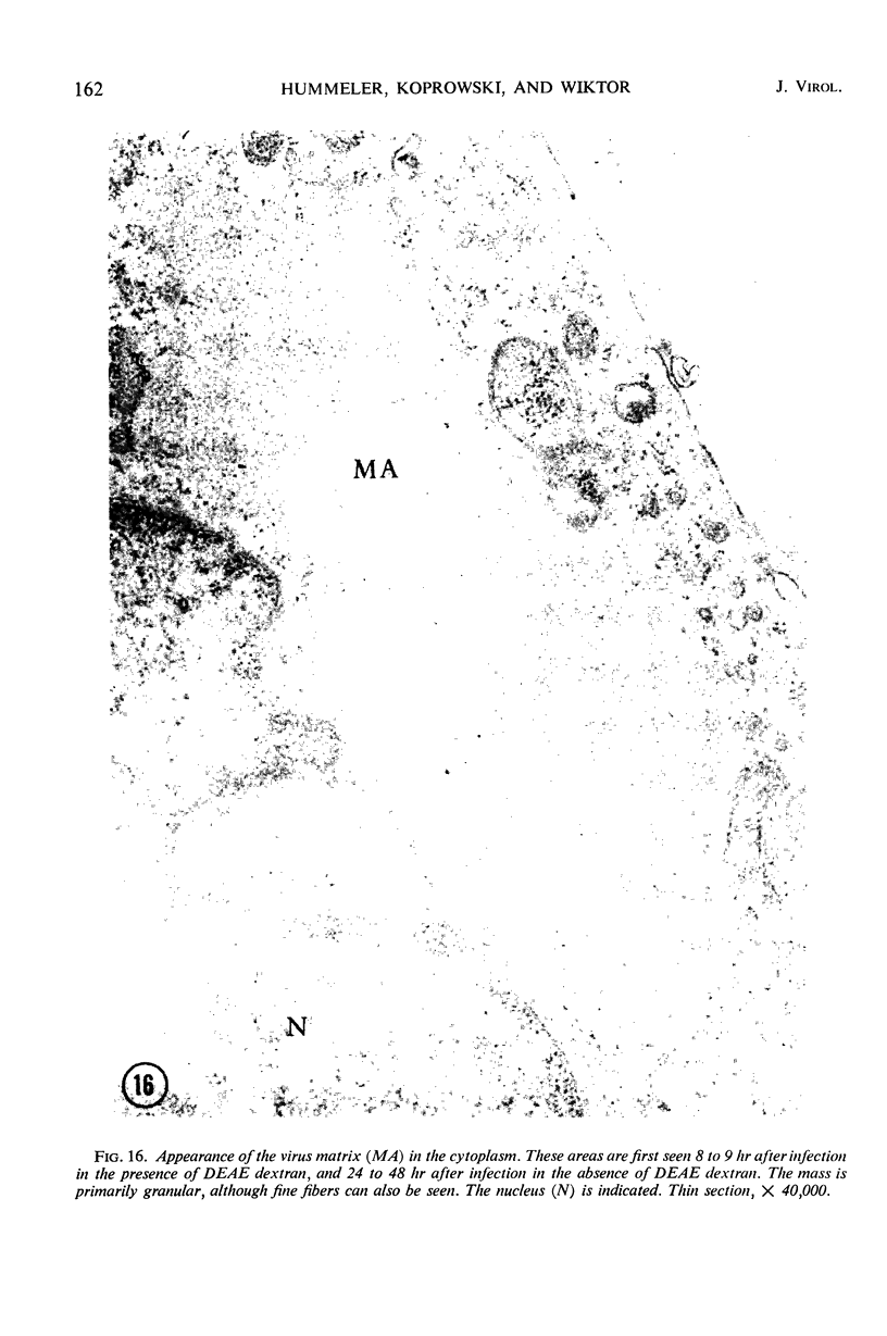
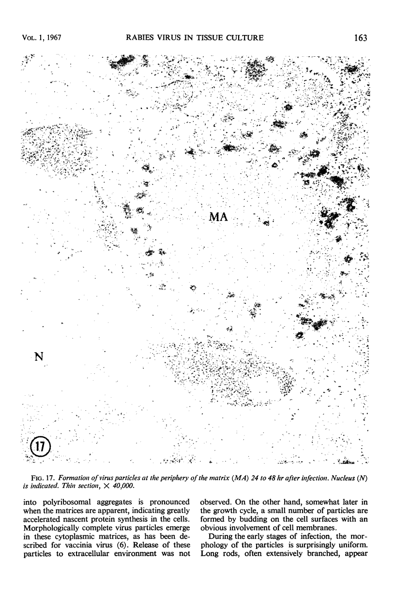
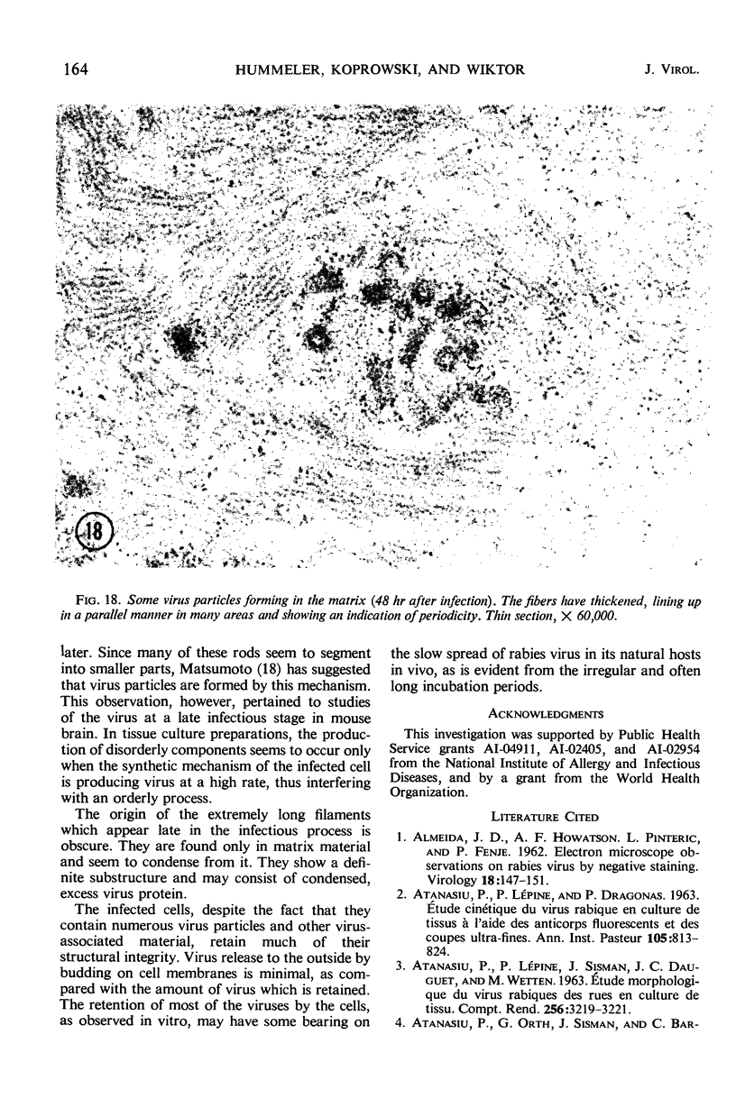
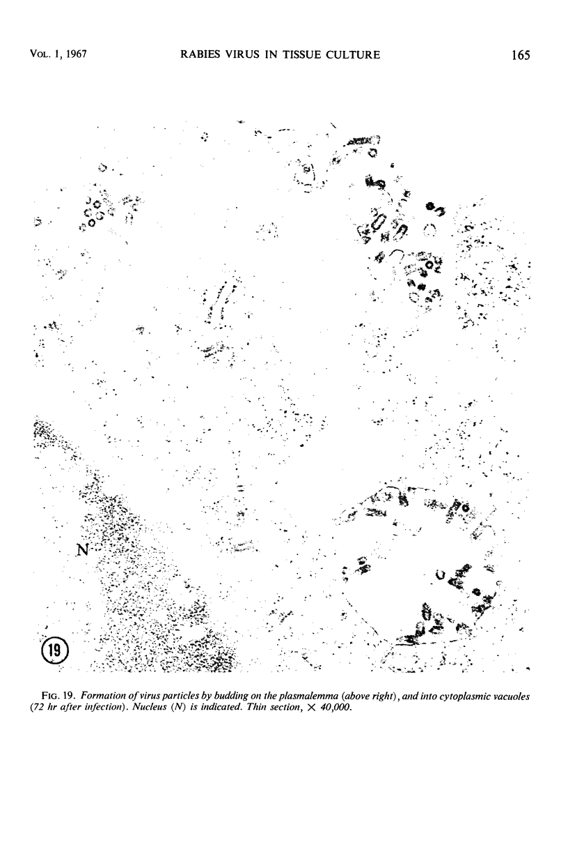
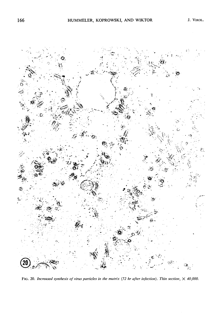
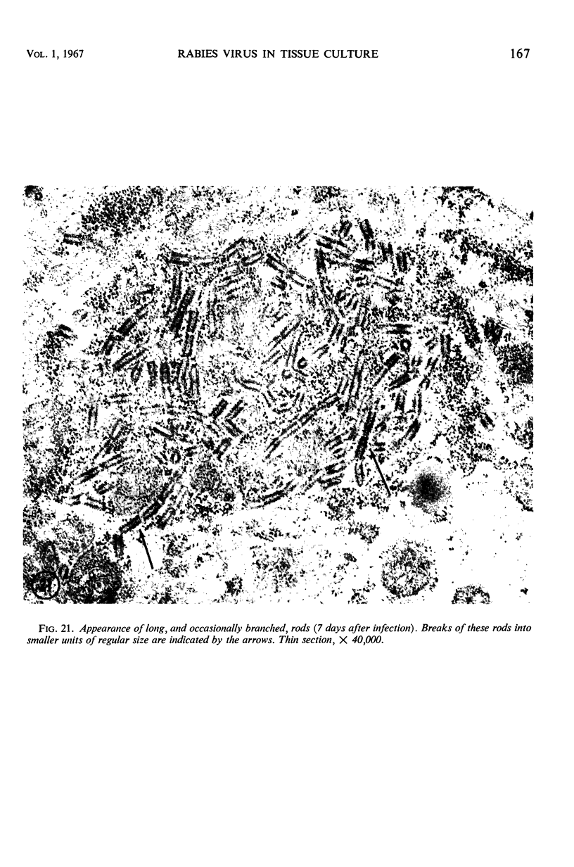
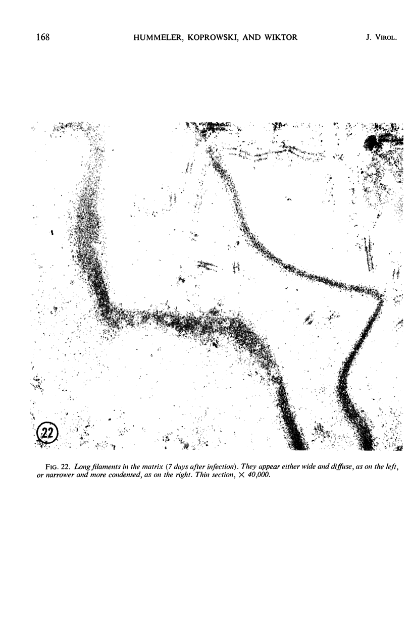
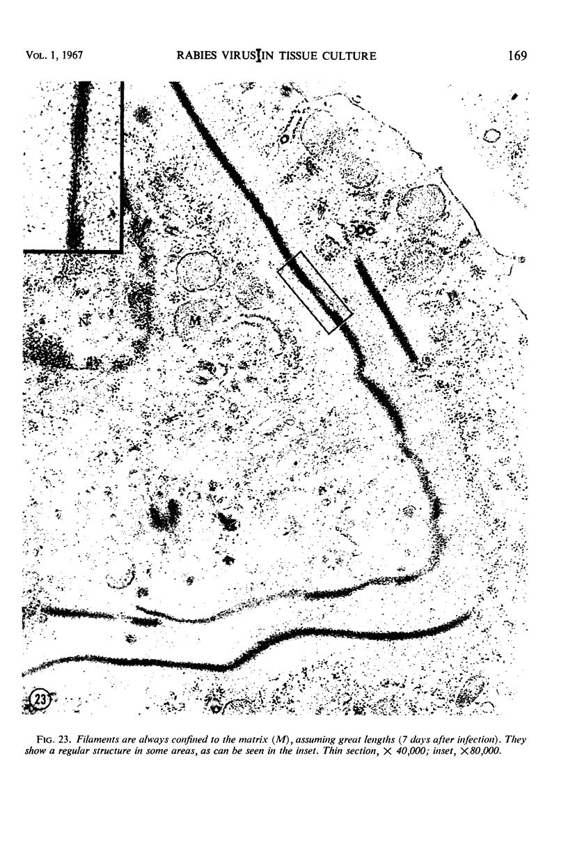
Structure and development of two fixed rabies virus strains in baby hamster kidney cells (BHK/21) were investigated by electron microscopy. The morphological development was correlated with fluorescent-antibody staining and infectivity titration. The uptake of virus was enhanced by addition of diethylaminoethyl dextran, and structural changes became apparent in the cytoplasm 8 to 9 hr after infection, when fluorescent-antibody staining was first discernible. These changes consisted of matrices containing fibers replacing normal cytoplasmic structures. Virus particles appeared at the edges of these matrices and inside them at 24 to 48 hr. This corresponded to significant rises in intracellular infectious virus. Formation of virus particles by budding from cell membranes was seen at 72 hr. Further incubation of the infected cells resulted in synthesis of bizarre structural elements. The complete virus particle was bullet-shaped with an average size of 180 by 75 mμ. It consisted of an inner core of filamentous material surrounded by two membranes of different densities. The surface showed a honeycomb arrangement with surface protrusions 60 to 70 A long having a knoblike structure at their distal end. These surface protrusions were absent at the flat end of the virus particle.
Full text
PDF
Images in this article

Selected References
These references are in PubMed. This may not be the complete list of references from this article.
- ATANASIU P., LEPINE P., DRAGONAS P. ETUDE CIN'ETIQUE DU VIRUS RABIQUE EN CULTURE DES TISSUS 'A L'AIDE DES ANTICORPS FLUORESCENTS ET DES COUPES ULTRA-FINES. Ann Inst Pasteur (Paris) 1963 Nov;105:813–824. [PubMed] [Google Scholar]
- DALES S., SIMINOVITCH L. The development of vaccinia virus in Earle's L strain cells as examined by electron microscopy. J Biophys Biochem Cytol. 1961 Aug;10:475–503. doi: 10.1083/jcb.10.4.475. [DOI] [PMC free article] [PubMed] [Google Scholar]
- DAVIES M. C., ENGLERT M. E., SHARPLESS G. R., CABASSO V. J. THE ELECTRON MICROSCOPY OF RABIES VIRUS IN CULTURES OF CHICKEN EMBRYO TISSUES. Virology. 1963 Dec;21:642–651. doi: 10.1016/0042-6822(63)90238-1. [DOI] [PubMed] [Google Scholar]
- DITCHFIELD J., ALMEIDA J. D. THE FINE STRUCTURE OF COCAL VIRUS. Virology. 1964 Oct;24:232–235. doi: 10.1016/0042-6822(64)90112-6. [DOI] [PubMed] [Google Scholar]
- HOWATSON A. F., WHITMORE G. F. The development and structure of vesicular stomatitis virus. Virology. 1962 Apr;16:466–478. doi: 10.1016/0042-6822(62)90228-3. [DOI] [PubMed] [Google Scholar]
- HUMMELER K., ARMSTRONG D., TOMASSINI N. CYTOPATHOGENIC MYCOPLASMAS ASSOCIATED WITH TWO HUMAN TUMORS. II. MORPHOLOGICAL ASPECTS. J Bacteriol. 1965 Aug;90:511–516. doi: 10.1128/jb.90.2.511-516.1965. [DOI] [PMC free article] [PubMed] [Google Scholar]
- Hitchborn J. H., Hills G. J., Hull R. Electron microscopy of viruslike particles found in diseased Plantago ianceolata in Britain. Virology. 1966 Apr;28(4):768–772. doi: 10.1016/0042-6822(66)90265-0. [DOI] [PubMed] [Google Scholar]
- Hummeler K., Henle G., Henle W. Fine structure of a virus in cultured lymphoblasts from Burkitt lymphoma. J Bacteriol. 1966 Mar;91(3):1366–1368. doi: 10.1128/jb.91.3.1366-1368.1966. [DOI] [PMC free article] [PubMed] [Google Scholar]
- JOHNSON R. T., MERCER E. H. THE DEVELOPMENT OF FIXED RABIES VIRUS IN MOUSE BRAIN. Aust J Exp Biol Med Sci. 1964 Aug;42:449–456. doi: 10.1038/icb.1964.42. [DOI] [PubMed] [Google Scholar]
- KOPROWSKI H. Biological modification of rabies virus as a result of its adaptation to chicks and developing chick embryos. Bull World Health Organ. 1954;10(5):709–724. [PMC free article] [PubMed] [Google Scholar]
- Kaplan M. M., Wiktor T. J., Maes R. F., Campbell J. B., Koprowski H. Effect of polyions on the infectivity of rabies virus in tissue culture: construction of a single-cycle growth curve. J Virol. 1967 Feb;1(1):145–151. doi: 10.1128/jvi.1.1.145-151.1967. [DOI] [PMC free article] [PubMed] [Google Scholar]
- MACPHERSON I., STOKER M. Polyoma transformation of hamster cell clones--an investigation of genetic factors affecting cell competence. Virology. 1962 Feb;16:147–151. doi: 10.1016/0042-6822(62)90290-8. [DOI] [PubMed] [Google Scholar]
- MATSUMOTO S. ELECTRON MICROSCOPE STUDIES OF RABIES VIRUS IN MOUSE BRAIN. J Cell Biol. 1963 Dec;19:565–591. doi: 10.1083/jcb.19.3.565. [DOI] [PMC free article] [PubMed] [Google Scholar]
- MATSUMOTO S. Electron microscopy of nerve cells infected with street rabies virus. Virology. 1962 May;17:198–202. doi: 10.1016/0042-6822(62)90099-5. [DOI] [PubMed] [Google Scholar]
- Miyamoto K., Matsumoto S. The nature of the Negri body. J Cell Biol. 1965 Dec;27(3):677–682. doi: 10.1083/jcb.27.3.677. [DOI] [PMC free article] [PubMed] [Google Scholar]