

Abstract
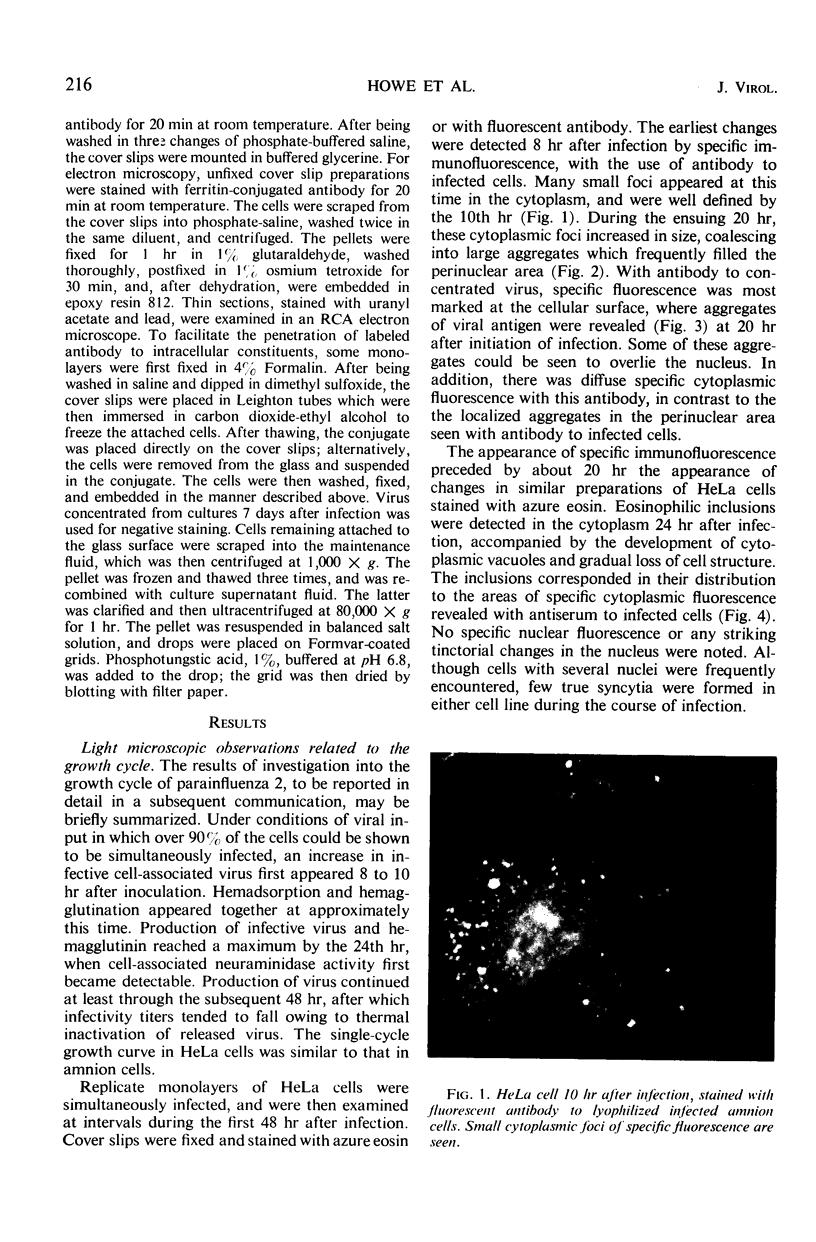
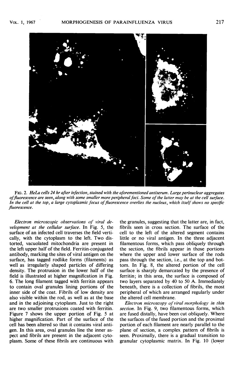
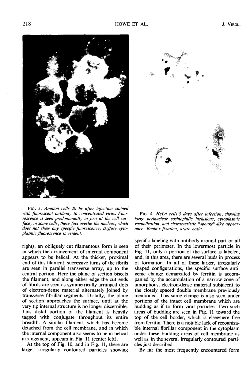
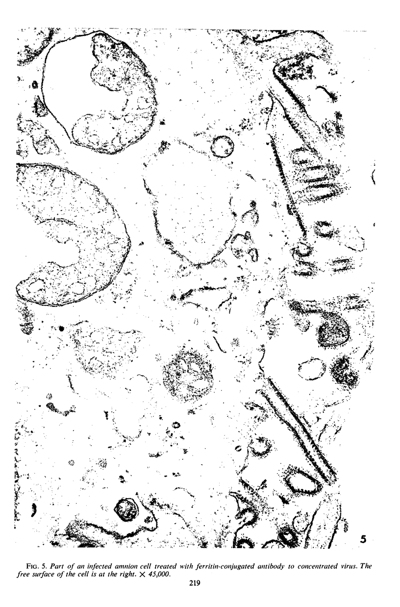
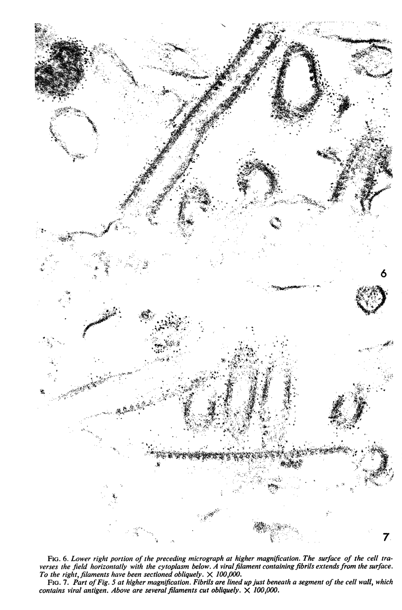
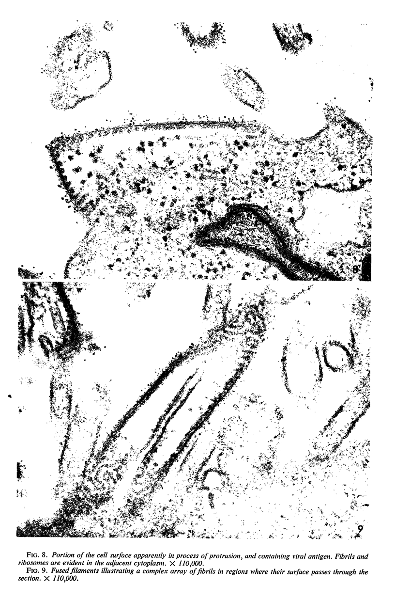
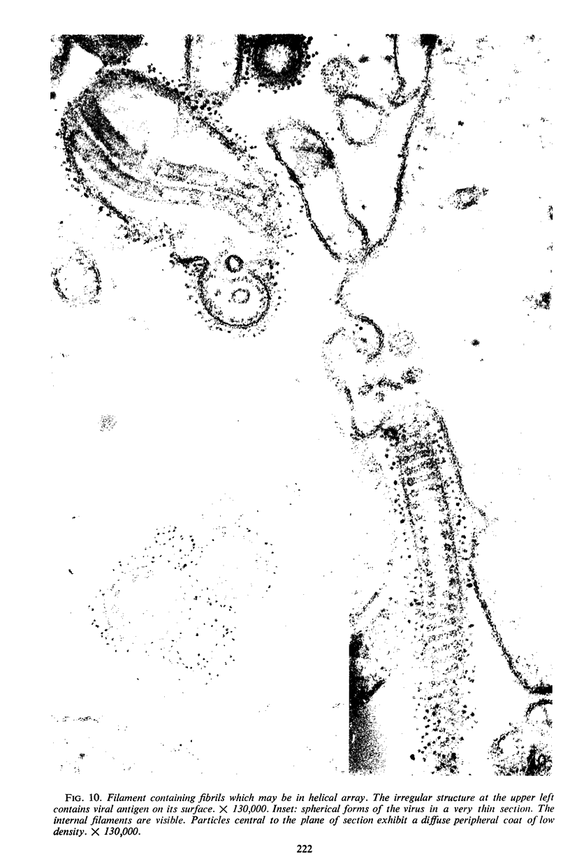
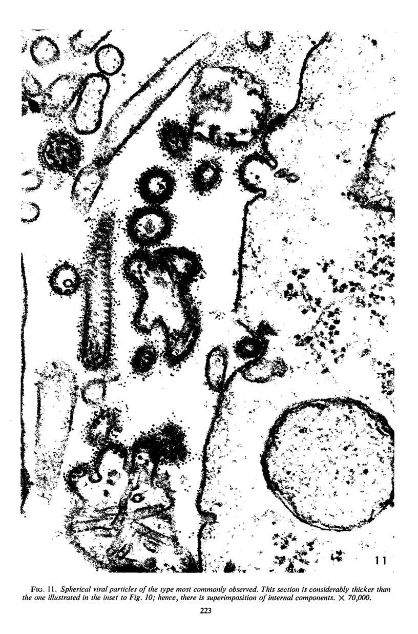
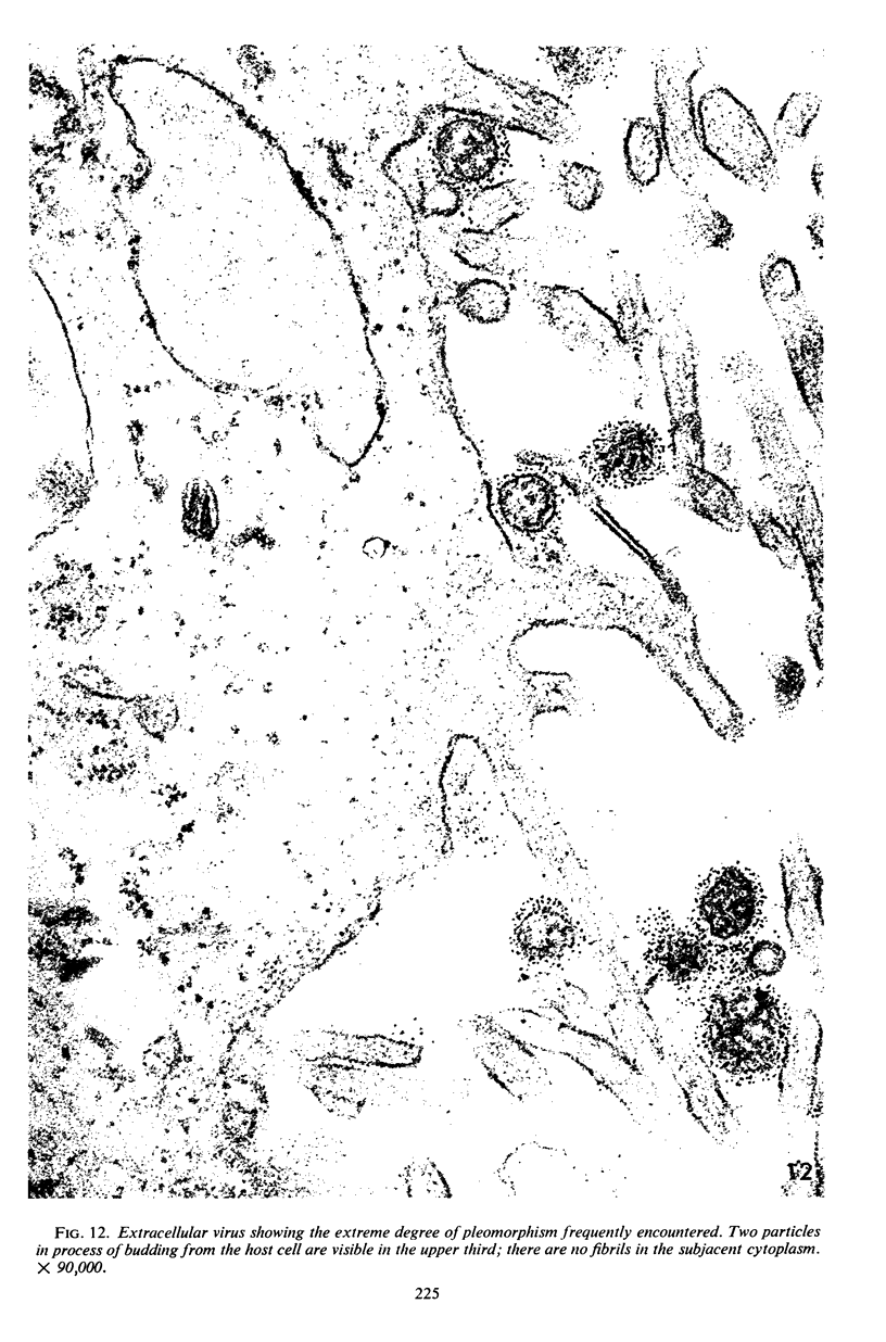
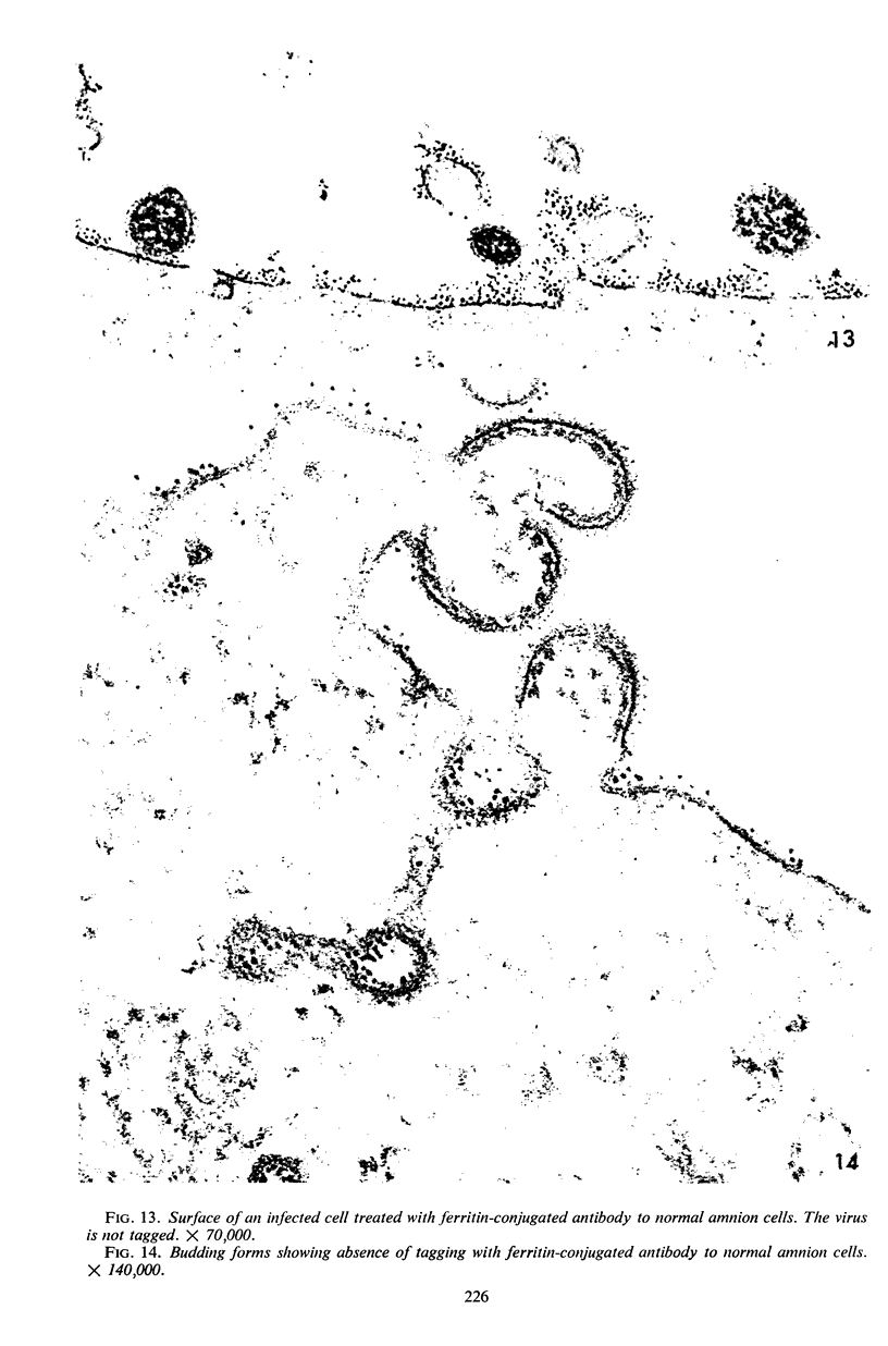
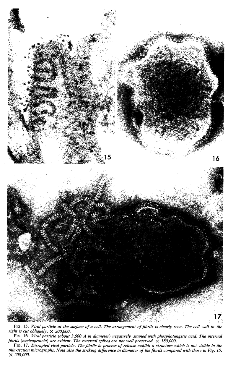
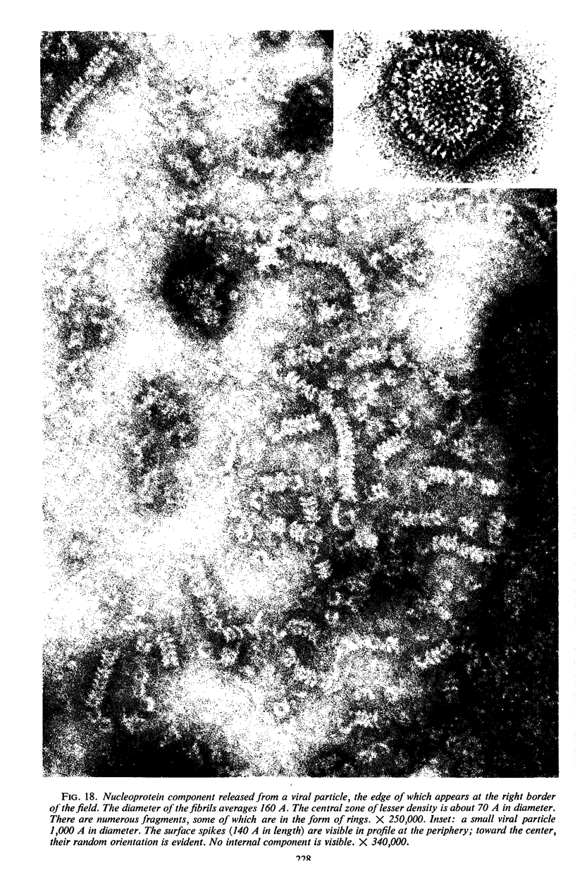
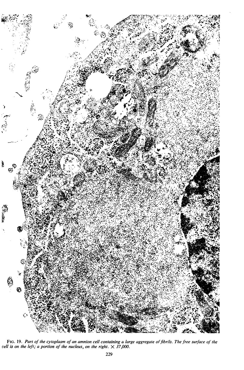
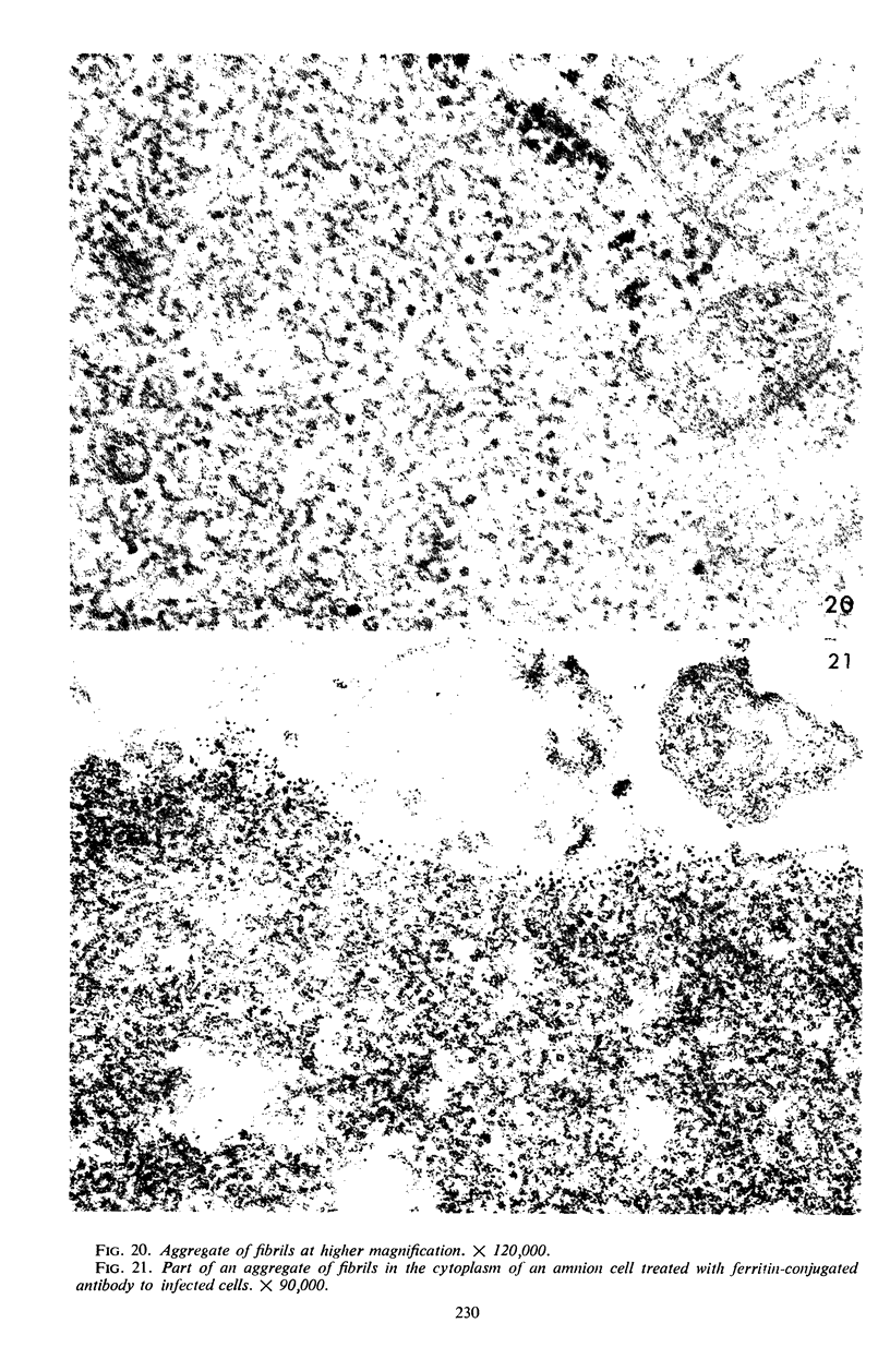
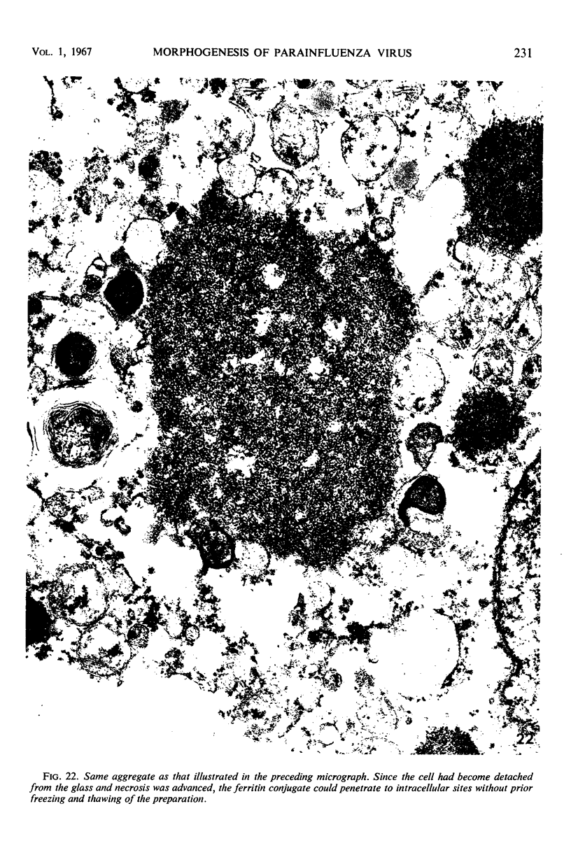
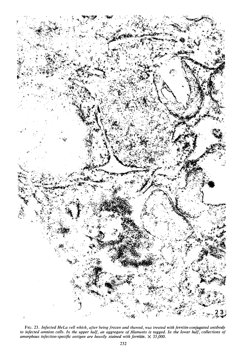
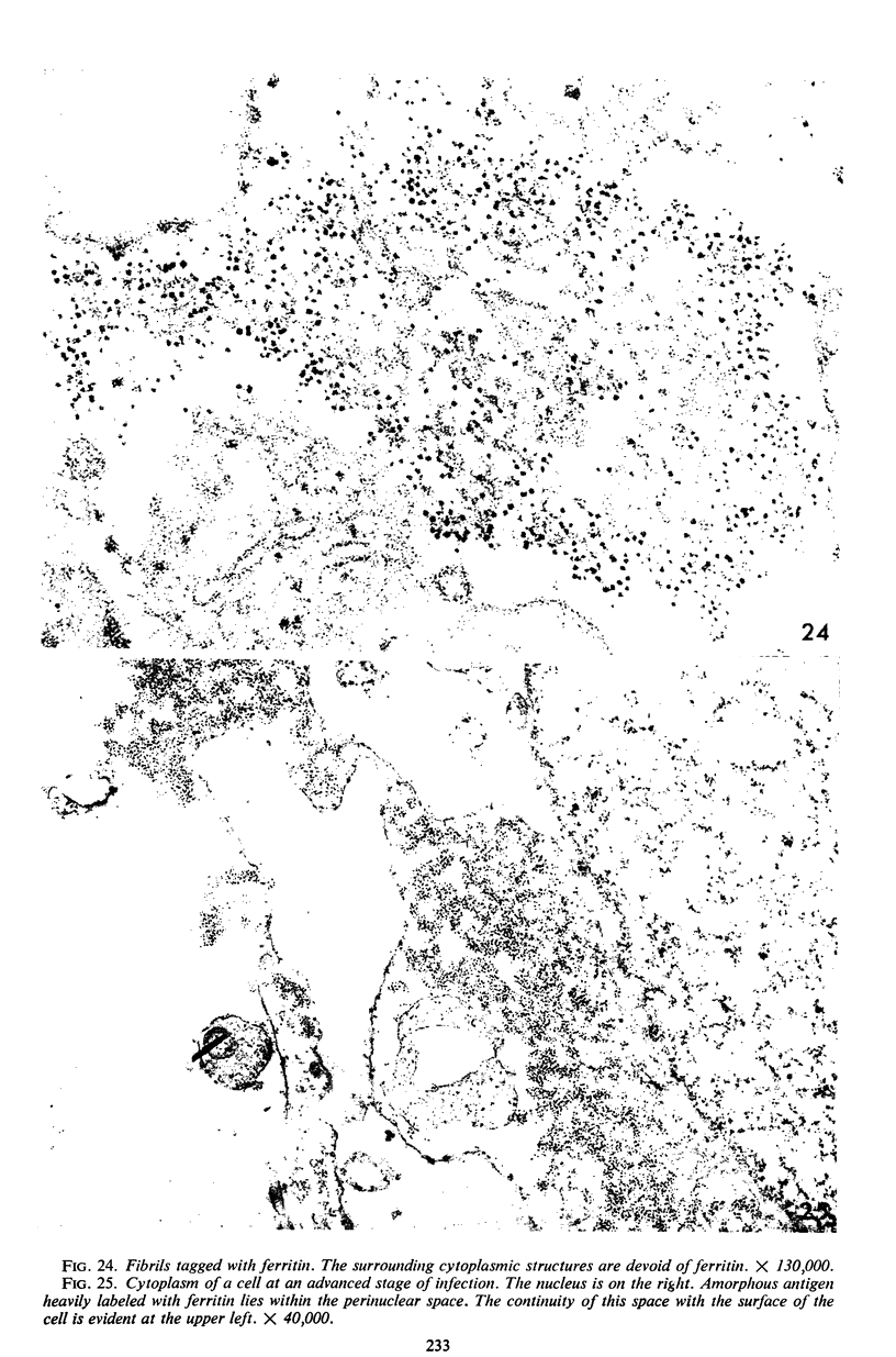
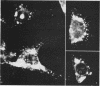
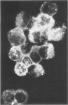
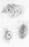
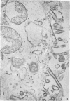
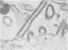
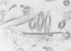
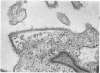
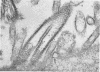
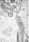
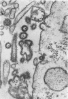
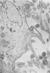
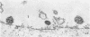
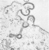
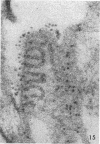
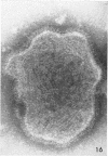
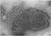
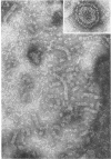
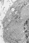
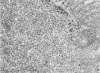
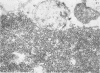
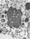
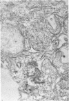
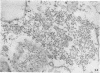
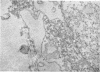
The development of type 2 parainfluenza virus in HeLa and stable human amnion cells was examined by use of antisera labeled with fluorescein and ferritin. Serum containing antibody predominantly to soluble viral antigen gave specific fluorescence which was first detectable in small cytoplasmic foci 8 to 10 hr after initiation of infection. By 20 to 24 hr, when the production of infective virus and hemagglutinin was maximal, large perinuclear aggregates of fluorescence were observed which corresponded in distribution and time of appearance to the eosinophilic inclusions seen in similar preparations stained with azure eosin. The inclusions, examined by electron microscopy, were composed of fibrils, presumably viral ribonucleoprotein, which specifically bound the antibody labeled with ferritin. With antiserum to concentrated virus, on the other hand, specific fluorescence was most marked at the surface of infected cells. Foci of fluorescence at the surface represented segments of membrane which had become differentiated morphologically and antigenically to resemble the viral envelope. These were the sites where mature virions appeared. The latter exhibited marked pleomorphism; in some instances, particles were formed which lacked recognizable internal fibrils but which possessed an enclosing membrane bearing viral antigen. Filamentous forms showing an organized internal structure were also observed at the cell surface, but were never encountered in negatively stained preparations. No clear relationship between these filaments and the spherical or oval forms could be established. In negatively stained preparations, nucleocapsid released by rupture of viral particles was similar in appearance to that reported for other paramyxoviruses. It seems probable that this component has a helical configuration.
Full text
PDF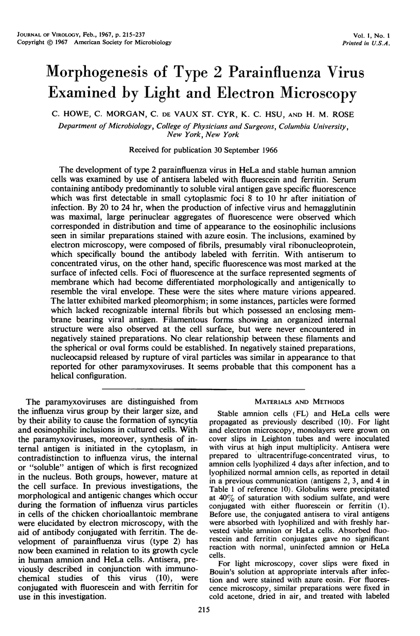
Images in this article
Selected References
These references are in PubMed. This may not be the complete list of references from this article.
- CHANOCK R. M. Association of a new type of cytopathogenic myxovirus with infantile croup. J Exp Med. 1956 Oct 1;104(4):555–576. doi: 10.1084/jem.104.4.555. [DOI] [PMC free article] [PubMed] [Google Scholar]
- CHOPPIN P. W., STOECKENIUS W. THE MORPHOLOGY OF SV5 VIRUS. Virology. 1964 Jun;23:195–202. doi: 10.1016/0042-6822(64)90282-x. [DOI] [PubMed] [Google Scholar]
- CHURCHILL A. E. Intranuclear inclusions produced by bovine para-influenza. Nature. 1963 Jan 26;197:409–409. doi: 10.1038/197409a0. [DOI] [PubMed] [Google Scholar]
- COHEN S. M., BULLIVANT S., EDWARDS G. A. A morphologic study of FL cells infected with para-influenza 3 virus. Arch Gesamte Virusforsch. 1962;11:493–515. doi: 10.1007/BF01241303. [DOI] [PubMed] [Google Scholar]
- COLOBERT L., BERKALOFF A. LIB'ERATION DU VIRUS SENDAUI PAR DES CELLULES PORTEUSES D'UNE INFECTION CHRONIQUE. Ann Inst Pasteur (Paris) 1964 Apr;106:581–587. [PubMed] [Google Scholar]
- Compans R. W., Holmes K. V., Dales S., Choppin P. W. An electron microscopic study of moderate and virulent virus-cell interactions of the parainfluenza virus SV5. Virology. 1966 Nov;30(3):411–426. doi: 10.1016/0042-6822(66)90119-x. [DOI] [PubMed] [Google Scholar]
- DINTER Z., HERMODSSON S., HERMODSSON L. STUDIES ON MYXOVIRUS YUCAIPA: ITS CLASSIFICATION AS A MEMBER OF THE PARAMYXOVIRUS GROUP. Virology. 1964 Mar;22:297–304. doi: 10.1016/0042-6822(64)90020-0. [DOI] [PubMed] [Google Scholar]
- De Vaux St Cyr C., Howe C. Immunochemical study of parainfluenza virus (type 2) in amnion cells. J Bacteriol. 1966 May;91(5):1911–1916. doi: 10.1128/jb.91.5.1911-1916.1966. [DOI] [PMC free article] [PubMed] [Google Scholar]
- GRESSER I., ENDERS J. F. A note on the presence of inclusion bodies in dividing human kidney cells infected with croup-associated virus. Virology. 1961 Mar;13:370–372. doi: 10.1016/0042-6822(61)90159-3. [DOI] [PubMed] [Google Scholar]
- HERMODSSON S., WESTMAN J. Ultrastructure of parainfluenza 3 virus. J Ultrastruct Res. 1962 Jun;6:499–510. doi: 10.1016/s0022-5320(62)80005-7. [DOI] [PubMed] [Google Scholar]
- HORNE R. W., WATERSON A. P., WILDY P., FARNHAM A. E. The structure and composition of the myxoviruses. I. Electron microscope studies of the structure of myxovirus particles by negative staining techniques. Virology. 1960 May;11:79–98. doi: 10.1016/0042-6822(60)90056-8. [DOI] [PubMed] [Google Scholar]
- Hosaka Y., Kitano H., Ikeguchi S. Studies on the pleomorphism of HVJ virons. Virology. 1966 Jun;29(2):205–221. doi: 10.1016/0042-6822(66)90027-4. [DOI] [PubMed] [Google Scholar]
- JOHNSON C. F., SCOTT A. D. CYTOLOGICAL STUDIES OF NEWCASTLE DISEASE VIRUS (NDV) IN HEP-2 CELLS. Proc Soc Exp Biol Med. 1964 Feb;115:281–286. doi: 10.3181/00379727-115-28891. [DOI] [PubMed] [Google Scholar]
- Kasten F. H., Churchill A. E. Cytochemistry of cytoplasmic and intranuclear inclusions induced by bovine parainfluenza 3 virus (SF-4) in human cell cultures. J Histochem Cytochem. 1966 Feb;14(2):187–195. doi: 10.1177/14.2.187. [DOI] [PubMed] [Google Scholar]
- LEPINE P., CHANY C., DROZ B., ROBBE-FOSSAT F. Cytopathogenic effect of two newly recognized myxovirus strains: mechanism of syncytial formation. Ann N Y Acad Sci. 1959 Jul 21;81:62–72. doi: 10.1111/j.1749-6632.1959.tb49295.x. [DOI] [PubMed] [Google Scholar]
- MAASSAB H. F., LOH P. C. Fluorescent antibody studies in tissue culture of parainfluenza 3 infection. Proc Soc Exp Biol Med. 1962 Apr;109:897–900. doi: 10.3181/00379727-109-27371. [DOI] [PubMed] [Google Scholar]
- Omar A. R. Cytopathic effects and immunofluorescence produced by the J 121 strain of bovine Parainfluenza 3 virus in tissue culture. J Comp Pathol. 1965 Jul;75(3):287–297. doi: 10.1016/0021-9975(65)90034-4. [DOI] [PubMed] [Google Scholar]
- Prose P. H., Balk S. D., Liebhaber H., Krugman S. Studies of a myxovirus recovered from patients with infectious hepatitis. II. Fine structure and electron microscopic demonstration of intracytoplasmic internal component and viral filament formation. J Exp Med. 1965 Dec 1;122(6):1151–1160. doi: 10.1084/jem.122.6.1151. [DOI] [PMC free article] [PubMed] [Google Scholar]
- RECZKO E., BOEGEL K. [Electron microscopy studies on the behavior of a parainfluenza-3 virus isolated from calves in calf kidney cell culture]. Arch Gesamte Virusforsch. 1962;12:404–420. [PubMed] [Google Scholar]
- REDA I. M., ROTT R., SCHAEFER W. FLUORESCENT ANTIBODY STUDIES WITH NDV-INFECTED CELL SYSTEMS. Virology. 1964 Mar;22:422–425. doi: 10.1016/0042-6822(64)90033-9. [DOI] [PubMed] [Google Scholar]
- REISINGER R. C. Parainfluenza 3 virus in cattle. Ann N Y Acad Sci. 1962 Nov 30;101:576–582. doi: 10.1111/j.1749-6632.1962.tb18898.x. [DOI] [PubMed] [Google Scholar]
- TRAVER M. I., NORTHROP R. L., WALKER D. L. Site of intracellular antigen production by myxoviruses. Proc Soc Exp Biol Med. 1960 Jun;104:268–273. doi: 10.3181/00379727-104-25803. [DOI] [PubMed] [Google Scholar]
- VERVILLE E., CONNOR J. D., SIGEL M. M. Double infection of tissue culture cells with CA virus and adenovirus. Virology. 1961 Feb;13:260–262. doi: 10.1016/0042-6822(61)90063-0. [DOI] [PubMed] [Google Scholar]
- WATERSON A. P., HURRELL J. M. The fine structure of the parainfluenza viruses. Arch Gesamte Virusforsch. 1962;12:138–142. doi: 10.1007/BF01258760. [DOI] [PubMed] [Google Scholar]
- WATERSON A. P., JENSEN K. E., TYRRELL D. A., HORNE R. W. The structure of parainfluenza 3 virus. Virology. 1961 Jul;14:374–378. doi: 10.1016/0042-6822(61)90323-3. [DOI] [PubMed] [Google Scholar]
- ZHDANOV V. M., AZADOVA N. B., URYVAYEV L. V. TOPOGRAPHY OF SYNTHESIS OF S AND V ANTIGENS OF SENDAI VIRUS. J Immunol. 1965 May;94:658–661. [PubMed] [Google Scholar]
- Zhdanov V. M., Azadova N. B., Uryvayev L. V. Topography and dynamics of synthesis of structural proteins of Newcastle disease virus. J Bacteriol. 1966 May;91(5):1902–1906. doi: 10.1128/jb.91.5.1902-1906.1966. [DOI] [PMC free article] [PubMed] [Google Scholar]