Dear Editor:
Bowen disease is a squamous cell carcinoma in situ and a common condition of the skin. It can occur at any location on the body. It usually presents as a red plaque with scales. The cause of Bowen disease includes solar damage, arsenic, immunosuppression and infection of human papillomavirus (HPV). The occurrence of Bowen disease in the nail unit has been previously reported1. Very rarely, longitudinal melanonychia associated with Bowen disease on the nail unit has been described2. Here, we report a case of Bowen disease presenting as longitudinal melanonychia of the lateral side of the fingernail, which is associated with HPV and melanocytic hyperplasia histopathologically.
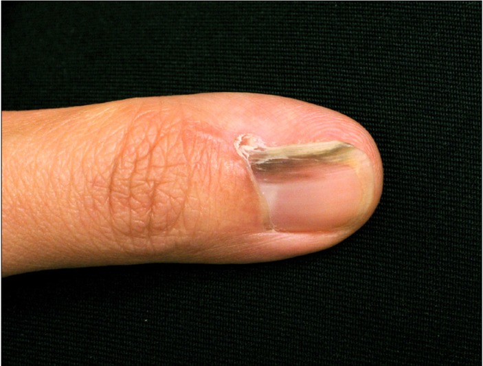
A 33-year-old Korean man was presented with a dark brown streak on the left thumbnail, which had been present for several years (Fig. 1). Although he received excision in the past, the streak recurred a year ago. On examination, a dark brown colored pigmentation was shown along with onycholysis and partial hyperkeratosis of the lateral side of the left 1st fingernail. Hyperkeratosis and onycholysis may be a secondary nonspecific change after the first excision. After the nail extraction, a longitudinal excision was performed. Histopathological findings revealed atypical dyskeratotic keratinocytes in the whole epidermis (Fig. 2A). Some nuclei of the atypical cells were large, pleomorphic and hyperchromatic. The number of melanocytes was increased by HMB 45 immunohistochemistry (Dako, Carpinteria, CA, USA) (Fig. 2B). HPV was identified in the horny layers via immunohistochemistry (Dako) (Fig. 2C). Finally, a diagnosis of Bowen's disease was made.
Fig. 1.
Dark brown colored longitudinal melanonychia on the left thumbnail.
Fig. 2.

(A) Many atypical dyskeratotic keratinocytes are observed in the epidermis (H&E, ×200). (B) Melanocytic hyperplasia is shown via HMB 45 immunohistochemistry (HMB-45, ×200). (C) Human papillomavirus is found in the horny layers through immunohistochemistry (HPV, ×200).
Previously, several longitudinal melanonychia cases associated with HPV infection have been reported in English-written literatures3-5. Several HPV types were discovered in the lesions. Among them, HPV-56 was the most common. In our case, although HPV typing HPV was not performed, HPV was found through immunohistochemisty. Like our case, characteristically, some cases which showed clinical pictures in the literatures involved the lateral side of the nail in longitudinal melanonychia. Thus, longitudinal melanonychia of the nail might be a sign of Bowen disease. In addition, based on our case and previous cases of Bowen disease which were manifested with longitudinal melanonychia, HPV infection was almost always associated, suggesting that HPV seems to be a cause of Bowen disease associated with longitudinal melanonychia. Bowen disease associated with HPV should be included in the differential diagnosis of longitudinal melanonychia.
References
- 1.Baran R, Dupré A, Sayag J, Letessier S, Robins P, Bureau H. Bowen disease of the nail apparatus. Report of 5 cases and review of the 20 cases of the literature (author's transl) Ann Dermatol Venereol. 1979;106:227–233. [PubMed] [Google Scholar]
- 2.Baran R, Simon C. Longitudinal melanonychia: a symptom of Bowen's disease. J Am Acad Dermatol. 1988;18:1359–1360. doi: 10.1016/s0190-9622(88)80115-4. [DOI] [PubMed] [Google Scholar]
- 3.Sass U, André J, Stene JJ, Noel JC. Longitudinal melanonychia revealing an intraepidermal carcinoma of the nail apparatus: detection of integrated HPV-16 DNA. J Am Acad Dermatol. 1998;39:490–493. doi: 10.1016/s0190-9622(98)70331-7. [DOI] [PubMed] [Google Scholar]
- 4.Lambiase MC, Gardner TL, Altman CE, Albertini JG. Bowen disease of the nail bed presenting as longitudinal melanonychia: detection of human papillomavirus type 56 DNA. Cutis. 2003;72:305–309. [PubMed] [Google Scholar]
- 5.Shimizu A, Tamura A, Abe M, Motegi S, Nagai Y, Ishikawa O, et al. Detection of human papillomavirus type 56 in Bowen's disease involving the nail matrix. Br J Dermatol. 2008;158:1273–1279. doi: 10.1111/j.1365-2133.2008.08562.x. [DOI] [PubMed] [Google Scholar]


