

Abstract
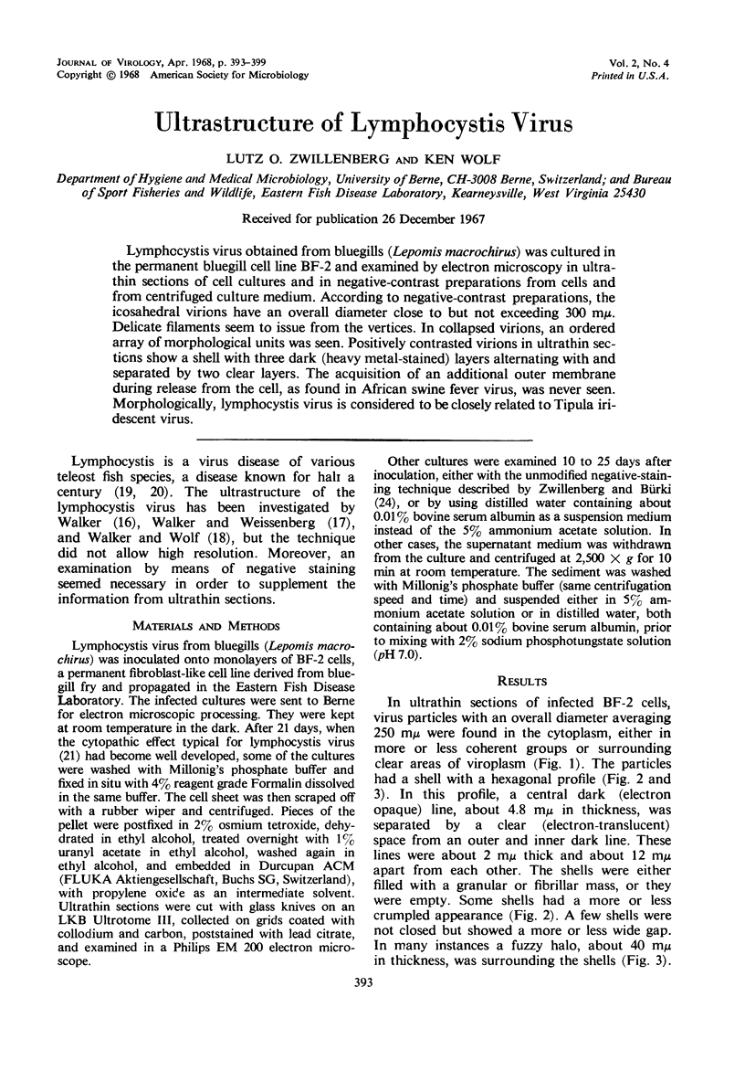
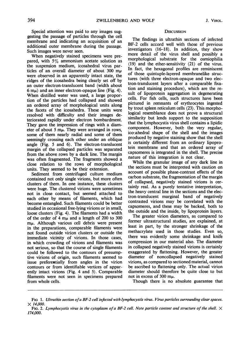
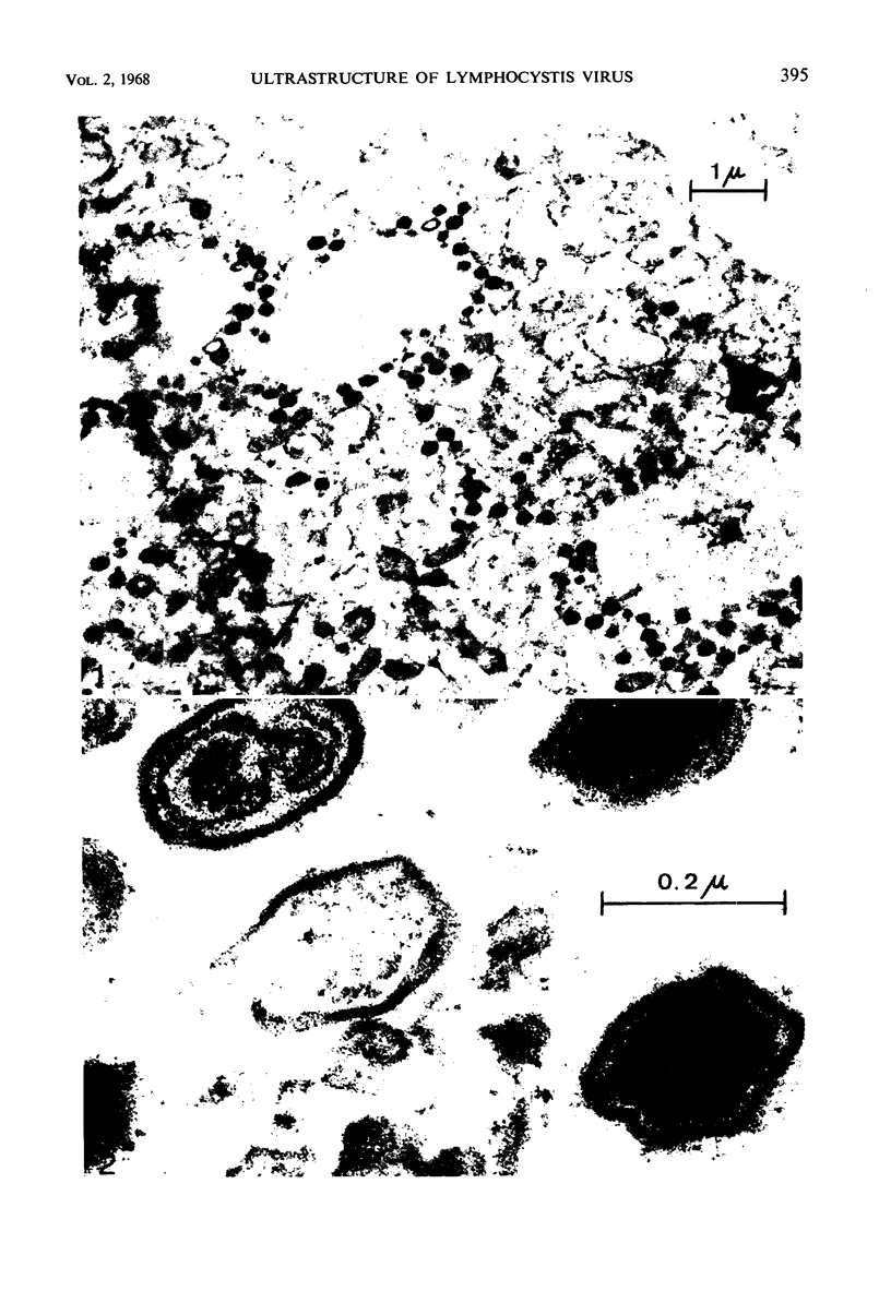
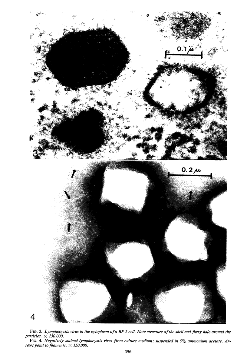
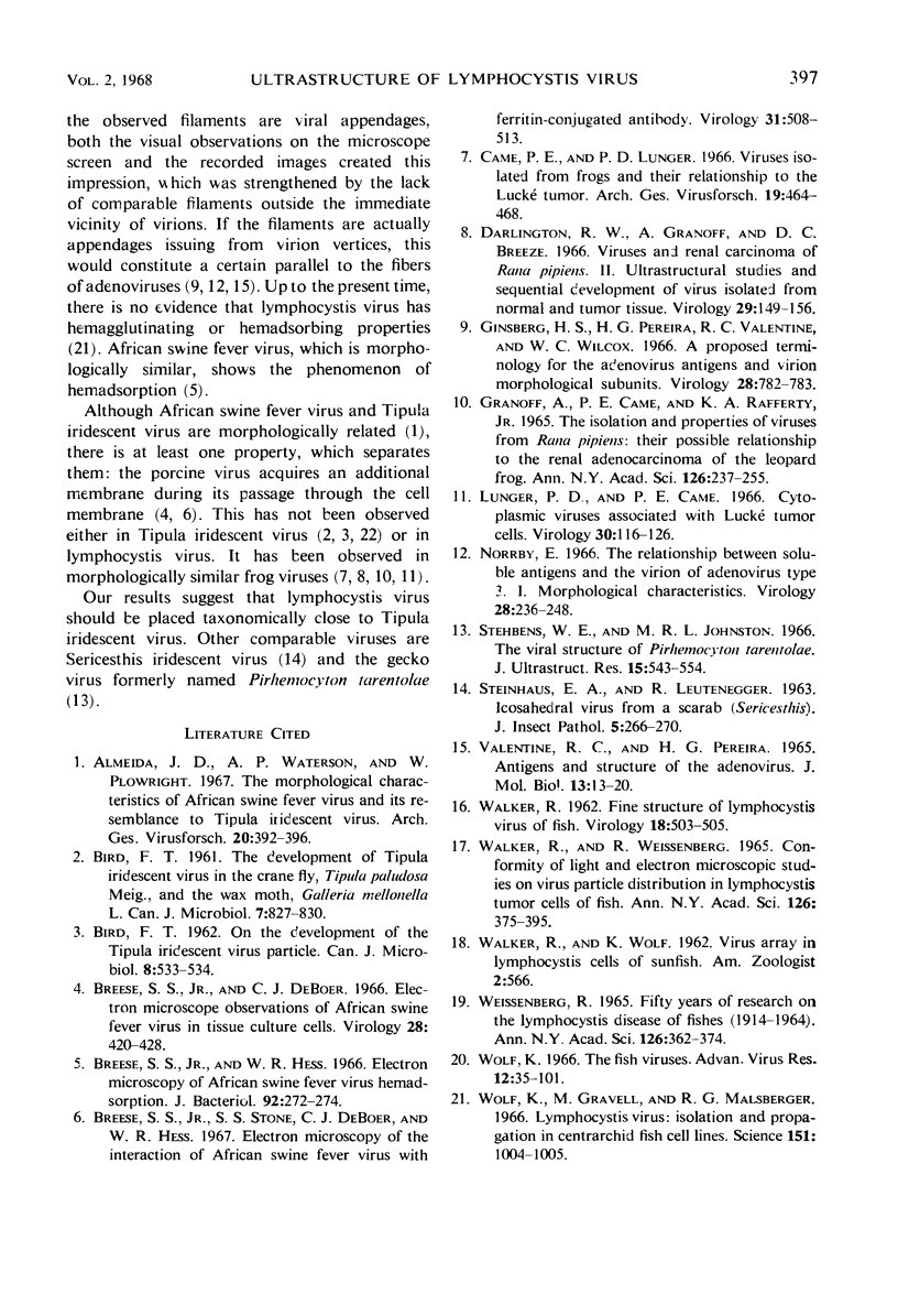
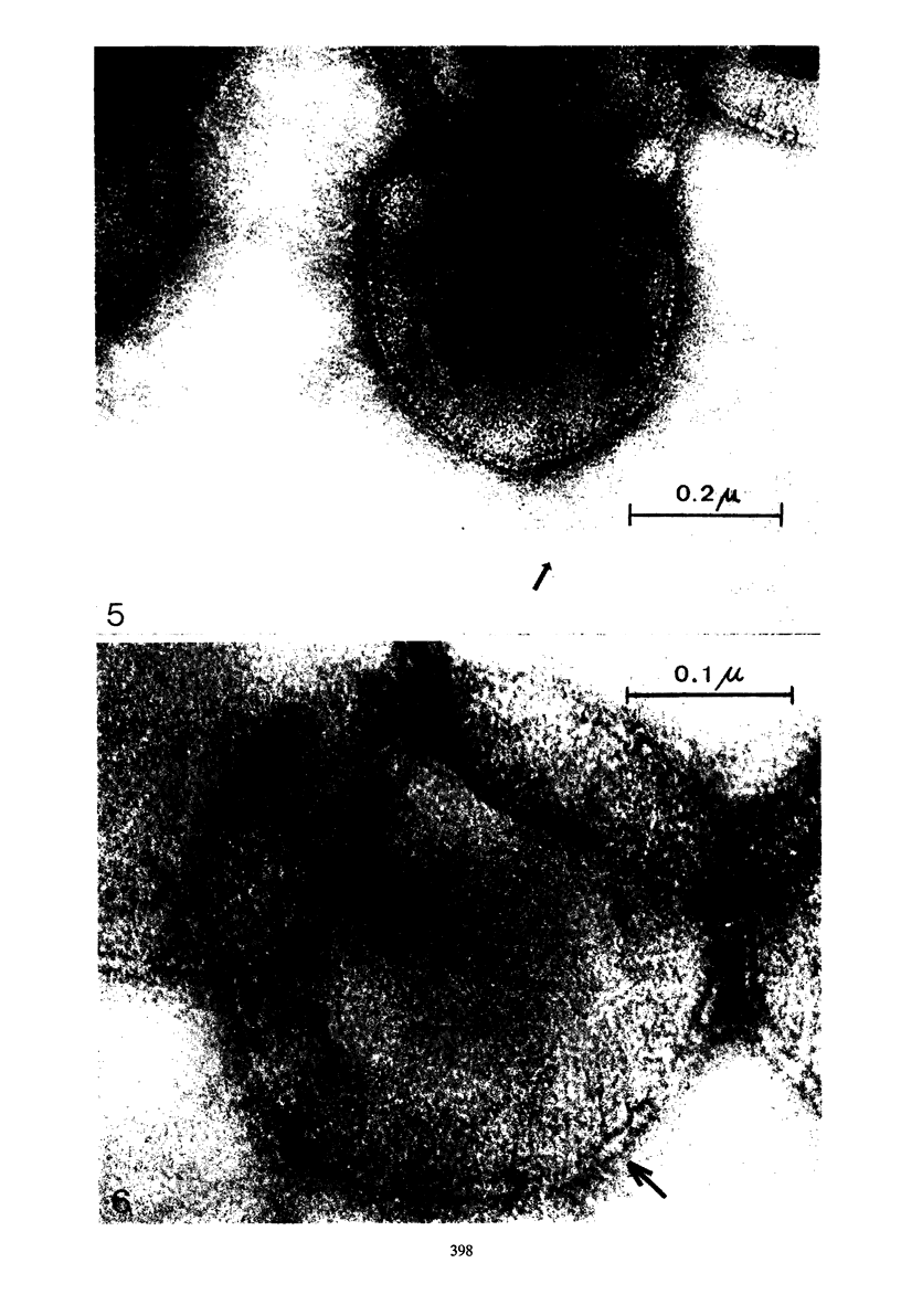
Lymphocystis virus obtained from bluegills (Lepomis macrochirus) was cultured in the permanent bluegill cell line BF-2 and examined by electron microscopy in ultrathin sections of cell cultures and in negative-contrast preparations from cells and from centrifuged culture medium. According to negative-contrast preparations, the icosahedral virions have an overall diameter close to but not exceeding 300 mμ. Delicate filaments seem to issue from the vertices. In collapsed virions, an ordered array of morphological units was seen. Positively contrasted virions in ultrathin sections show a shell with three dark (heavy metal-stained) layers alternating with and separated by two clear layers. The acquisition of an additional outer membrane during release from the cell, as found in African swine fever virus, was never seen. Morphologically, lymphocystis virus is considered to be closely related to Tipula iridescent virus.
Full text
PDF
Images in this article
Selected References
These references are in PubMed. This may not be the complete list of references from this article.
- Almeida J. D., Waterson A. P., Plowright W. The morphological characteristics of African swine fever virus and its resemblance to tipula iridescent virus. Arch Gesamte Virusforsch. 1967;20(3):392–396. doi: 10.1007/BF01241958. [DOI] [PubMed] [Google Scholar]
- BIRD F. T. The development of Tipula iridescent virus in the crane fly, Tipula paludosa Meig., and the wax moth, Galleria mellonella L. Can J Microbiol. 1961 Oct;7:827–830. doi: 10.1139/m61-098. [DOI] [PubMed] [Google Scholar]
- Breese S. S., Jr, Hess W. R. Electron microscopy of African swine fever virus hemadsorption. J Bacteriol. 1966 Jul;92(1):272–274. doi: 10.1128/jb.92.1.272-274.1966. [DOI] [PMC free article] [PubMed] [Google Scholar]
- Breese S. S., Jr, Stone S. S., DeBoer C. J., Hess W. R. Electron microscopy of the interaction of African swine fever virus with ferritin-conjugated antibody. Virology. 1967 Mar;31(3):508–513. doi: 10.1016/0042-6822(67)90232-2. [DOI] [PubMed] [Google Scholar]
- Came P. E., Lunger P. D. Viruses isolated from frogs and their relationship to the Lucké tumor. Arch Gesamte Virusforsch. 1966;19(4):464–468. doi: 10.1007/BF01250614. [DOI] [PubMed] [Google Scholar]
- Darlington R. W., Granoff A., Breeze D. C. Viruses and renal carcinoma of Rana pipiens. II. Ultrastructural studies and sequential development of virus isolated from normal and tumor tissue. Virology. 1966 May;29(1):149–156. doi: 10.1016/0042-6822(66)90204-2. [DOI] [PubMed] [Google Scholar]
- Ginsberg H. S., Pereira H. G., Valentine R. C., Wilcox W. C. A proposed terminology for the adenovirus antigens and virion morphological subunits. Virology. 1966 Apr;28(4):782–783. doi: 10.1016/0042-6822(66)90271-6. [DOI] [PubMed] [Google Scholar]
- Granoff A., Came P. E., Rafferty K. A., Jr The isolation and properties of viruses from Rana pipiens: their possible relationship to the renal adenocarcinoma of the leopard frog. Ann N Y Acad Sci. 1965 Aug 10;126(1):237–255. doi: 10.1111/j.1749-6632.1965.tb14278.x. [DOI] [PubMed] [Google Scholar]
- Lunger P. D., Came P. E. Cytoplasmic viruses associated with Lucké tumor cells. Virology. 1966 Sep;30(1):116–126. doi: 10.1016/s0042-6822(66)81015-2. [DOI] [PubMed] [Google Scholar]
- Norrby E. The relationship between the soluble antigens and the virion of adenovirus type 3. I. Morphological characteristics. Virology. 1966 Feb;28(2):236–248. doi: 10.1016/0042-6822(66)90148-6. [DOI] [PubMed] [Google Scholar]
- Stehbens W. E., Johnston M. R. The viral nature of Pirhemocyton tarentolae. J Ultrastruct Res. 1966 Aug;15(5):543–554. doi: 10.1016/s0022-5320(66)80127-2. [DOI] [PubMed] [Google Scholar]
- Valentine R. C., Pereira H. G. Antigens and structure of the adenovirus. J Mol Biol. 1965 Aug;13(1):13–20. doi: 10.1016/s0022-2836(65)80076-6. [DOI] [PubMed] [Google Scholar]
- WALKER R. Fine structure of lymphocystis virus of fish. Virology. 1962 Nov;18:503–505. doi: 10.1016/0042-6822(62)90047-8. [DOI] [PubMed] [Google Scholar]
- Walker R., Weissenberg R. Conformity of light and electron microscopic studies on virus particle distribution in lymphocystis tumor cells of fish. Ann N Y Acad Sci. 1965 Aug 10;126(1):375–385. doi: 10.1111/j.1749-6632.1965.tb14287.x. [DOI] [PubMed] [Google Scholar]
- Weissenberg R. 50 years of research on the lymphocystis virus disease of fishes (1914-1964). Ann N Y Acad Sci. 1965 Aug 10;126(1):362–374. doi: 10.1111/j.1749-6632.1965.tb14286.x. [DOI] [PubMed] [Google Scholar]
- Wolf K., Gravell M., Malsberger R. G. Lymphocystis virus: isolation and propagation in centrarchid fish cell lines. Science. 1966 Feb 25;151(3713):1004–1005. doi: 10.1126/science.151.3713.1004. [DOI] [PubMed] [Google Scholar]
- Wolf K. The fish viruses. Adv Virus Res. 1966;12:35–101. doi: 10.1016/s0065-3527(08)60846-5. [DOI] [PubMed] [Google Scholar]
- ZWILLENBERG H. H., ZWILLENBERG L. O. [On the erythrocyte decomposition in the trout spleen with special reference to the erythrocyte fine structure]. Z Zellforsch Mikrosk Anat. 1963;60:313–324. [PubMed] [Google Scholar]
- Zwillenberg L. O., Bürki F. On the cpsid structure of some small feline and bovine RNA viruses. Arch Gesamte Virusforsch. 1966;19(4):373–384. doi: 10.1007/BF01250606. [DOI] [PubMed] [Google Scholar]