Abstract
A novel composite kidney stone phantom has been developed. This stone phantom is producible with mechanical properties mimicking the range of tensile fracture strength and acoustic properties of human kidney stones and is an inorganic/organic composite material, as are natural kidney stones. Diametral compression testing was used to measure tensile fracture strength, which determines the acoustic comminution behavior of kidney stones. Ultrasound transmission tests were made to characterize the acoustic properties of these stone phantoms. Both the tensile fracture strength (controllable from 1 to ~ 5 MPa) and acoustic properties (CL = 2700 to 4400m/s and CT = 1600 – 2300 m/s) of these composite phantom stones match those of a wide variety of human kidney stones. These artificial stone phantoms should have wide utility in lithotripsy research.
Keywords: lithotripsy, kidney stones, fracture
1. Introduction
Renal calculi (kidney stones) occur in 12% of men and 5% of women by the age of 70 and that number is rising (Coe 2005). Since the early 1980’s, the widespread clinical application of shock wave lithotripsy (SWL) has increased the importance of understanding the physical properties and comminution behavior of human kidney stones (Khan et al, 1986; Zhu et al, 2002; Zohdi and Szeri, 2005). The mechanical properties of human kidney stones vary significantly depending on stone composition (Chuong, et al, 1993a; Zhong, et al, 1993).
A number of different stone phantom compositions have been described. Liu and Zhong (2002), for example, measured the Vickers harness of Begostone phantoms and determined their hardness to be about 200 MPa. In the Vickers test, hardness is determined by the size of the indentation produced by a given load applied to a pyramid-shaped indenter, and hence is not directly interpretable in terms of tensile fracture stress. Previous studies of phantom stone mechanical properties have generally reported the crushing strength of phantom stones in various compression tests. Wang and Yip (2002) reported the compressive strength of human kidney stones to range from 6.17 to 3.2 MPa, but did not give the tensile fracture strength, which for brittle materials is usually lower than the compressive fracture strength. Yet it is the tensile fracture strength that determines the comminution of kidney stones in SWL.
Heimbach et al. (2000) produced nearly spherical phantom stones using the mineral carbonate apatite and measured their fragmentation behavior as observed in a clinical acoustic lithotripter, but did not report either compressive or tensile fracture strength. Vakil et al, (1991) produced plate-like kidney stone phantoms from Plaster of Paris and determined their surface spallation behavior in a clinical acoustic lithotripter, but did not report their mechanical properties. Blitz et al, (1995) reported the number of acoustic shocks required to produce a given degree of comminution of phantom stones produced from Iceland Spar (calcium carbonate). Ebrahimi and Wang (1989) used rectangular samples of a soft commercial brick (Z-brick) containing vermiculite and found its fracture strength under compression to be about half that of most struvite human kidney stones. In this paper, we present for the first time the tensile fracture strength of both human stones and composite stone phantoms, as well as those of all the main classes of human kidney stones, and show that these new stone phantoms can be produced with tensile fracture strength that mimics the range of tensile fracture strength of natural kidney stones.
BegoStone is a commercially available material (BEGO USA, Smithfield, RI) that has long been used in dental applications, and the use of this inorganic material as a kidney stone phantom has been described (Liu and Zhong, 2002). All natural kidney stones contain a few percent of insoluble complex organic matter that is primarily protein, embedded in a number of different inorganic materials. Existing kidney stone phantoms do not contain insoluble organic material in combination with an inorganic matrix. The inorganic component of the present composite phantom stones was BegoStone Plus, which is essentially a strengthened gypsum plaster. The organic component used in this novel composite phantom stone was polymerized albumen, a complex, organic material, mainly protein. This albumen was polymerized as part of the phantom stone preparation procedure so that it was insoluble, thereby allowing this stone phantom to be used in a variety of lithotripsy investigations, including animal experiments. The composition and resulting properties of these new composite inorganic/organic kidney stone phantoms have been found to be controllable via their water, inorganic, and organic content. The range of tensile strength properties that can be produced with these phantom stones mimics the observed range of tensile fracture strength, as well as the acoustic properties of human kidney stones.
Experimental Methods
The composition of Begostone Plus has not been reported in the literature, and therefore both x-ray diffraction and spectroscopic analyses were carried out to detail the chemical nature of this material. A commercial service (St. Louis Testing Laboratories, St. Louis, Missouri) carried out the spectroscopic analysis. A Phillips diffractometer using 0.154 nm (Cu Kα) radiation x-ray diffraction was used for the x-ray analysis.
Because the acoustic properties, as well as the tensile fracture strength properties, are important in lithotripsy, ultrasonic, acoustic wave speeds, both longitudinal and transverse, of stone phantoms were measured over a range of compositions using the ultrasound pulse transmission method developed by Zhong et al 1993a.
The stone phantoms were prepared in the form of right circular cylinders using a polytetrafluoroethylene mold so that the stones, as fabricated, could be released from this mold directly without the need for a mold lubricant. The mold was filled with BegoStone Plus, water, and albumen that have been mixed uniformly together. Since albumen was only about 15% organic material, the remainder being water, this water must be accounted for in computing the final Begostone-water-albumen composition. After initial setting, the stone phantom was slowly heated to 90°C and held at this temperature for 12 hours to polymerize the albumen. The average diameter and length of the phantom stones were 6.2 mm and 13.1 mm, respectively.
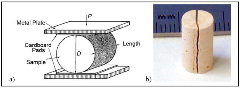
The tensile fracture strength of both natural and artificial kidney stones have been determined using the diametral compression testing method (Mellor and Hawkes, 1971). This test involves compression of a right circular cylinder specimen across its diameter. The specimen then splits in half along the diameter in which the compressive force was applied. The elastic constraint imposed by the material not directly in line with the direction of the applied compressive force results in a tensile stress normal to the direction of the applied compressive force (Mellor and Hawkes 1971). Diametral compression testing is uniquely applicable to the determination of the tensile fracture strength of brittle materials because it only requires samples that are right circular cylinders, not the normal machined tensile bar geometry used for ductile, metallic materials. Diametral compression tests of 15 human kidney stones were prepared and tested in our laboratory over an extended period as large stones became available. The required right circular cylinder shape was achieved by the technique of center-less grinding.
Most brittle materials are not tested for their tensile fracture strength, which is usually lower than their corresponding compressive fracture strength (Hayden, Moffatt and Wulff, 1965). In shock wave lithotripsy, it is the tensile fracture strength of kidney stones that dominates their fracture behavior during the comminution process, not their compressive fracture behavior.
The mechanical testing procedure used and a sample as fractured by this test are shown in Fig. 1(a) and (b).
Fig. 1.

(a) a schematic drawing of the diametral compression procedure. (b) A photograph of a kidney stone phantom as-fractured during this test.
Results and Discussion
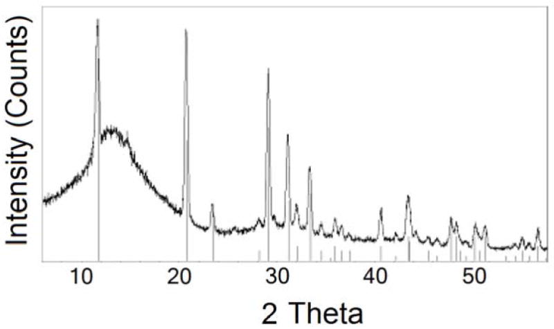
Fig. 2 shows a portion of the diffraction pattern of a typical kidney stone phantom as a function of the 2θ (two theta) diffraction angle. With this data and the Jade x-ray diffraction database (MDI, Inc., Livermore, California), the match with gypsum is evident.
Fig. 2.

A partial diffractometer pattern of a phantom stone. The straight lines rising from the abscissa give the positions of the x-ray diffraction peaks expected from gypsum.
Spectroscopic analysis of Begostone Plus identified its major elements as calcium and sulfur with minor amounts of potassium and iron. Converting these detected elements to CaSO4.2H2O, Fe2O3, and K2O yields the mineral composition as approximately 99% gypsum modified by the addition of approximately equal amounts of iron and potassium oxide, amounting to approximately 1% in total. Begostone Plus sets and hardens by the well known CaSO4 / H2O hydration reaction, forming CaSO4.2H2O (gypsum). Iron and potassium oxide are presumably used to enhance the strength of this gypsum.
The tensile fracture stress, σmax, was calculated from the diametral compression data using Eq. 1 (Mellor and Hawkes, 1971),
| (1) |
in which P is the applied load which caused fracture, D is the diameter and L is the length of the right circular cylinder. Under these compression conditions, brittle solids fracture in tension along the diametral line in which the compressive force is applied, as shown in Fig. 1b, thereby giving the tensile fracture stress. These tests yielded a range of fracture strength that varied with stone composition in the range of 0.6 to 5.2 MPa (Table I). All of these human stone fracture strength measurements, as well as those of the phantom stones were made using the same equipment.
Table I.
Range of tensile fracture strength for human kidney stones as a function of the predominant stone composition.
| Predominant Stone type | Tensile Fracture Strength (MPa) |
|---|---|
| MAPH/CA | 0.6 - 1.3 |
| UA | 1.2 - 3.6 |
| Cystine | 1.3 - 3.7 |
| Oxalate | 3.1 - 5.2 |
| CHPD | 3.0 – 4.8 |
MAPH = Magnesium ammonium phosphate hexahydrate, CA = Calcium apatite, UA = Uric acid, CHPD = Calcium hydrogen phosphate dihydrate, Oxalate = Calcium oxalate monohydrate and Calcium oxalate dihydrate. This table includes data on natural stones published by Johrde and Cocks (1985) and as presented in the thesis of Akers (1990), in addition to those determined subsequently in our laboratory.
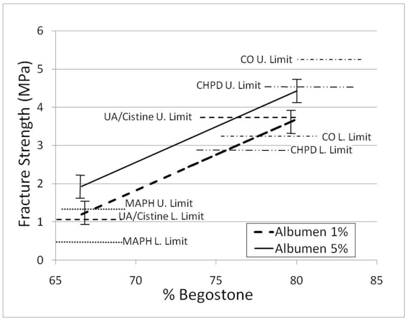
The results of approximately 100 diametral compression tests of phantom stones having a variety of different proportions of the ternary BegoStone Plus, water, and albumen system allowed the generation of an equation relating phantom stone composition to mechanical properties using three variable least squares analysis with Gauss-Jordan reduction. This ternary data reduction technique has been used previously to relate lattice parameter to composition in ternary alloy systems (Cocks, 1972). This reference provides details of the mathematics of this method. As seen from Fig. 3, the tensile fracture properties that can be produced using these composite stone phantoms mimic the range of tensile fracture properties for human kidney stones. The strength of the major types of kidney stones, also determined by the diametral compression method, are shown horizontally. Begostone content of the phantom stones is plotted along the absissa, and the two sloping lines shown give the calculated tensile strength for stones containing either 1% or 5% albumen. The error bracket in these calculated strength levels is indicated by showing one standard deviation for these lines in Fig. 3.
Fig. 3.

The calculated relationship between fracture strength (ordinate) and weight percent BegoStone (abscissa) including two polymerized albumen compositions shown by the sloped solid and dashed lines. For a ternary system A + B + C = 100%. Therefore the weight percent water can be calculated from the Begostone plus albumen content. The tensile strength limits for different stone types are shown by horizontal dashed lines. The error brackets shown on the sloping lines for the calculated tensile fracture strength as a function of Begostone and albumen content are about +/-0.3 Mpa.
Phantom stone compositions suitable for mimicking the mid-range properties of different types of human stones are given in Table II.
Table II.
Compositions for stone phantoms that mimic the mid-range fracture stresses of common human kidney stone types.
| Stone type | BegoStone | Water | Protein | Tensile Strength |
|---|---|---|---|---|
| MAPH/CA | 66 wt % | 31.5 wt % | 2.5 wt % | 1.2 ± 0.08 MPa |
| UA | 72 wt % | 25.5 wt% | 2.5 wt% | 2.3 ± 0.16 MPa |
| Cystine | 73 wt % | 24.5 wt % | 2.5 wt % | 2.5 ± 0.18 MPa |
| CHPD | 81 wt % | 16.5 wt % | 2.5 wt % | 4.0 ± 0.28 MPa |
| Oxalate | 82 wt % | 15.5 wt % | 2.5 wt % | 4.2 ± 0.29 MPa |
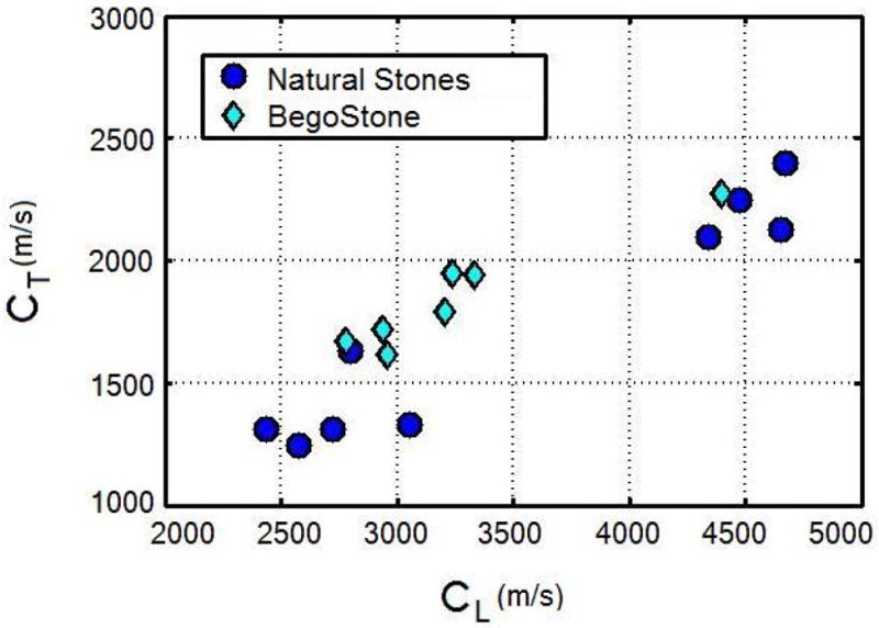
The acoustic measurements show that these composite kidney stone phantoms exhibit longitudinal wave speeds ranging from about 2700 m/s to 4400 m/s and transverse wave speeds ranging from about 1600 meters per second to 2300 m/s. These stone phantoms therefore approximate the wave speeds that have been measured for natural kidney stones (Chuong, Zhong, and Preminger, 1993b). For nine natural stones, they found the average longitudinal wave speed to be 3525 m/s and the average transverse wave speed to be 1744 m/s. The Begostone phantom data shown was measured as part of this present work. The data for natural kidney stones is that of Chuong, Zhong, and Preminger (1993b).
Conclusion
The new inorganic/organic composite kidney stone phantoms described here can be produced with both mechanical and acoustic properties that mimic the range of these properties measured for human renal calculi. These ternary composite kidney stone phantoms should have wide utility in a variety of lithotripsy studies.
Figure 4.

The longitudinal (CL) and transverse (CT) wave speeds of natural kidney stones and phantom stones.
Acknowledgments
This work was supported in part by NIH through grant RO1 DK052985. The assistance of B. Dieckman, W. Young, D. Bartlett, and E. Esch is gratefully acknowledged.
References
- Akers SR. Ph D Dissertation. Duke University; 1990. Extracorporeal Shock Wave Lithotripsy; An Experimental Lithotripter and Enhanced Kidney Stone Comminution. [Google Scholar]
- Blitz BF, Lyon ES, Gerber GS. Applicability of Iceland spar as a stone model standard for lithotripsy devices. J Endourol. 1995;9(6):449–452. doi: 10.1089/end.1995.9.449. [DOI] [PubMed] [Google Scholar]
- Cocks FH. A lattice parameter method for the investigation of solid state precipitation. J Mat Sci. 1972;7(4):771–780. [Google Scholar]
- Chuong CJ, Zhong P, Preminger GM. Acoustic and mechanical properties of renal calculi: implication in shock wave lithotripsy. J Endourol. 1993;7(6):437–444. doi: 10.1089/end.1993.7.437. [DOI] [PubMed] [Google Scholar]
- Coe F, Evan A, Worcester E. Kidney Stone Disease. J Clin Invest. 2005;115(10):2598–2608. doi: 10.1172/JCI26662. [DOI] [PMC free article] [PubMed] [Google Scholar]
- Ebrahimi F, Wang F. Fracture behavior of urinary stones under compression. J Biomed Mater Res. 1989;23(5):507–521. doi: 10.1002/jbm.820230505. [DOI] [PubMed] [Google Scholar]
- Hayden HW, Moffatt WG, Wulff J. The Structure and Properties of Materials: Vol 3: Mechanical Behavior. Wiley; New York: 1964. pp. 148–149. [Google Scholar]
- Heimbach D, Munver R, Zhong P, Jacobs J, Hesse A, Muller SC, Preminger GM. Acoustic and mechanical properties of artificial kidney stones in comparison to natural kidney stones. J Urol. 2000;164(2):537–544. [PubMed] [Google Scholar]
- Johrde LG, Cocks FH. Fracture strength of renal calculi. J Mater Sci Lett. 1985;4(10):1264–1265. [Google Scholar]
- Khan SR, Finlayson B, Hackett RL. Morphology of urinary stone particles resulting from ESWL treatment. J Urol. 1986;136:1367–1372. doi: 10.1016/s0022-5347(17)45340-7. [DOI] [PubMed] [Google Scholar]
- Liu Y, Zhong P. BegoStone - a new stone phantom for shock wave lithotripsy research. J Acoust Soc Am. 2002;112(4):1265–1268. doi: 10.1121/1.1501905. [DOI] [PubMed] [Google Scholar]
- Mellor MI, Hawkes I. Measurement of tensile strength by diametral compression of disks and annuli. Eng Geol. 1971;5:173–225. [Google Scholar]
- Vakil N, Everbach EC, Sheryl M. Relationship of model stone properties to fragmentation mechanisms during lithrotripsy. J Lithotr Stone Dis. 1991;3(4):304–310. [Google Scholar]
- Wang SJ, Yip MC. The modulus and toughness of urinary calculi. J Biomech Eng. 2002;24(18):133–134. doi: 10.1115/1.1431264. [DOI] [PubMed] [Google Scholar]
- Zhong P, Chuong CJ, Preminger GM. Characterization of fracture toughness of renal calculi using a microindentation technique. J Mater Sci Lett. 1993a;12(18):1460–1462. [Google Scholar]
- Zhong P, Chuong CJ, Preminger GM. Propagation of elastic shock waves in elastic solids caused by the impact of cavitation microjets: Part II Application to extracorporeal shock wave lithotripsy. J Acoust Soc Am. 1993b;94:119–134. doi: 10.1121/1.407088. [DOI] [PubMed] [Google Scholar]
- Zhu SL, Cocks FH, Zhong P, Preminger GM. The role of stress waves and cavitation in stone comminution in shock wave lithotripsy. Ultrasound Med Biol. 2002;28(5):661–671. doi: 10.1016/s0301-5629(02)00506-9. [DOI] [PubMed] [Google Scholar]
- Zohdi TI, Szeri AJ. Fatigue of kidney stones with heterogeneous microstructure subjected to shock wave lithotripsy. J Biomed Mater Res: B - App Biomater. 2005;57B(2):35358. doi: 10.1002/jbm.b.30307. [DOI] [PubMed] [Google Scholar]


