Abstract
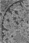
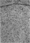
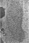
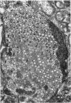
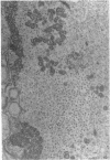
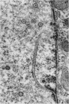
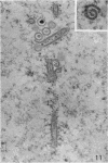
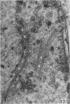
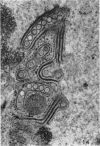
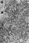
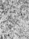
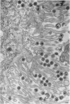
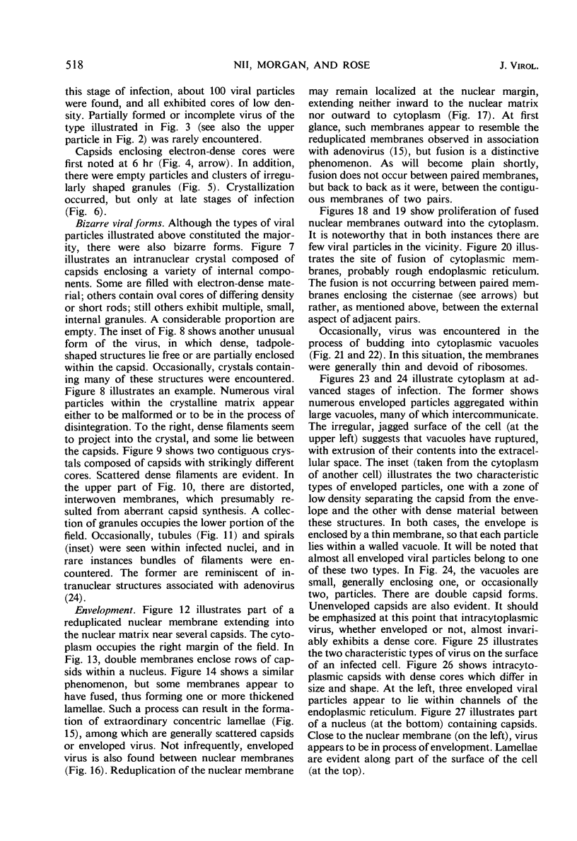
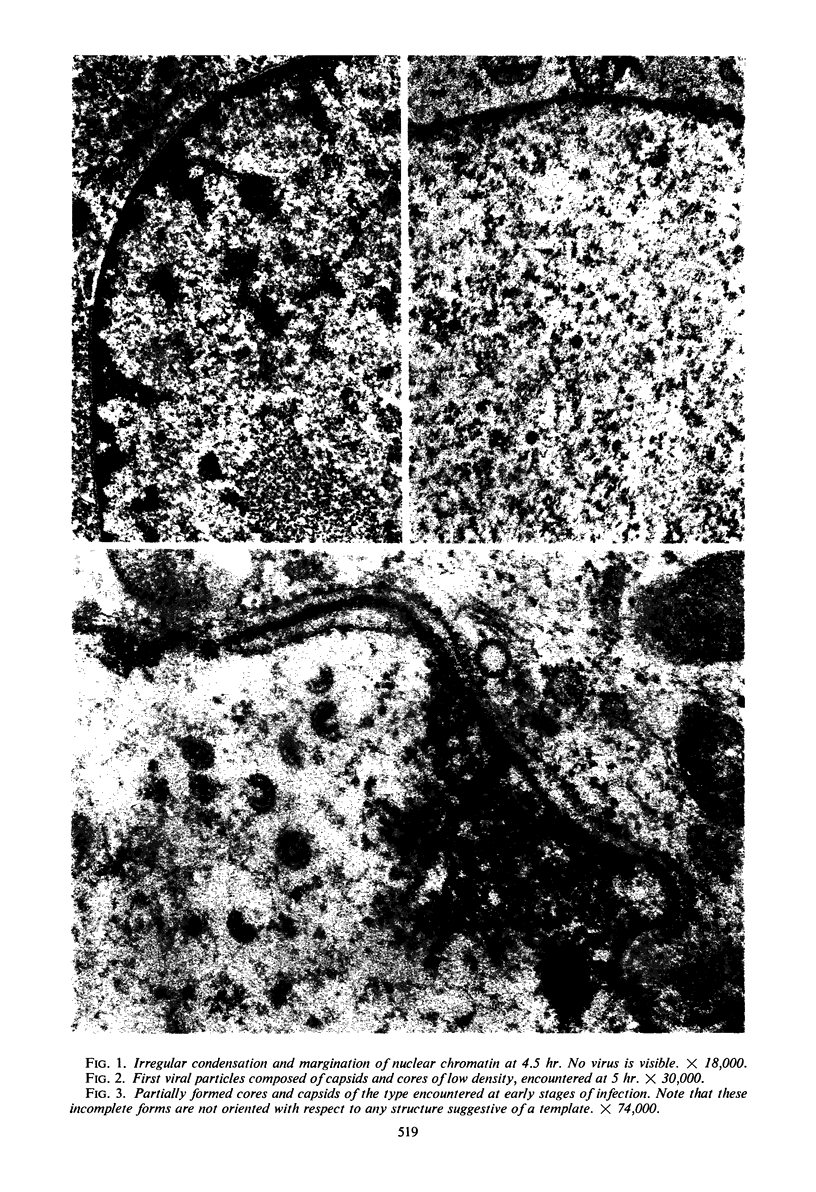
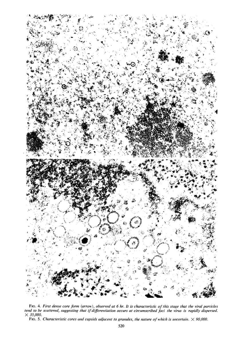
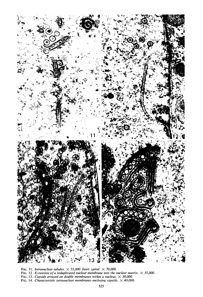
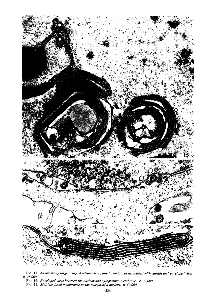
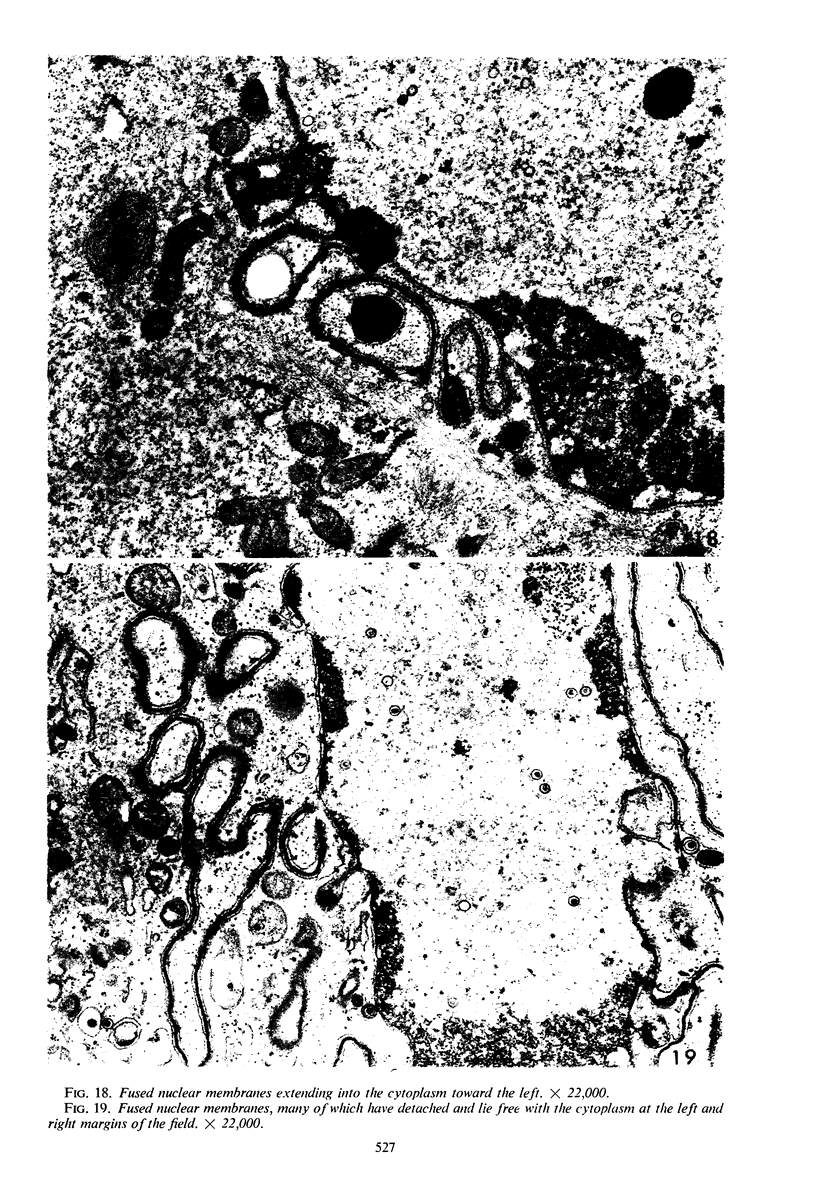
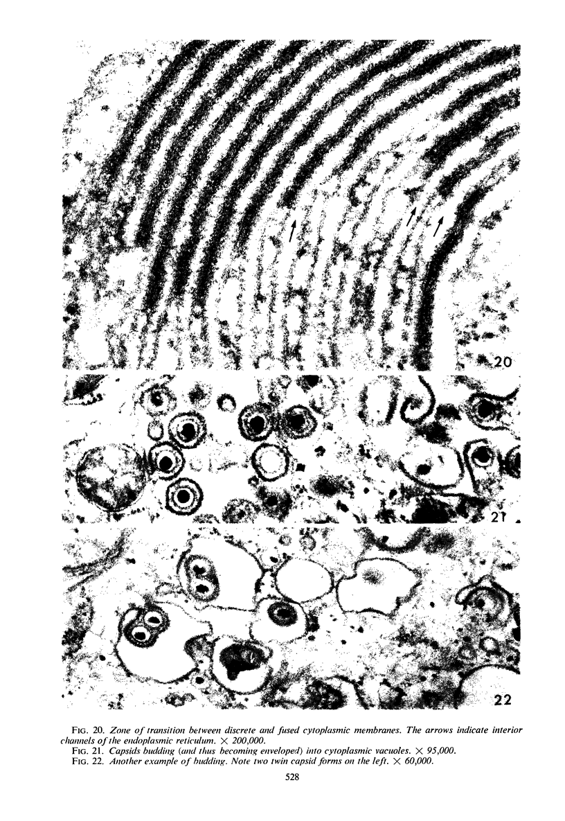
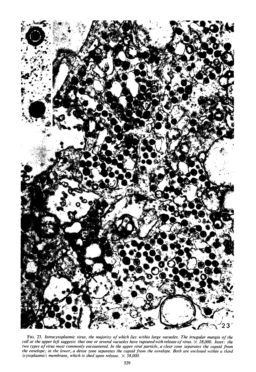
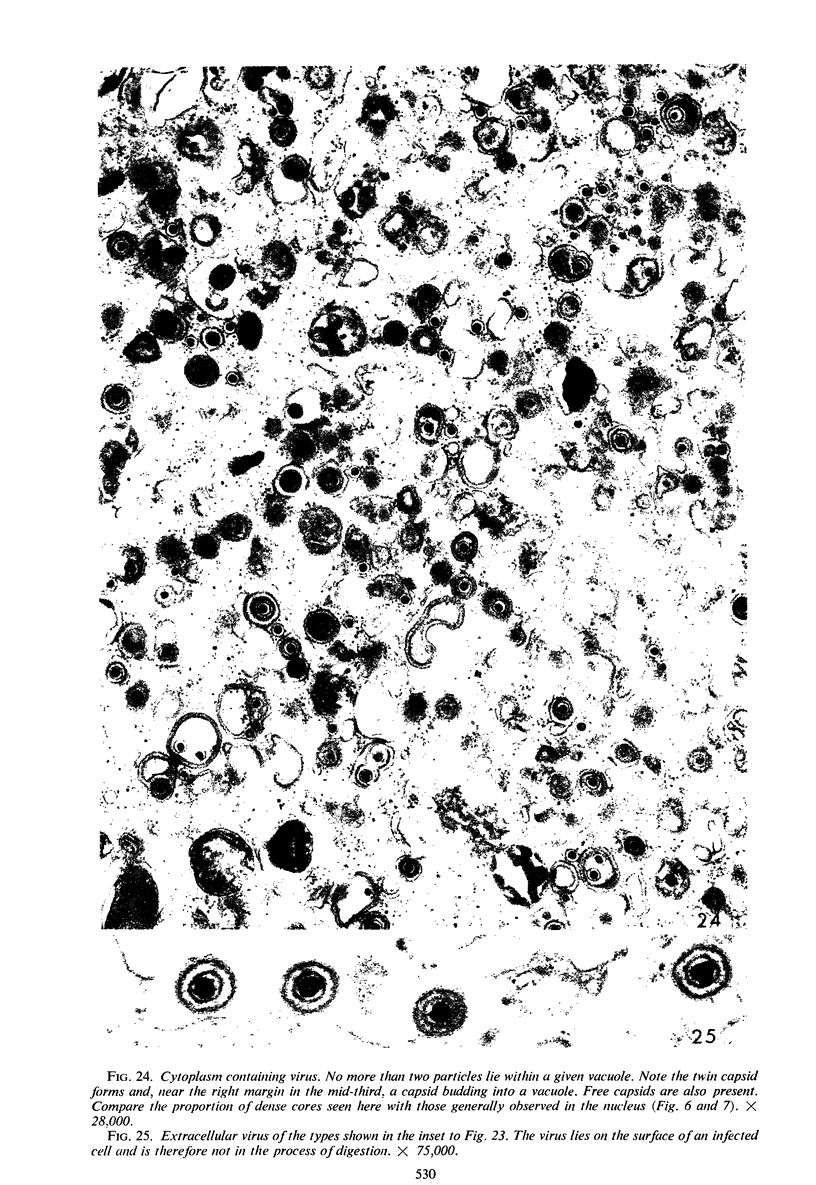
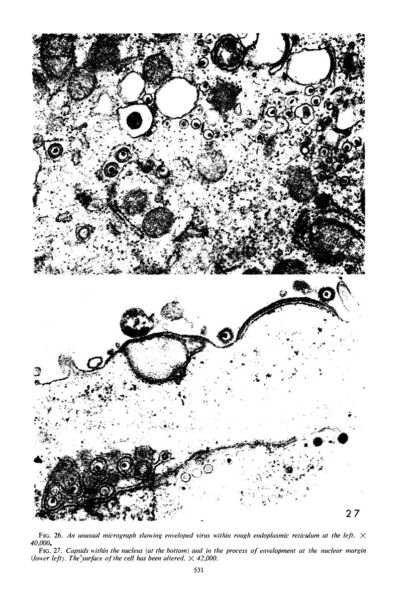
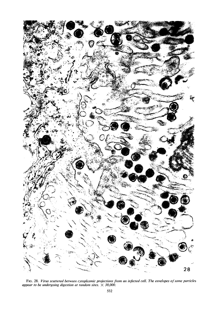
Examination of infected cells at sequential intervals after infection revealed that the first viral forms to appear were capsids enclosing cores of low density. Not until the 6th hr were dense cores encountered, and at approximately the same time enveloped virus was seen. Envelopment occurred most frequently in close proximity to the nuclear surface, although the process was also encountered within the nuclear matrix and in the cytoplasm. There was often extensive proliferation of the nuclear membrane. Envelopment of the virus by budding from the cell surface was not observed. It was concluded that enveloped virus consitutes the infectious particle and that the unenveloped capsid is unstable outside the cell. Nevertheless, it is likely that capsids enclosing infectious nucleic acid can pass directly from one cell to another after fusion has taken place.
Full text
PDF
Images in this article
Selected References
These references are in PubMed. This may not be the complete list of references from this article.
- ARMSTRONG J. A., PEREIRA H. G., ANDREWES C. H. Observations on the virus of infectious bovine rhinotracheitis, and its affinity with the Herpesvirus group. Virology. 1961 Jun;14:276–285. doi: 10.1016/0042-6822(61)90204-5. [DOI] [PubMed] [Google Scholar]
- BECKER P., MELNICK J. L., MAYOR H. D. A MORPHOLOGIC COMPARISON BETWEEN THE DEVELOPMENTAL STAGES OF HERPES ZOSTER AND HUMAN CYTOMEGALOVIRUS. Exp Mol Pathol. 1965 Feb;76:11–23. doi: 10.1016/0014-4800(65)90020-1. [DOI] [PubMed] [Google Scholar]
- BLACK F. L., MELNICK J. L. Microepidemiology of poliomyelitis and herpes-B infections: spread of the viruses within tissue cultures. J Immunol. 1955 Mar;74(3):236–242. [PubMed] [Google Scholar]
- BUTHALA D. A., MATHEWS J. STUDIES ON HERPESVIRUS SIMIAE (B-VIRUS). INTRACELLULAR DEVELOPMENT. Cornell Vet. 1963 Oct;53:494–505. [PubMed] [Google Scholar]
- Darlington R. W., James C. Biological and morphological aspects of the growth of equine abortion virus. J Bacteriol. 1966 Jul;92(1):250–257. doi: 10.1128/jb.92.1.250-257.1966. [DOI] [PMC free article] [PubMed] [Google Scholar]
- Donnellan W. L., Chantra-Umporn S., Kidd J. M. The cytomegalic inclusion cell. An elctron microscopic study. Arch Pathol. 1966 Oct;82(4):336–348. [PubMed] [Google Scholar]
- EPSTEIN M. A., HOLT S. J. ELECTRON MICROSCOPE OBSERVATIONS ON THE SURFACE ADENOSINE TRIPHOSPHATASE-LIKE ENZYMES OF HELA CELLS INFECTED WITH HERPES VIRUS. J Cell Biol. 1963 Nov;19:337–347. doi: 10.1083/jcb.19.2.337. [DOI] [PMC free article] [PubMed] [Google Scholar]
- EPSTEIN M. A. Observations on the mode of release of herpes virus from infected HeLa cells. J Cell Biol. 1962 Mar;12:589–597. doi: 10.1083/jcb.12.3.589. [DOI] [PMC free article] [PubMed] [Google Scholar]
- Epstein M. A., Achong B. G., Barr Y. M., Zajac B., Henle G., Henle W. Morphological and virological investigations on cultured Burkitt tumor lymphoblasts (strain Raji). J Natl Cancer Inst. 1966 Oct;37(4):547–559. [PubMed] [Google Scholar]
- FALKE D., SIEGERT R., VOGELL W. [Electron microscopic findings on the problem of double membrane formation in herpes simplex virus]. Arch Gesamte Virusforsch. 1959;9:484–496. [PubMed] [Google Scholar]
- FARNHAM A. E. The formation of microscopic plaques by herpes simplex virus in HeLa cells. Virology. 1958 Oct;6(2):317–327. doi: 10.1016/0042-6822(58)90085-0. [DOI] [PubMed] [Google Scholar]
- FAWCETT D. W. Electron microscope observations on intracellular virus-like particles associated with the cells of the Lucké renal adenocarcinoma. J Biophys Biochem Cytol. 1956 Nov 25;2(6):725–741. doi: 10.1083/jcb.2.6.725. [DOI] [PMC free article] [PubMed] [Google Scholar]
- GREGG M. B., MORGAN C. Reduplication of nuclear membranes in HeLa cells infected with adenovirus. J Biophys Biochem Cytol. 1959 Dec;6:539–540. doi: 10.1083/jcb.6.3.539. [DOI] [PMC free article] [PubMed] [Google Scholar]
- HOLMES I. H., WATSON D. H. AN ELECTRON MICROSCOPE STUDY OF THE ATTACHMENT AND PENETRATION OF HERPES VIRUS IN BHK21 CELLS. Virology. 1963 Sep;21:112–123. doi: 10.1016/0042-6822(63)90309-x. [DOI] [PubMed] [Google Scholar]
- Hummeler K., Henle G., Henle W. Fine structure of a virus in cultured lymphoblasts from Burkitt lymphoma. J Bacteriol. 1966 Mar;91(3):1366–1368. doi: 10.1128/jb.91.3.1366-1368.1966. [DOI] [PMC free article] [PubMed] [Google Scholar]
- LUSE S. A., SMITH M. G. Electron microscopy of salivary gland viruses. J Exp Med. 1958 May 1;107(5):623–632. doi: 10.1084/jem.107.5.623. [DOI] [PMC free article] [PubMed] [Google Scholar]
- Lunger P. D., Darlington R. W., Granoff A. Cell-virus relationships in the Lucké renal adenocarcinoma: an ultrastructure study. Ann N Y Acad Sci. 1965 Aug 10;126(1):289–314. doi: 10.1111/j.1749-6632.1965.tb14281.x. [DOI] [PubMed] [Google Scholar]
- MORGAN C., ELLISON S. A., ROSE H. M., MOORE D. H. Internal structure in virus particles. Nature. 1954 Jan 30;173(4396):208–208. doi: 10.1038/173208a0. [DOI] [PubMed] [Google Scholar]
- MORGAN C., ELLISON S. A., ROSE H. M., MOORE D. H. Structure and development of viruses as observed in the electron microscope. I. Herpes simplex virus. J Exp Med. 1954 Aug 1;100(2):195–202. doi: 10.1084/jem.100.2.195. [DOI] [PMC free article] [PubMed] [Google Scholar]
- MORGAN C., ROSE H. M., HOLDEN M., JONES E. P. Electron microscopic observations on the development of herpes simplex virus. J Exp Med. 1959 Oct 1;110:643–656. doi: 10.1084/jem.110.4.643. [DOI] [PMC free article] [PubMed] [Google Scholar]
- Middelkamp J. N., Patrizi G., Reed C. A. Light and electron microscopic studies of the guinea pig cytomegalovirus. J Ultrastruct Res. 1967 Apr;18(1):85–101. doi: 10.1016/s0022-5320(67)80233-8. [DOI] [PubMed] [Google Scholar]
- ORNSTEIN L. Mitochondrial and nuclear interaction. J Biophys Biochem Cytol. 1956 Jul 25;2(4 Suppl):351–352. doi: 10.1083/jcb.2.4.351. [DOI] [PMC free article] [PubMed] [Google Scholar]
- PATRIZI G., MIDDELKAMP J. N., HERWEG J. C., THORNTON H. K. HUMAN CYTOMEGALOVIRUS: ELECTRON MICROSCOPY OF A PRIMARY VIRAL ISOLATE. J Lab Clin Med. 1965 May;65:825–838. [PubMed] [Google Scholar]
- Patrizi G., Middelkamp J. N., Reed C. A. Reduplication of nuclear membranes in tissue-culture cells infected with guinea-pig cytomegalovirus. Am J Pathol. 1967 May;50(5):779–790. [PMC free article] [PubMed] [Google Scholar]
- RUEBNER B. H., HIRANO T., SLUSSER R. J., MEDEARIS D. N., Jr HUMAN CYTOMEGALOVIRUS INFECTION. ELECTRON MICROSCOPIC AND HISTOCHEMICAL CHANGES IN CULTURES OF HUMAN FIBROBLASTS. Am J Pathol. 1965 Mar;46:477–496. [PMC free article] [PubMed] [Google Scholar]
- RUEBNER B. H., MIYAI K., SLUSSER R. J., WEDEMEYER P., MEDEARIS D. N., Jr MOUSE CYTOMEGALOVIRUS INFECTION. AN ELECTRON MICROSCOPIC STUDY OF HEPATIC PARENCHYMAL CELLS. Am J Pathol. 1964 May;44:799–821. [PMC free article] [PubMed] [Google Scholar]
- Ruebner B. H., Hirano T., Slusser R., Osborn J., Medearis D. N., Jr Cytomegalovirus infection. Viral ultrastructure with particular reference to the relationship of lysosomes to cytoplasmic inclusions. Am J Pathol. 1966 Jun;48(6):971–989. [PMC free article] [PubMed] [Google Scholar]
- SCOTT T. F., McLEOD D. L., TOKUMARU T. A biologic comparison of two strains of Herpesvirus hominis. J Immunol. 1961 Jan;86:1–12. [PubMed] [Google Scholar]
- SMITH K. O. RELATIONSHIP BETWEEN THE ENVELOPE AND THE INFECTIVITY OF HERPES SIMPLEX VIRUS. Proc Soc Exp Biol Med. 1964 Mar;115:814–816. doi: 10.3181/00379727-115-29045. [DOI] [PubMed] [Google Scholar]
- STOKER M. G. Mode of intercellular transfer of herpes virus. Nature. 1958 Nov 29;182(4648):1525–1526. doi: 10.1038/1821525a0. [DOI] [PubMed] [Google Scholar]
- STOKER M. G., SMITH K. M., ROSS R. W. Electron microscope studies of HeLa cells infected with herpes virus. J Gen Microbiol. 1958 Oct;19(2):244–249. doi: 10.1099/00221287-19-2-244. [DOI] [PubMed] [Google Scholar]
- Shipkey F. H., Erlandson R. A., Bailey R. B., Babcock V. I., Southam C. M. Virus biographies. II. Growth of herpes simplex virus in tissue culture. Exp Mol Pathol. 1967 Feb;6(1):39–67. doi: 10.1016/0014-4800(67)90005-6. [DOI] [PubMed] [Google Scholar]
- Siegert R., Falke D. Elektronenmikroskopische Untersuchungen über die Entwicklung des Herpesvirus hominis in Kulturzellen. Arch Gesamte Virusforsch. 1966;19(2):230–249. [PubMed] [Google Scholar]
- Siminoff P., Menefee M. G. Normal and 5-bromodeoxyuridine-inhibited development of herpes simplex virus. An electron microscope study. Exp Cell Res. 1966 Nov-Dec;44(2):241–255. doi: 10.1016/0014-4827(66)90429-0. [DOI] [PubMed] [Google Scholar]
- Sirtori C., Bosisio-Bestetti M. Nucleolar changes in KB tumor cells infected with herpes simplex virus. Cancer Res. 1967 Feb;27(2):367–376. [PubMed] [Google Scholar]
- Swanson J. L., Craighead J. E., Reynolds E. S. Electron microscopic observations on Herpesvirus hominis (herpes simplex virus) encephalitis in man. Lab Invest. 1966 Dec;15(12):1966–1981. [PubMed] [Google Scholar]
- WATSON D. H., WILDY P., RUSSELL W. C. QUANTITATIVE ELECTRON MICROSCOPY STUDIES ON THE GROWTH OF HERPES VIRUS USING THE TECHNIQUES OF NEGATIVE STAINING AND ULTRAMICROTOMY. Virology. 1964 Dec;24:523–538. doi: 10.1016/0042-6822(64)90204-1. [DOI] [PubMed] [Google Scholar]
- WATSON D. H., WILDY P. SOME SEROLOGICAL PROPERTIES OF HERPES VIRUS PARTICLES STUDIED WITH THE ELECTRON MICROSCOPE. Virology. 1963 Sep;21:100–111. doi: 10.1016/0042-6822(63)90308-8. [DOI] [PubMed] [Google Scholar]
- Zambernard J., Mizell M. Viral particles of the frog renal adenocarcinoma: causative agent or passenger virus? I. Fine structure of primary tumors and subsequent intraocular transplants. Ann N Y Acad Sci. 1965 Aug 10;126(1):127–145. doi: 10.1111/j.1749-6632.1965.tb14272.x. [DOI] [PubMed] [Google Scholar]


