Abstract
Background
The controversy regarding the outcome of total knee arthroplasties after high tibial osteotomy may relate to malalignment secondary to overcorrection after high tibial osteotomy (HTO) [1, 2] and to the type of arthroplasty itself (posterior-stabilized arthroplasty or posterior cruciate ligament-retaining prosthesis).
Questions/Purpose
We asked two questions: (1) Would a posterior-stabilized arthroplasty provide sufficient constrain and improve pain and function in patients with severe malalignment due to a previous HTO? (2) Will malalignment of the previous HTO jeopardize the long-term results of a total knee reconstruction with a posterior-stabilized implant?
Patients and Methods
We retrospectively reviewed 25 posterior-stabilized TKAs in 25 patients with severe valgus deformity after HTO (ranging from 10° to 20° of valgus) and compared the results with a series of matched 25 posterior-stabilized TKAs in 25 patients with normocorrection after HTO ranging from 5° of valgus to 5° of varus. Clinical, operative, and radiographic data were reviewed. Minimum follow-up was 10 years after the arthroplasty (average, 15 years; range, 10–20 years).
Results
All the knees had standard posterior-stabilized total knee arthroplasty implants. Patients with an overcorrected HTO were more likely to require a soft tissue release to balance the knee. However, Average Knee Society and Function Score improved, respectively, from 48 to 85 and from 50 to 90 points in the severely overcorrected group, versus, respectively, 50 to 89 and 52 to 97 in the normocorrected group, but the range of mobility was superior for patients with normal alignment. Fifteen-year survivorship after the arthroplasty comparison showed no significant difference between the two groups (one revision in each group).
Conclusions
Patients with an overcorrected HTO are more likely to require a soft tissue release to balance the knee. However, both groups show improvements in function and pain. With a posterior-stabilized arthroplasty, the degree of deformity has no impact on the longevity of the TKA.
Electronic supplementary material
The online version of this article (doi:10.1007/s11420-013-9344-x) contains supplementary material, which is available to authorized users.
Keywords: total knee arthroplasty, high tibial osteotomy, posterior-stabilized arthroplasty
Introduction
The published clinical results of total knee arthroplasty (TKA) after high tibial osteotomy (HTO) are controversial [10–14]: some studies report no clinical or radiographic difference in TKAs with or without a previous osteotomy [7, 12] while others see substandard outcomes in TKAs after a prior HTO [10, 13, 14]. This controversy may be in relation with malalignment secondary to overcorrection after HTO [1, 4, 8, 9] and with the type of arthroplasty (posterior-stabilized arthroplasty vs. posterior cruciate ligament-retaining prosthesis). However, no series has compared TKA performed on knees with malalignment secondary to large overcorrection after HTO with TKA performed on knees with normocorrection after HTO, using only a posterior-stabilized arthroplasty.
To address this controversy, we therefore retrospectively reviewed 25 posterior-stabilized arthroplasties in 25 patients with severe valgus deformity (ranging from 10° to 20° of valgus, i.e., hip–knee–ankle angle ranging from 190° to 200°) after closed wedge HTO and compared the results with a series of the same matched 25 TKAs in 25 patients with normocorrection (ranging from 5° of valgus to 5° of varus after closed wedge HTO, i.e., HKA from 175° to 185°). The hip–knee–ankle angle of 180° is considered as a straight line going through the center of the femoral head, the center of the knee and the center of the ankle as we previously reported [8]. We asked two questions: (1) Would a posterior-stabilized arthroplasty provide sufficient constrain and improve pain and function in patients with severe malalignment due to a previous HTO? (2) Will malalignment of the previous HTO jeopardize the long-term results of a total knee reconstruction with a posterior-stabilized implant?
Patients and Methods
We retrospectively identified all the patients with severely overcorrected HTO who underwent TKAs between January 1990 and December 2000. The preoperative deformities were measured on hip to ankle standing films. A severely overcorrected knee was a knee with a hip–knee–ankle angle (HKA) superior to 190°, which means more than 10° of valgus for the mechanical axis. There were 10 men and 15 women who underwent 25 TKAs in this overcorrected group. These patients were compared to a randomly chosen control group of 25 patients (matched for age at the time of arthroplasty and sex, but not for weight) who underwent a TKA after HTO with a HKA angle between 175° and 185° (between 5° of varus and 5° of valgus for the mechanical axis) during the same period. The mean age at revision of TKA was 65 years (range, 48–75 years). In the group with severe overcorrection, one patient was lost to follow-up and two died before the 10 mark. In the group with normocorrection, two patients were lost to follow-up and three died before 10 years. The minimum follow-up was 10 years in both groups (mean, 15 years; range, 10–20 years). Complications included resolved peroneal nerve palsies in two knees with preoperative overcorrection.
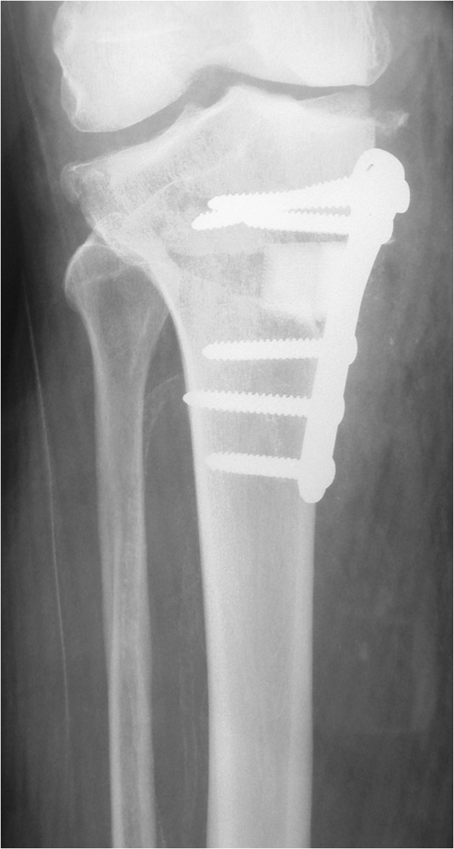
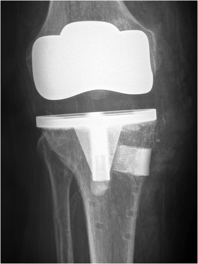
All the knees received the same implant (posterior-stabilized Ceraver Hermes TKA—Ceraver Osteal, Roissy, France) which incorporates a femoral cam mechanism that articulates with a tibial post to act as a functional substitute for the PCL (Figs. 1 and 2).
Fig. 1.

Osteotomy with severe overcorrection anteroposterior view of the knee with the patient standing.
Fig. 2.

Correction of the knee after arthroplasty; anteroposterior view of the knee with the patient standing.
All the patients were assessed postoperatively (2 weeks, 6 weeks, 3 months, 12 months, and every year thereafter) and at follow-up using Knee Society Scores and Function Scores, and any complication was recorded. Operative records were analyzed and cases that necessitated special techniques during the procedure were identified: i.e., extensile approaches and exposure (tibial tuberosity osteotomy or a quadriceps snip [18]), management of deformity with navigation, soft tissue releases or lateral epicondyle sliding osteotomies to achieve balanced gaps, and lateral retinacular releases to obtain central patellar tracking when necessary. Radiographic assessment of the prosthesis was performed (each year and at the most recent follow-up) according to alignment by the HKA angle, the number of radiolucent lines and the presence of loosening [5]. Patella height was also assessed radiographically on a lateral view using the Caton Index [3]; the normal ratio is 1 ± 0.2; a ratio >1.1 indicates a high patella, whereas a ratio <0.6 indicates a low patella. All data were obtained from the medical records, and no patients were called specifically for this study.
Statistical Analysis
Data collection and analysis were performed using SPSS version 13.0. Differences between the two groups were examined and compared using the Mann–Whitney test and one-way analysis of variance (ANOVA) and Pearson chi-square test.
Results
There was no statistical difference in postoperative alignment between the two groups (overcorrected and normocorrected; p = 0.32; Table 1). Patella baja (p = 0.05) and the use of special surgical techniques were more frequent (30 cases versus 4 cases; p = 0.01) when performing TKA in patients of the group with severe overcorrection. The average Knee Society Score and Function Score showed no significant difference; (p = 0.34) between both groups, (from 48 to 85 and from 50 to 90 points, respectively, in the severely overcorrected group, versus 50 to 89, and 52 to 97, respectively, in the normocorrected group). Only postoperative flexion improved significantly (p < 0.01) from 115° to 127° in the control group versus 83° to 108° in the overcorrected HTO group.
Table 1.
Differences between the two groups of total knee arthroplasties with surgery after HTO
| Group with severe overcorrection n = 25 | Control group n = 25 | |
|---|---|---|
| Operative records (number) | ||
| Quadriceps snip | 1 | 0 |
| Tibial tuberosity detachment | 5 | 0 |
| Soft tissue expander | 1 | 0 |
| Computer assistance navigation | 6 | 0 |
| Soft tissue release | 9 | 1 |
| Epicondyle osteotomy | 2 | 0 |
| Lateral retinacular release | 6 | 3 |
| Radiological results (mean, range) | ||
| Alignment (HKA angle) | 181° (179–184°) | 180° (177–183°) |
| Caton index | 0.62 (0.52–0.91) | 0.75 (0.59–1.05) |
Malalignment of the previous HTO did not jeopardize the longevity of the posterior-stabilized arthroplasty. No radiolucent lines were observed around the femoral component suggesting loosening or requiring close surveillance in either of the groups. Three of the 25 TKAs after overcorrected HTO had suspected tibial radiolucencies, whereas no suspected radiolucencies were seen in the control group. Comparison of 15-year survivorship rates after arthroplasty showed no significant difference (p = 0.24) between the two groups (one revision in each group).
Discussion
The controversy regarding the outcome of total knee arthroplasty after high tibial osteotomy may relate to the degree of malalignment secondary to overcorrection after HTO and with the type of arthroplasty (posterior-stabilized arthroplasty or posterior cruciate ligament-retaining prosthesis).
We asked two questions: (1) Would a posterior-stabilized arthroplasty provide sufficient constrain and improve pain and function in patients with severe malalignment due to a previous HTO; (2) will malalignment of the previous HTO jeopardize the long-term results of a total knee reconstruction with a posterior-stabilized implant.
We found that patients with an overcorrected HTO had improvements in function and pain after a posterior-stabilized arthroplasty and that the degree of deformity has no impact on the longevity of the TKA. However we observed technical difficulties, inferior clinical results on flexion, and resolved peroneal palsies
Our study has some limitations as it is a retrospective study and the number of patients is small. Matching was only possible to a certain extent, limited by patient numbers and availability of data. Other factors that might have influenced the outcome were not matched such as weight and patella height at the time of arthroplasty, and age of the patient at the time of osteotomy. Although surgically challenging, TKA in the context of a previous HTO is a reasonable treatment option to provide pain relief and improved function even when the previous HTO was severely overcorrected. In this study where a posterior-stabilized TKA was always used, alignment was obtained and clinical results revealed consistent improvements in pain and function, but with inferior clinical results on mobility as compared with those observed in patients with a previous osteotomy without severe overcorrection.
As described in other series [13, 15, 16], we confirm with our series of patients that after a closed osteotomy with severe overcorrection technical difficulties needing release [2, 8], and sometimes snipping of the quadriceps [6] or osteotomy of the anterior tibial tuberosity were more common. A reduced rate of satisfactory results and increased difficulty in exposing the knee was reported by Windsor et al. [18] as early as the mid-1980s. The great variability reported in the literature [16–18] with respect to the results of knee joint arthroplasty after failure of a tibial osteotomy is, in our opinion, due to the wide heterogeneity of patients undergoing joint arthroplasty after osteotomy who have varying degrees of deformity on the frontal and sagittal planes, bone loss, ligament imbalance, low patella, and damaged soft tissues. All the types of prostheses implanted cannot possibly resolve all of these problems. In our series, it was also difficult to maintain the appropriate tension of the medial and lateral ligaments and to balance the flexion and extension gap because of the tibial bone defect, but the posterior-stabilized TKA appears more forgiving for this problem than a posterior cruciate ligament-retaining prosthesis [10–12].
The degree of deformity had no impact on the longevity of the posterior-stabilized TKA. Mode of failure in TKA includes osteolysis, malalignment, or malpositioning. In the present study, radiolucencies were only present in three tibial components in the overcorrected group. Given the extended follow-up of the present study group (average 15 years), drawing a conclusion about loosening is therefore possible to a certain extent.
As observed by some authors [11, 14] we were unable to show significant differences in migration, alignment, or positioning of the TKA components between the two groups. In contrast to our results, other authors [12, 13, 17, 18] noticed a higher rate of femoral and tibial component loosening after previous HTO when using posterior cruciate ligament-retaining prostheses.
Electronic supplementary materials
(PDF 510 kb)
(PDF 510 kb)
(PDF 510 kb)
(PDF 510 kb)
(PDF 510 kb)
(PDF 510 kb)
Disclosures
Conflict of Interest:
Philippe Hernigou, MD, Pascal Duffiet, MD, Didier Julian, MD, Issac Guissou, MD, Alexandre Poignard, MD, and Charles Henri Flouzat-Lachaniette, MD have declared that they have no conflict of interest.
Human/Animal Rights:
All procedures followed were in accordance with the ethical standards of the responsible committee on human experimentation (institutional and national) and with the Helsinki Declaration of 1975, as revised in 2008 (5).
Informed Consent:
Informed consent was obtained from all patients for being included in the study.
Required Author Forms
Disclosure forms provided by the authors are available with the online version of this article.
References
- 1.Aglietti P, Buzzi R, Vena LM, Baldini A, Mondaini A. High tibial valgus osteotomy for medial gonarthrosis: a 10- to 21-year study. J Knee Surg. 2003;16(1):21–26. [PubMed] [Google Scholar]
- 2.Buechel FF. A sequential three-step lateral release for correcting fixed valgus knee deformities during total knee arthroplasty. Clin Orthop Relat Res. 1990;260:170–175. [PubMed] [Google Scholar]
- 3.Caton J. Patella Infera. Apropos of 128 cases. Rev Chir Orthop Réparatrice Appar Mot. 1982;68(5):317–325. [PubMed] [Google Scholar]
- 4.Coventry MB. Osteotomy about the knee for degenerative and rheumatoid arthritis: indications, operative technique, and results. J Bone Joint Surg Am. 1973;55(1):23–48. [PubMed] [Google Scholar]
- 5.Ewald FC. The Knee Society total arthroplasty roentgenographic evaluation and scoring system. Clin Orthop Relat Res. 1989;248:9–12. [PubMed] [Google Scholar]
- 6.Garvin KL, Scuderi GR, Insall JN. Evolution of the quadriceps snip. Clin Orthop Relat Res. 1995;321:131–137. [PubMed] [Google Scholar]
- 7.Haddad FS, Bentley G. Total Knee arthroplasty after high tibial osteotomy. A medium term review. J Arthroplasty. 2000;15(5):597–603. doi: 10.1054/arth.2000.6621. [DOI] [PubMed] [Google Scholar]
- 8.Hernigou P, Medevielle D, Debeyre J, Goutallier D. Proximal tibial osteotomy for osteoarthritis with varus deformity. A ten to thirteen-year follow-up study. J Bone Joint Surg Am. 1987;69(3):332–354. [PubMed] [Google Scholar]
- 9.Insall JN, Joseph DM, Msika C. High tibial osteotomy for varus gonarthrosis. A long-term follow-up study. J Bone Joint Surg Am. 1984;66(7):1040–1048. [PubMed] [Google Scholar]
- 10.Katz MM, Hungerford DS, Krackow KA, Lennox DE. Results of knee arthroplasty after failed proximal tibial osteotomy for osteoarthritis. J Bone Joint Surg Am. 1987;69(2):225–233. [PubMed] [Google Scholar]
- 11.KrackowKA HJL. Experience with a new technique for managing severely overcorrected valgus high tibial osteotomy at total knee arthroplasty. Clin Orthop Relat Res. 1990;258:213–224. [PubMed] [Google Scholar]
- 12.Meding JB, Keating EM, Ritter MA, Faris PM. Total knee arthroplasty after high tibial osteotomy. A comparison study in patients who had bilateral total knee replacement. J Bone Joint Surg Am. 2000;82(9):1252–1259. doi: 10.2106/00004623-200009000-00005. [DOI] [PubMed] [Google Scholar]
- 13.Mont MA, Alexander N, Krackow KA, Hungerford DS. Total knee arthroplasty after failed proximal tibial valgus osteotomy. A comparison with a matched group. Clin Orthop Relat Res. 1994;299:125–130. [PubMed] [Google Scholar]
- 14.Nelson CL, Haas SB. Total knee arthroplasty following high tibial osteotomy. In: Sculco TP, Martucci EA, editors. Knee arthroplasty. Wien: Springer; 2002. pp. 91–10123. [Google Scholar]
- 15.Scuderi GR, Windsor RE, Insall JN. Observation on patellar height after proximal tibial osteotomy. J Bone Joint Surg Am. 1989;71(2):245–248. [PubMed] [Google Scholar]
- 16.Whiteside LA, Ohl MD. Tibial tubercle osteotomy for exposure of the difficult total knee arthroplasty. Clin Orthop Relat Res. 1990;260:6–9. [PubMed] [Google Scholar]
- 17.Whiteside LA. Selective ligament release in total knee arthroplasty of the knee in valgus. Clin Orthop Relat Res. 1999;367:130–140. doi: 10.1097/00003086-199910000-00016. [DOI] [PubMed] [Google Scholar]
- 18.Windsor RE, Insall JN, Vince KG. Technical considerations of total knee arthroplasty after proximal tibial osteotomy. J Bone Joint Surg Am. 1988;70(4):547–555. [PubMed] [Google Scholar]
Associated Data
This section collects any data citations, data availability statements, or supplementary materials included in this article.
Supplementary Materials
(PDF 510 kb)
(PDF 510 kb)
(PDF 510 kb)
(PDF 510 kb)
(PDF 510 kb)
(PDF 510 kb)


