Sir,
Hypoxia in intensive care unit (ICU) is not uncommon and if managed well in time has no sequelae. But if it goes unnoticed, it has serious consequences. There are multiple causes of hypoxia in ICU like bronchospasm, secretions, tube obstruction, circuit misconnection, low inspired oxygen, ventilation perfusion mismatch due to lung pathology, severe hypoperfusion, etc.
We report a case of old male patient weighing 72 kg ASA (American Society of Anaesthesiologists physical status classification) 2 who underwent maxillectomy for maxillary carcinoma. He was a known hypertensive from last one year, controlled on tab amlodipine once daily. All other investigations were within normal limits. He was posted in elective operation theatre with consent for ventilation. Airway examination showed mallampatti I and three finger breadth mouth opening. Though the airway did not seem to be difficult but extubation could be a problem owing to airway edema post-operatively. General anesthesia was induced with inj. propofol, fentanyl and vecuronium, flexometallic tube 7.0 mm i.d. was inserted and it was maintained with O2, air and isoflurane. Patient's trachea was not extubated and patient was shifted to ICU with re-inforced tube for ventilation and further management.
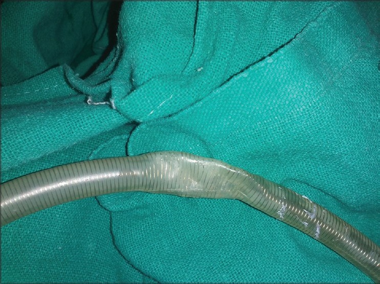
Patient was kept on controlled mode of ventilation for initial 2 hours in ICU. After that sedation was continued and paralysis was stopped. The mode of ventilation was switched to SIMV (synchronized intermittent mandatory ventilation). After thirty minutes, the patient's saturation started dropping. The circuit's integrity was checked and was found to be intact. On auscultation there were markedly reduced breath sounds. Thinking it in terms of bronchospasm nebulization was started and theophyllin and hydrocortisone were given stat. Then endotracheal suction was attempted. To our surprise suction catheter could not be negotiated through the endotracheal tube. Then it became evident that the tube was completely blocked at the level of incisors [Figure 1]. The tube was immediately withdrawn followed by mask ventilation with 100% oxygen. The patient's arterial saturation increased and now his trachea was re-intubated with portex cuffed endotracheal tube. Next morning laryngoscopy revealed minimal edema and his trachea was extubated. The patient was transferred back to the respective ward in the evening.
Figure 1.

Kink at the level of the incisors
Flexomettalic tubes are not recommended in intensive care settings. But there are many cases like hemiglossectomy, oral surgeries involving tongue, palate, and cheek which mandate the use of such tubes intra-operatively. At the end of the surgery airway becomes so edematous that replacing the tube with portex becomes difficult. Fear of losing the airway is such a night mare that these patients are shifted to ICU with reinforced tubes.
Obstruction of re-inforced endotracheal tube has been reported many a times in the literature. Most common cause of obstruction being repeated usage and use of nitrous oxide that may cause dissection of re-inforced tube.[1,2] There may be development of bubbles in the wall that expand on exposure to nitrous oxide. The bubbles may appear in the tube owing to ethylene oxide sterilization or repeated usage.[3,4,5]
Obstruction of such tubes has also been seen after prone positioning but it has never been reported in critical care settings.[4] In our case the patient was shifted to ICU with flexomettalic tube in view of edematous airway. The development of this critical obstruction was due to inadequate sedation that made the patient to bite the tube. There should be a bite block or an airway or even a rolled gauge that would have avoided this situation.
One should be aware of such complications in the ICU when we receive such a patient. Always take care of reinforced tubes with proper bite blocks and sedation. Consider change of endotracheal tube as soon as edema subsides if the patient needs prolonged ventilation. Still always remember this complication with reinforced tubes being used.
Once a suction catheter cannot be passed one can go for fiber-optic evaluation of the tube but that is possible only if the patient's saturation and condition permits. Other causes such as oxygen supply, ventilator malfunction, circuit disconnection, pneumothorax, etc., need to be checked in cases of sudden desaturation.
REFERENCES
- 1.Balakrishna P, Shetty A, Bhat G, Raveendra U. Ventilatory obstruction from kinked armoured tube. Indian J Anaesth. 2010;54:355–6. doi: 10.4103/0019-5049.68380. [DOI] [PMC free article] [PubMed] [Google Scholar]
- 2.Populaire C, Robard S, Souron R. An armoured endotracheal tube obstruction in a child. Can J Anaesth. 1989;36:331–2. doi: 10.1007/BF03010775. [DOI] [PubMed] [Google Scholar]
- 3.Tose R, Kubota T, Hirota K, Sakai T, Ishihara H, Matsuki A. Obstruction of an reinforced endotracheal tube due to dissection of internal tube wall during total intravenous anesthesia. Masui. 2003;52:1218–20. [PubMed] [Google Scholar]
- 4.Rajkumar A, Bajekal R. Intraoperative airway obstruction due to dissection of a reinforced endotracheal tube in a prone patient. J Neurosurg Anesthesiol. 2011;23:377. doi: 10.1097/ANA.0b013e31822cf882. [DOI] [PubMed] [Google Scholar]
- 5.Rouco Martínez I, Catalá Puigbó E, Baxarias Gascón P. Internal blistering of armored latex endotracheal tubes. Rev Esp Anestesiol Reanim. 1984;31:208–10. [PubMed] [Google Scholar]


