Abstract
Pulpal necrosis in young permanent teeth often results in teeth with open apex, thin root walls and poor crown root ratio. Out of the available treatment options maturogenesis has been the most conservative option that exploits full potential of pulp for dentin deposition. Maturogenesis involves disinfecting the root canal system followed by stimulation of blood clot from the periapical tissue, which provides a matrix into which the cell could grow and sealing the coronal excess. In the present case report, tri antibacterial paste (3 Mix) was used as an intracanal medicament that proved successful in stimulating vital pulp cells of the periapical region for maturogenesis. Five months radiograph follow-up showed thickening of lateral dentinal walls, which progress until 15 months resulting in apical closure, thickening of lateral dentinal walls and increase root length.
Keywords: Apexification, apical papilla, maturogenesis, non-vital immature, permanent teeth, tri antibacterial paste
Introduction
Pulpal necrosis is a frequent sequale of trauma to the anterior teeth and if it occurs in young permanent teeth, this will result in the cessation of root development.[1] Cessation of root development results in teeth with open apex, thin root walls and poor crown root ratio that are difficult to instrument and impossible to seal. Various treatment options exist to manage non-vital young permanent teeth which includes non-surgical root canal treatment (apexification), single visit apexification, apical surgery and extraction.[1] Traditionally customized gutta-percha cone was used to obturate immature canal space, but there is a danger of root fracture during lateral condensation. Long-term calcium hydroxide therapy was considered as the ideal treatment for such teeth, but this therapy has its own disadvantages, like multiple visits, relatively long period of time and alteration of mechanical properties of dentin.[2] Recently, single visit apexification using mineral trioxide aggregate (MTA) has gained popularity. Although this technique is faster compared to traditional apexification, but leaves the tooth with poor crown root ratio and prone to fracture.[3] Recently maturogenesis or pulpal revascularization is considered as the ideal treatment for non-vital immature permanent teeth. Maurogenesis stimulates regeneration of a functional pulp dentin complex that allows continued root development, thickening of dentinal walls and apical closure.[4] Obtaining and maintaining sterile root canal is one of the most important steps during maturogenesis. Combination of antibiotic drugs, i.e., ciprofloxacin, metronidazole and minocycline (3 Mix) has been used to maintain sterile root canal due to its broad spectrum antibacterial property, low toxicity and biocompatibility.[5] The present case reports describe successful maturogenesis of permanent maxillary right central incisor in a 10-year-old child, which sets another example to the mainstream of such treatment option.
Case Report
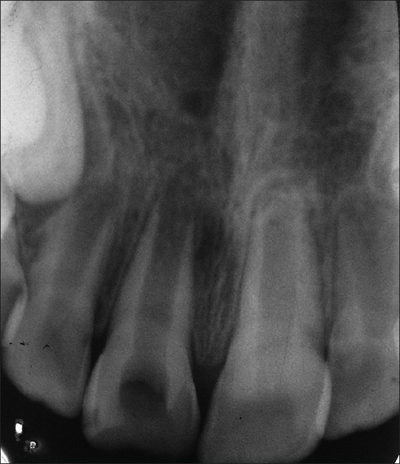
A 10-year-old girl with non-contributory medical history reported to the Department of Pediatric Dentistry, Government Dental College and Hospital, Nagpur for the treatment of pain and swelling in maxillary anterior region. History revealed trauma to maxillary anterior region 6 month back while playing in school. Clinical examination revealed fractured and discolored permanent maxillary right central incisor. Soft-tissue examination in relation to permanent maxillary right central incisor showed soft-tissue swelling of 1 × 1 cm. The tooth was tender to vertical percussion and showed negative response to thermal (cold test [Polfofluorange Pharma Dental Handelsges]) and electric pulp testing (Parkell Farmingdate). Intraoral periapical radiograph of permanent maxillary right central incisor revealed incompletely formed apex with periapical rarefaction [Figure 1]. The tooth was diagnosed with a pulpal necrosis and chronic periapical abscess. After rubber dam isolation and gaining access to the root canal system, the canal debris was loosen with minimal instrumentation and the root canal system was slowly irrigated with 0.5% sodium hypochlorite to prevent further damage to the survived apical papilla. Minimal instrumentation within the root canal system prevents further weakening of the root canal system. A light plain cotton pellet was placed in the pulp chamber for 48 h to facilitate purulent drainage. At the next visit working length was measured using diagnostic radiograph, followed by very minimal instrumentation and copious irrigation with normal saline and the canal was dried with sterile paper points. Mixture of ciprofloxacin, metronidazole and minocycline (3 Mix) was placed in the canal and coronal access was sealed with zinc oxide eugenol cement (Dentifiss India Ltd. Mumbai). Patient was recalled after 1 week, the tooth was asymptomatic and the canal was found to be dry.
Figure 1.

Intraoral periapical radiograph of 11 showing incompletely formed apex with periapical rarefaction
Maturogenesis procedure
A 23 gauge sharp sterile needle was pushed with a sharp stroke beyond the working length of the canal into the periapical tissue to intentionally induce bleeding into the canal. When frank bleeding was evident at the cervical portion of the root canal system, a tight sterile dry cotton pellet was inserted at a depth of 3-4 mm into the canal and held there for 10 min to allow clot formation. Thick paste of calcium hydroxide base (Deepti Dental Product, Raigad) was placed over the clot and the excess opening was sealed with Zinc oxide eugenol cement (Dentifiss India Ltd. Mumbai). In the subsequent visit the excess cavity was sealed with glass ionomer cement.
Results
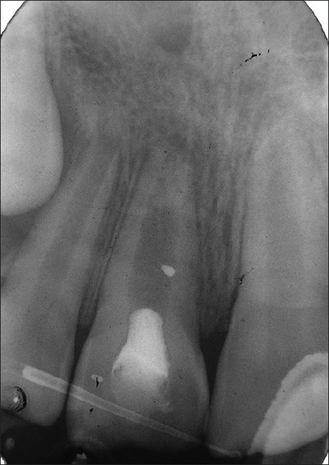
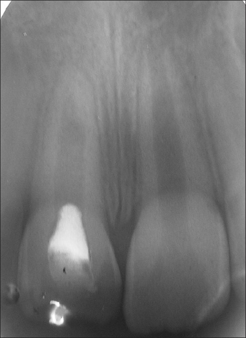
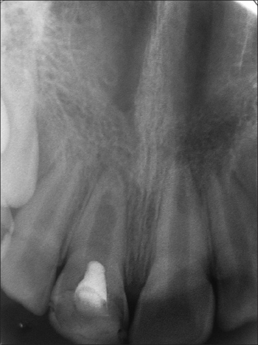
The child was called after every 3 months for clinical examination and radiographic evaluation. Five months radiograph showed thickening of lateral dentinal walls, increased root length [Figure 2]. Nine months radiograph showed increased root length, complete apical closure and thickening of lateral dentinal walls [Figure 3]. Thereafter, the tooth is under follow-up of 15 months without any signs of endodontic failure clinically and radiographically [Figure 4].
Figure 2.

Five months periapical radiograph showed thickening of lateral dentinal walls and increased root length
Figure 3.

Nine months periapical radiograph showing maturogenesis
Figure 4.

Fifteen months follow-up periapical radiograph showing no signs of endodontic failure
Discussion
Maturogenesis is a relatively new treatment modality for non-vital necrotic immature permanent teeth, which allows continued thickening of root dentin and apical closure. This biologically based endodontic procedure has several advantages over conventional methods such as; (1) continued root development increases the crown root ratio, (2) continued thickening of root dentin strengthens the thin and week root, (3) reduces the risk of root fracture during lateral condensation and (4) easy to obtain apical seal. Although the exact mechanism of action of maturogenesis is not known, different hypotheses have been proposed: (a) Presence of few vital pulp cells apically, (b) abundance of multipotent dental pulp cell in immature permanent teeth, (c) stem cells from periodontal ligament or bone marrow, (d) stem cell from apical papilla (SCAP) and (e) growth factors releasing from the blood clot.[3,5,6,7] Of the above hypothesis SCAP shows promising results. It has been experimentally shown that the removal of an apical papilla halted the development of that particular root despite the pulp tissue being intact. In contrast, the root of the tooth containing apical papilla showed normal growth and development.[5]
So far, different treatment protocols have been presented for creating a favorable disinfecting condition within the root canal system of non-vital immature permanent teeth. Most of the authors recommended the use of tri-antibacterial paste (ciprofloxacin, metronidazole and minocycline) to creating a favorable disinfecting condition in the root canal system.[5,8] In the present case, complete disinfection was obtained using tri-antibacterial paste that resulted in successful maturogenesis. Studies have shown the tri antibacterial paste is effective in killing the bacteria in the deep layers of root canal dentin and are almost effective against all the pathogens responsible for endodontic failure.[8,9] It is assumed that ones the infection is subsided survived SCAP under the influence of the Hertwig's epithelial root sheath give rise to formation primary odontoblast to complete root formation.[5] We also noticed that maturogenesis in permanent maxillary right central incisor was faster than the physiologic root development of permanent maxillary left central incisor. The exact reason for rapid maturogenesis is not known but following hypothesis can be made; (1) wide root canals allow rapid transfer of small blood vessels which aid in pulp revascularization by stimulating undifferentiated mesenchymal cells to odontoblast, (2) young children have high healing potential with more stem cell regenerative potential; thus, maximized the chances of faster and successful maturogenesis.
Maturogenesis protocol says placement of MTA over the blood clot. However, in the present case, we placed thick paste of calcium hydroxide over the blood clot. The reasons are (1) calcium hydroxide is time tasted medicament for pulpotomy of primary and young permanent tooth; (2) secondly calcium hydroxide is economical compared to MTA; (3) in the developing countries like India where majority of population can not afford MTA.
Although the calcium hydroxide can be used for placement over blood clot, it should not be used as root canal disinfectant because; (1) it might damage the cells of apical papilla due to its high pH and tissue dissolving property, (2) it might alters the mechanical properties of dentin which further increases the chance of tooth fracture, (3) tightly filled calcium hydroxide reduces the space available for SCAP to proliferate into the canal.[2,10] Although the clinical and radiographic findings (continued root development, thickening of dentinal walls and apical closure) are suggestive of regeneration of pulp dentin complex, human histologic studies are still needed to further evaluate whether maturogenesis procedure truly replicate the pulp dentin complex.
Conclusion
(1) Tri antibacterial paste (3 Mix) can be used as an intra-canal medicament to obtain sterile root canal system during maturogenesis. Calcium hydroxide can be used as an alternative medicament over blood clot. Maturogenesis was faster compared to physiologic root development of adjacent normal young permanent tooth.
Footnotes
Source of Support: Nil
Conflict of Interest: None declared
References
- 1.Webber RT. Apexogenesis versus apexification. Dent Clin North Am. 1984;28:669–97. [PubMed] [Google Scholar]
- 2.Andreasen JO, Farik B, Munksgaard EC. Long-term calcium hydroxide as a root canal dressing may increase risk of root fracture. Dent Traumatol. 2002;18:134–7. doi: 10.1034/j.1600-9657.2002.00097.x. [DOI] [PubMed] [Google Scholar]
- 3.Chueh LH, Ho YC, Kuo TC, Lai WH, Chen YH, Chiang CP. Regenerative endodontic treatment for necrotic immature permanent teeth. J Endod. 2009;35:160–4. doi: 10.1016/j.joen.2008.10.019. [DOI] [PubMed] [Google Scholar]
- 4.Jung IY, Lee SJ, Hargreaves KM. Biologically based treatment of immature permanent teeth with pulpal necrosis: A case series. J Endod. 2008;34:876–87. doi: 10.1016/j.joen.2008.03.023. [DOI] [PubMed] [Google Scholar]
- 5.Huang GT, Sonoyama W, Liu Y, Liu H, Wang S, Shi S. The hidden treasure in apical papilla: The potential role in pulp/dentin regeneration and bioroot engineering. J Endod. 2008;34:645–51. doi: 10.1016/j.joen.2008.03.001. [DOI] [PMC free article] [PubMed] [Google Scholar]
- 6.Wang Q, Lin XJ, Lin ZY, Liu GX, Shan XL. Expression of vascular endothelial growth factor in dental pulp of immature and mature permanent teeth in human. Shanghai Kou Qiang Yi Xue. 2007;16:285–9. [PubMed] [Google Scholar]
- 7.Weisleder R, Benitez CR. Maturogenesis: Is it a new concept? J Endod. 2003;29:776–8. doi: 10.1097/00004770-200311000-00022. [DOI] [PubMed] [Google Scholar]
- 8.Windley W, 3rd, Teixeira F, Levin L, Sigurdsson A, Trope M. Disinfection of immature teeth with a triple antibiotic paste. J Endod. 2005;31:439–43. doi: 10.1097/01.don.0000148143.80283.ea. [DOI] [PubMed] [Google Scholar]
- 9.Sato I, Ando-Kurihara N, Kota K, Iwaku M, Hoshino E. Sterilization of infected root-canal dentine by topical application of a mixture of ciprofloxacin, metronidazole and minocycline in situ. Int Endod J. 1996;29:118–24. doi: 10.1111/j.1365-2591.1996.tb01172.x. [DOI] [PubMed] [Google Scholar]
- 10.Iwaya SI, Ikawa M, Kubota M. Revascularization of an immature permanent tooth with apical periodontitis and sinus tract. Dent Traumatol. 2001;17:185–7. doi: 10.1034/j.1600-9657.2001.017004185.x. [DOI] [PubMed] [Google Scholar]


