Abstract
Inactivation of CDH1, encoding E-cadherin, promotes cancer initiation and progression. According to a newly proposed molecular mechanism, loss of E-cadherin triggers an upregulation of the anti-apoptotic oncoprotein BCL2. Conversely, reconstitution of E-cadherin counteracts overexpression of BCL2. This reciprocal regulation is thought to be critical for early tumor development. We determined the relevance of this new concept in human infiltrating lobular breast cancer (ILBC), the prime tumor entity associated with CDH1 inactivation. BCL2 expression was examined in human ILBC cell lines (IPH-926, MDA-MB-134, SUM-44) harboring deleterious CDH1 mutations. To test for an intact regulatory axis between E-cadherin and BCL2, wild-type E-cadherin was reconstituted in ILBC cells by ectopic expression. Moreover, BCL2 and E-cadherin were evaluated in primary invasive breast cancers and in synchronous lobular carcinomas in situ (LCIS). MDA-MB-134 and IPH-926 showed little or no BCL2 expression, while SUM-44 ILBC cells were BCL2-positive. Reconstitution of E-cadherin failed to impact on BCL2 expression in all cell lines tested. Primary ILBCs were almost uniformly E-cadherin-negative (97%) and were frequently BCL2-negative (46%). When compared with an appropriate control group, ILBCs showed a trend towards an increased frequency of BCL2-negative cases (P = 0.064). In terminal duct-lobular units affected by LCIS, the E-cadherin-negative neoplastic component showed a similar or a reduced BCL2-immunoreactivity, when compared with the adjacent epithelium. In conclusion, upregulation of BCL2 is not involved in lobular breast carcinogenesis and is unlikely to represent an important determinant of tumor development driven by CDH1 inactivation.
Introduction
E-cadherin is a transmembrane glycoprotein that mediates calcium-dependent cell-cell adhesion in epithelial tissues. Loss of E-cadherin has mainly been implicated in cancer progression [1]. Experimental animal models have shown that loss of E-cadherin induces epithelial-mesenchymal transition (EMT) and thereby promotes metastatic dissemination [2]. However, E-cadherin has a much wider implication in human cancer biology. The CDH1 gene, which encodes for E-cadherin, functions as a tumor suppressor gene and CDH1 germline mutations are associated with a hereditary tumor syndrome [3], [4]. Thus, loss of E-cadherin can initiate tumor development. The molecular mechanisms that drive tumor formation following CDH1 inactivation are controversial and may include aberrant activation of the WNT signaling pathway through β-catenin, induction of anoikis-resistance through p120-catenin and cytoplasmic mislocalization of Kaiso, a transcriptional modulator [5], [6], [7]. A new molecular mechanism involving the BCL2 oncoprotein has recently been proposed by Ferreira and colleagues [8]. According to this new concept, loss of E-cadherin triggers an upregulation of the anti-apoptotic oncoprotein BCL2, a process mediated by upregulation of Notch-1 expression and activity, and thereby increases cell survival [8]. Conversely, reconstitution of E-cadherin has been reported to counteract overexpression of BCL2 [8]. This reciprocal regulation may be a critical determinant of early tumor development following CDH1 inactivation or loss of E-cadherin expression [8].
Infiltrating lobular breast cancer (ILBC) is a special breast cancer subtype and accounts for 5 - 15% percent of all mammary carcinomas [9]. ILBCs consist of small, discohesive epithelial cells, which are individually dispersed or arranged in single file linear cords [10]. From a clinical point of view, ILBC is an indolent, hormone-responsive and slowly-progressive malignancy [11]. From a cell biology perspective, ILBC is the most important model disease for studying carcinogenesis driven by CDH1 inactivation [2], [12], [13], [14], [15], [16], [17]. In fact, ILBCs are almost uniformly E-cadherin-negative and harbor deleterious CDH1 mutations [12], [13], [16], [17]. Loss of E-cadherin is otherwise rare in breast cancer. Complete absence of E-cadherin or a full-blown EMT phenotype are encountered in less than 5% of infiltrating ductal breast cancers (IDBC), which account for the vast majority of all mammary carcinomas [18], [19]. Of note, ILBCs can arise from a non-obligate intraepithelial precursor lesion termed lobular carcinoma in situ (LCIS) [10]. In LCIS, the tumor cells are confined to terminal duct-lobular units (TDLUs) and have not yet infiltrated the basement membrane [10]. E-cadherin is already lost in the neoplastic cellular component of TDLUs affected by LCIS [15]. Hence, LCIS provides the opportunity for studying gene expression alterations associated with the inactivation of E-cadherin in comparison to the immediately adjacent, non-neoplastic epithelium [15].
Using human ILBC cell lines, primary tumors and pre-invasive lesions as a model, the present study aimed to determine the relevance of the newly proposed relationship between E-cadherin and BCL2 for tumor development driven by CDH1 inactivation.
Materials and Methods
Cell Culture
The human breast cancer cell lines MDA-MB-134 and IPH-926 have been described previously [20], [21], [22], [23], [24], [25]. BT-549 and MDA-MB-435/M14 cells were obtained by American Type Culture Collection (ATCC, Manassas, VA, U.S.A.). SUM-44-PE cells were kindly provided by D. Derksen [26]. Cell line characteristics are summarized below ( Table 1 ). All cell lines were authenticated by short tandem repeat (STR) profiling. MDA-MB-134, IPH-926 and SUM-44-PE cells were additionally authenticated by detection of their unique, homozygous CDH1 mutations ( Table 1 ) [5], [20], [24]. All cell lines were routinely cultured in RPMI-1640 medium supplemented with 10% FCS, 10 µg/ml bovine insulin, 2.5 g/l glucose, 1 mM sodium pyruvate, 2 mM glutamine, and 10 mM HEPES, in a water-saturated atmosphere containing 5% CO2 at 37.5°C.
Table 1. Tumor cell line characteristics.
| CDH1 status, cell line | CDH1 status, | ||||||
| cell line | origin | established | nucleotide | meth. | protein | primary | ref. |
| IPH-926 | ILBC1 | 2006 | 241ins4 (homo) | neg | neg | 241ins4 (homo) | [20]–[22] |
| MDA-MB-134 | ILBC2 | 1973 | 688del145 (homo) | neg | neg | na | [23]–[25] |
| SUM-44-PE | ILBC2 | 1993 | 1269delT (homo) | neg | neg | na | [5]; [26] |
| MDA-MB-435/M14 | melanoma | 1976 | wt | pos | neg | na | [37]–[39] |
| BT-549 | PABC | 1978 | wt | pos | neg | na | [36]; [37] |
origin from ILBC proven by genetic comparison with the corresponding primary ILBC.
origin from ILBC proposed ex post based on molecular features, corresponding primary tumor remained uncharacterized ILBC; infiltrating lobular breast cancer, PABC; papillary breast cancer, homo; homozygous, meth.; aberrant methylation of the CDH1 promoter, neg; negative, pos; positive, na; not assessed.
Reconstitution of E-cadherin
Cells were transiently transfected either with vector pEGFP-N2 (Clontech Laboratories, Mountain View, CA, U.S.A.) or with the p-wtEcad-EGFP-N2 expression construct encoding for full-length wild-type human E-cadherin fused to the cDNA sequence of EGFP, which was kindly provided by B. Luber [27]. Transfection reactions were carried out using Lipofectamine™ 2000 (Invitrogen, Darmstadt, Germany) according to manufacturer’s recommendations.
Fluorescent Imaging
Cells were grown on LAB-TEK-II chamber slides (Thermo Fisher Scientific, Waltham, MA, U.S.A.) for 24 h after the transfection. Next, cells were fixed in 4% paraformaldehyde, and were incubated with a mouse monoclonal anti-p120-catenin antibody (clone 98, 2.5 ng/µl, BD Transduction Laboratories, Heidelberg, Germany) or with a mouse monoclonal anti-Kaiso antibody (clone 6F, 2 ng/µl, Merck Millipore, Darmstadt, Germany). Then, cells were incubated with a secondary goat anti-mouse antibody labeled with Alexa Fluor 647 (10 ng/µl, Invitrogen, Darmstadt, Germany). Finally, cells were counterstained with DAPI (2 µg/ml, Sigma-Aldrich, St. Louis, MI, U.S.A) and were mounted in ProLong Gold cover medium (Invitrogen, Darmstadt, Germany). Fluorescent imaging was performed with a Leica Inverted-2 confocal laser scanning microscope (Leica Microsystems, Wetzlar, Germany).
Cell Sorting
Pre-analytical enrichment of EGFP- or Ecad-EGFP-positive cells (minimum 70% purity) was carried out by FACS sort using a MoFlo cell sorter (Cytomation, Fort Collins, CO, U.S.A.) [28]. Subsequent expression analyses and cell aggregation assays were performed immediately after the FACS sort, without any interim expansion in cell culture.
Cell Aggregation Assay
Single cell sorted EGFP- or Ecad-EGFP-positive cell preparations were incubated in either normal growth medium or medium supplemented with 2.5 mM EDTA for 45 minutes at 37°C. Relative cell aggregation was assessed by flow cytometry-based detection of aggregates on a MoFlo cytometer. Relative cell cohesion was expressed as a cell aggregation index (CAI), representing the percentage of aggregated cells in the test condition (Ecad-EGFP, with or without EDTA) divided by the percentage of aggregated cells in the control condition (EGFP), as described previously [29], [30].
Western Blot
Cells were lysed in RIPA buffer and 20 µg total cellular protein were separated by SDS-PAGE and were transferred to nitrocellulose membranes. Membranes were probed with anti-E-cadherin (clone 36, BD Transduction Laboratories, Heidelberg, Germany), anti-BCL2 (clone 124, Dako, Glostrup, Denmark), anti-Notch-1 (clone c20, Santa Cruz Biotechnology, Heidelberg, Germany) and anti–β-actin antibodies (clone AC15, Acris, Hiddenhausen, Germany), as described previously [31]. 293T cells transfected with a Notch-1 expression plasmid (sc-110326, Santa Cruz Biotechnology) served as a positive control for Notch-1.
Tumor Specimen and Tissue Microarrays
Formalin-fixed paraffin-embedded (FFPE) human primary invasive breast cancer specimens (n = 172) were retrieved from the tissue archive of the Institute of Pathology of the Hannover Medical School according to the guidelines of the local ethics committee ("Ethik-Kommission der Medizinischen Hochschule Hannover", head: Prof. Dr. Tröger). This study was exempted from review from the local ethics committee since the specimens used in this study were left-over samples from diagnostic procedures and thereby retrieved retrospectively. All specimens were made anonymous and waived the need for consent. They were compiled on tissue microarrays (TMAs), as described previously [32], [33]. Tumor characteristics are summarized below ( Table 2 ). Tumor characteristics of the subset of estrogen receptor (ER)-positive cases are provided in Table S2. From the primary ILBCs represented on the TMAs, we selected n = 11 cases with synchronous LCIS in separate FFPE tissue blocks for analysis of pre-invasive tumor cells.
Table 2. Characteristics of primary tumors.
| number of cases | percent | |
| cases | 172 | 100 |
| age | ||
| >60 | 72 | 42 |
| ≤60 | 100 | 58 |
| histological type | ||
| ILBC | 37 | 22 |
| IDBC | 135 | 78 |
| pT status | ||
| pT1/pT2 | 149 | 86 |
| pT3/pT4 | 22 | 13 |
| pTx | 1 | 1 |
| pN status | ||
| pN0 | 94 | 55 |
| pN1+ | 57 | 33 |
| pNx | 21 | 12 |
| histological grade | ||
| G1 | 15 | 9 |
| G2 | 97 | 56 |
| G3 | 60 | 35 |
| estrogen receptor | ||
| Positive | 139 | 81 |
| negative | 33 | 19 |
| progesterone receptor | ||
| Positive | 100 | 58 |
| negative | 72 | 42 |
| c-erbB2 expression | ||
| 0, 1+ | 161 | 94 |
| 2+ | 3 | 2 |
| 3+ | 8 | 4 |
| E-cadherin (in ILBC) | ||
| Positive | 1 | 3 |
| negative | 36 | 97 |
| E-cadherin (in IDBC) | ||
| Positive | 130 | 96 |
| negative | 5 | 4 |
| Ki67 LI | ||
| ≤10 | 30 | 17 |
| >10, <24 | 89 | 52 |
| ≥25 | 53 | 31 |
Immunohistochemistry
Four micrometer sections of TMAs or FFPE breast tissue were mounted on Superfrost slides (Thermo Fisher Scientific). Slides were deparaffinized and rehydrated conventionally. Staining was performed on a Benchmark Ultra (Ventana, Tucson, AZ, U.S.A.) automated stainer using the CC1 mild protocol for antigen retrieval, monoclonal anti-E-cadherin (clone 4A2C7, Invitrogen) or anti-BCL2 (clone 124, Dako) antibodies and the ultraView DAB kit for signal detection. Evaluation of BCL2 immunoreactivity was carried out using an immunoreactivity score (IRS) as described by Remmele and Stegener [34]. Tumors with an IRS≤2 were considered as BCL2-negative, whereas those with an IRS≥3 were considered as BCL2-positive. The same IRS score was implemented for evaluation of BCL2 expression and LCIS and at least three TDLU were considered per individual case. For E-cadherin, only cases with complete absence of any membranous E-cadherin immunoreactivity were considered as E-cadherin-negative [33]. Detection and evaluation of estrogen receptor (ER), progesterone receptor (PR), c-erbB2 and Ki67 expression were performed as described previously [33]. Ki67 labeling index cutoffs for definition of low, intermediate and high proliferation were 10 and 25% [33]. Statistical analyses were performed with GraphPads Prism software. Fisher’s exact test or the Chi square test for trends were used to assess associations between clinicopathological variables and the BCL2-status. P values <0.05 were considered statistically significant.
Results
Reconstitution of E-cadherin Fails to Impact on BCL2 in Human ILBC Cells in vitro
According to a newly proposed molecular mechanism, loss of E-cadherin triggers an upregulation of the oncoprotein BCL2 [8]. Conversely, reconstitution of E-cadherin has been reported to counteract overexpression of BCL2 [8]. We sought to study this reciprocal regulation in cell lines derived from ILBC, a tumor entity associated with CDH1 inactivation. Few ILBC cell lines have been established so far [35]. For our analyses, we selected three cell lines, which are of proven (IPH-926), or proposed (MDA-MB-134 and SUM-44-PE) origin from ILBC and harbor deleterious CDH1 mutations ( Table 1 ). We also included BT-549 cells, which were derived from a papillary carcinoma of the breast and display an EMT phenotype in vitro [36], [37]. Moreover, we included MDA-MB-435 cells, which were formerly known as breast cancer cells, but were derived from the M14 melanoma cell line instead [38], [39]. MDA-MB-435/M14 was included because this cell line had been used in the original publication describing the reciprocal regulation of E-cadherin and BCL2 [8].
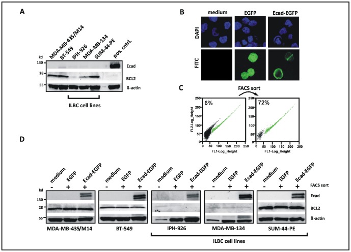
Western blot analysis confirmed lack of E-cadherin in all cell lines studied ( Figure 1A ). Interestingly, MDA-MB-134 and IPH-926 ILBC cells showed little or no BCL2 expression, while SUM-44-PE, BT-549 and MDA-MB-435/M14 were BCL2-positive ( Figure 1A ). Increased expression and activation of the Notch receptor family member Notch-1 has been proposed to mediate the upregulation of BCL2 in response to inactivation of E-cadherin [8]. However, all E-cadherin-deficient cells tested also lacked detectable Notch-1 expression, regardless of their BCL2 expression status (Figure S1). To test whether reconstitution of E-cadherin downregulates BCL2, as proposed previously [8], cells were transiently transfected with expression plasmids encoding for EGFP-tagged full-length wild-type E-cadherin (Ecad-EGFP) or EGFP alone ( Figure 1B–D ). To compensate for limited transfection efficiency, positive cells were enriched by FACS sort ( Figure 1C ). Subsequent expression analyses were performed immediately after the FACS sort, without any interim expansion in cell culture. However, reconstitution of E-cadherin failed to impact on BCL2 expression in all cell lines tested, including BCL2-positive MDA-MB-435/M14 and SUM-44-PE ILBC cells ( Figure 1D ).
Figure 1. Reconstitution of E-cadherin fails to impact on BCL2 expression in human ILBC cells.
(A) Analysis of E-cadherin (Ecad) and BCL2 protein expression by Western Blot. Pos cntrl; positive control for E-cadherin. (B) Fluorescent imaging of cells subjected to ectopic expression of EGFP or EGFP-tagged E-cadherin (Ecad-EGFP) by transient transfection. Representative photomicrographs show the IPH-926 ILBC cell line. Note the membranous localization of Ecad-EGFP. (C) Pre-analytical enrichment of EGFP- or Ecad-EGFP-positive cells by FACS sort. Cells within the EGFP-positive gate are colored in green. Representative dot blots show IPH-926 ILBC cells transfected with the Ecad-EGFP expression construct. (D) Analysis of E-cadherin and BCL2 protein expression by Western blot. Cells were subjected to ectopic expression of EGFP or Ecad-EGFP for 24 h and subsequent pre-analytical enrichment of EGFP-positive cells by FACS sort (minimum 70% purity). Untreated cells were included as controls. Ecad-EGFP presented as a double band of approximately 150 kd, as reported previously [27]. Similar results were obtained 48 h after transfection (not shown).
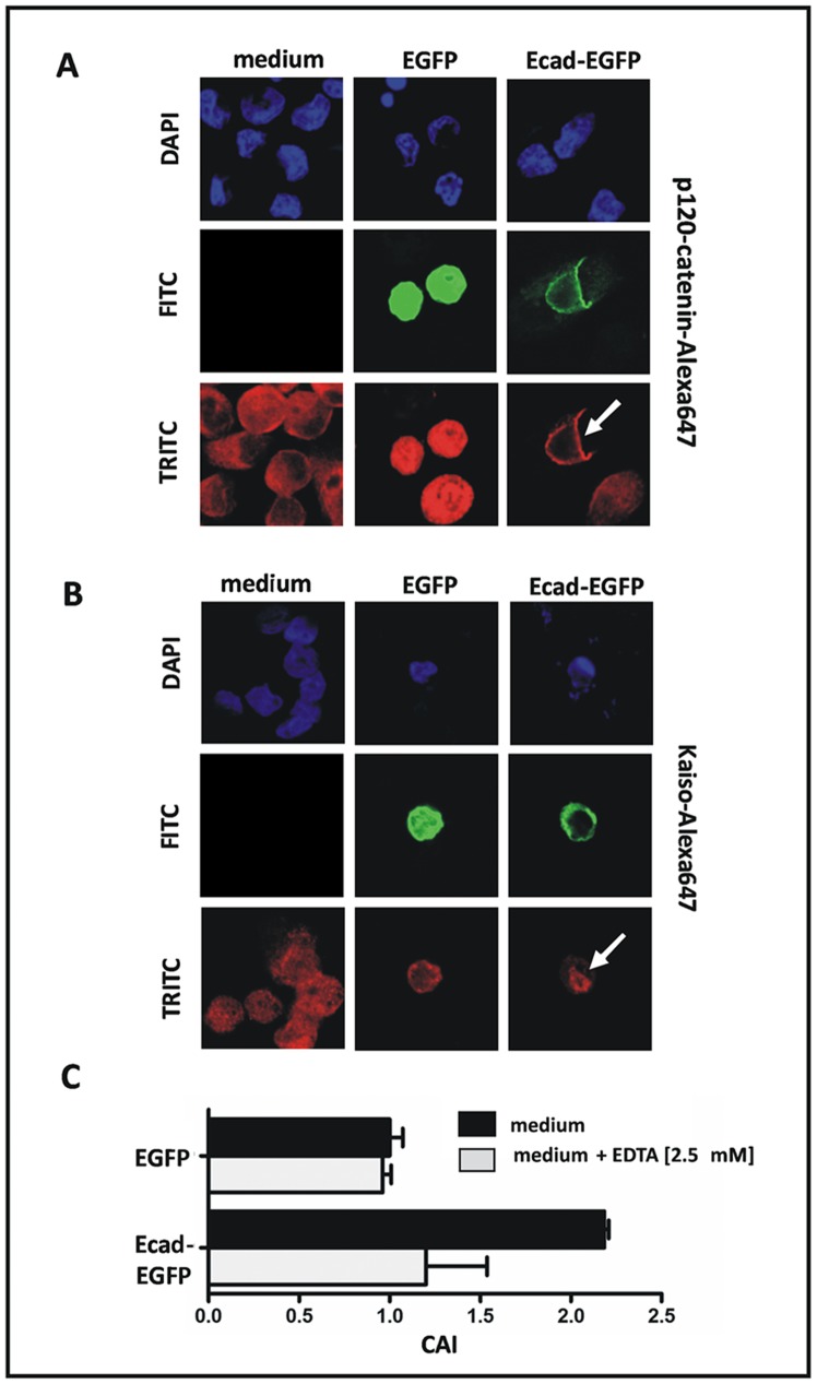
This prompted us to test the functionality of the E-cadherin construct. The E-cadherin binding protein p120-catenin shows an aberrant cytoplasmic localization and triggers anoikis resistance in E-cadherin-deficient tumor cells [6]. Besides that, cytoplasmic p120-catenin sequesters the nuclear transcriptional modulator Kaiso, which thus exhibits a cytoplasmic mislocalization, once E-cadherin is lost [7]. Hence, we utilized the subcellular localization of p120-catenin and Kaiso, as detected by immunofluorescence, as a read-out system for the functionality of the E-cadherin construct. E-cadherin-deficient cells transfected with Ecad-EGFP showed a relocation of p120-catenin to the cell membrane ( Figure 2A ) and a relocation of Kaiso to the nucleus, arguing for the intact functionality of this expression construct ( Figure 2B ). In addition to the immunofluorescence read-out system, which demonstrated a restored intracellular signaling, we also assessed whether the E-cadherin construct re-activates calcium-dependent cell-cell adhesion. E-cadherin-deficient cells transfected with Ecad-EGFP showed an increased cell aggregation ( Figure 2C ). Notably, this effect was abrogated by the calcium chelating agent EDTA, arguing for the correct functionality of this E-cadherin expression construct ( Figure 2C ).
Figure 2. Validation of the functional activity of the E-cadherin construct.
(A) Fluorescent imaging of cells subjected to ectopic expression of EGFP- or Ecad-EGFP showing the relocation of p120-catenin to the cell membrane in Ecad-EGFP-positive cells. (B) Fluorescent imaging showing the relocation of Kaiso to the nucleus. Representative photomicrographs were taken from experiments with the E-cadherin-deficient IPH-926 ILBC cell line. (C) Increased calcium-dependent cell-cell adhesion in E-cadherin-deficient IPH-926 ILBC cells transiently transfected Ecad-EGFP. The cell aggregation index (CAI) was determined by FACS analysis as described in the “Material and Methods” section.
High Frequency of BCL2-negative Cases in Human Primary ILBCs
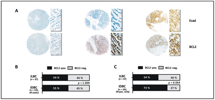
Reconstitution of E-cadherin in human ILBC cells in vitro failed to substantiate a reciprocal regulation between E-cadherin and BCL2. For several reasons, which are discussed below, this does not necessarily rule out such a regulatory interrelationship in human tumors in vivo. The human mammary epithelium is constitutively positive for BCL2-expression [40]. In breast cancer, loss of BCL2 has repeatedly been associated with high grade tumors and other aggressive carcinoma entities, which shows that BCL2 acts as a differentiation marker in the mammary gland [41], [42], [43]. In view of the recently proposed concept, that loss of E-cadherin propels malignant transformation by upregulation of BCL2, we hypothesized that primary ILBCs, which typically lack E-cadherin, might preferentially be BCL2-positive or retain BCL2 expression. Accordingly, BCL2 and E-cadherin were evaluated in a series of n = 172 human primary invasive breast cancers compiled on TMAs using immunohistochemical stainings. Consistent with previous findings [41], [42], [43], loss of BCL2 expression was associated with high histological grade, lack of ER, lack of PR, and high Ki67 labeling index (P<0.001, each) (Table S1). ILBCs were almost uniformly E-cadherin-negative (97%), while IDBCs were almost exclusively E-cadherin-positive (96%) ( Table 2 and Figure 3A ). Despite this difference in E-cadherin expression in ILBCs versus IDBCs, the proportion of BCL2-positive/-negative cases was similar in the two entities (P = 1.000) ( Figure 3B ). Variation of the IRS cutoff defining BCL2-positive/-negative cases yielded similar results (data not shown). As a matter of fact, a large fraction of ILBCs (46%) showed essentially no BCL2 immunoreactivity ( Figure 3A and B ). As ILBCs are typically ER-positive, we decided to restrict our tumor series to ER-positive cases for a refined analysis. A total of n = 139 ER-positive carcinomas entered this refined evaluation and these cases included all ILBCs (Table S2 and S3). In this more focused tumor series, ILBCs even showed a trend towards a higher frequency of BCL2-negative cases (P = 0.064) ( Figure 1C ). Hence, primary ILBCs were almost uniformly E-cadherin-negative, but showed a comparatively high frequency of BCL2-negative cases, which indicates that BCL2 expression is dispensable during the development of these tumors.
Figure 3. Primary ILBCs lack E-cadherin expression and are frequently BCL2-negative.
(A) Representative immunohistochemical stainings of two primary ILBCs (left, middle) and one primary IDBC (right) for E-cadherin (Ecad) and BCL2, as performed on TMAs. An overview of the tumor cores embedded in the TMAs is shown on the left side of each cases and a detail photomicrograph (magnification, ×400) is shown on the right side of each case. (B) Comparison of the frequency of BCL2-positive/−negative cases in ILBCs versus IDBCs. Statistical significance was determined with Fisher’s exact test. (C) Comparison of BCL2 positive/−negative cases in ILBCs versus ER-positive IDBCs.
Similar or Reduced BCL2 Expression in LCIS Compared with the Adjacent Epithelium
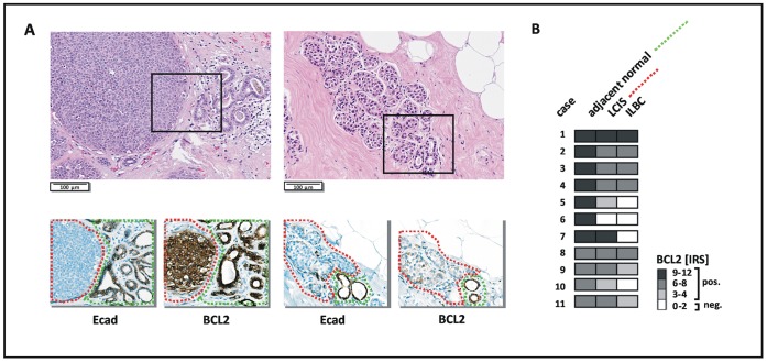
Analysis of gene expression in tumor cohorts is an indirect way to assess potential regulatory interrelationships. Relevant associations may be obscured by inter-individual tumor heterogeneity. To circumvent this methodological limitation and to rule out the possibility that upregulation of BCL2 is restricted to the pre-invasive stage of tumor development driven by CDH1 inactivation, we evaluated E-cadherin and BCL2 immunoreactivity in LCIS lesions. This provides the opportunity to use the normal mammary epithelium of affected TDLUs as an intra-individual reference for baseline expression of the gene of interest [15]. From the ILBCs represented in the TMA series, we selected n = 11 cases with synchronous LCIS lesions on separate FFPE tissue blocks for analysis of pre-invasive tumor cells. In line with previous findings [42], the normal mammary epithelium was always E-cadherin-positive and BCL2-positive ( Figure 4A ). Also, there was essentially no variation in BCL2 immunoreactivity between different TDLUs within the same patient (data not shown). However, the E-cadherin-negative, neoplastic cellular component of TDLUs affected by LCIS showed a similar, a reduced or a complete loss of BCL2-immunoreactivity, when compared with the adjacent normal epithelium ( Figure 4A and B ). No increased immunoreactivity for BCL2 was observed in any of the LCIS lesions evaluated ( Figure 4B ).
Figure 4. Similar or reduced BCL2 immunoreactivity in LCIS compared with the adjacent mammary epithelium.
(A) Representative photomicrographs showing two LCIS lesions characterized by either similar (left, case #1) or reduced (right, case #10) BCL2 immunoreactivity compared with the adjacent E-cadherin-positive epithelium. HE stained sections are shown on top. Serial sections subjected to immunohistochemical staining of E-cadherin (Ecad) and BCL2 are shown below. (B) Overview on BCL2 immunoreactivity in LCIS lesions of n = 11 patients. At least three TDLUs affected by LCIS were considered per case, but showed essentially the same staining characteristics (not shown). A color scale indicative of the IRS is included on the right side.
Discussion
Loss of E-cadherin can initiate tumor development, but the molecular mechanisms that mediate this tumor suppressive function are less well understood than the role of E-cadherin in cancer progression and metastasis [5],[6]. The recently described reciprocal regulation between E-cadherin and the anti-apoptotic oncoprotein BCL2, which is supposed to be mediated by Notch-1, provides an attractive new explanation how CDH1 inactivation might propel neoplastic transformation [8]. According to this new model, loss of E-cadherin triggers an upregulation of Notch-1 expression and activity, which in turn induces increased BCL2 expression. BCL2 for its part is believed to increase cell survival in E-cadherin-deficient tumor cells [8]. Conversely, reconstitution of E-cadherin has been reported to counteract overexpression of BCL2 [8]. However, the relevance of this new model for human tumor development has remained uncertain, as previous studies have only focused on a limited number of in vitro cell models and few clinical tumor specimens [8]. ILBC is a special breast cancer entity associated with mutational CDH1 inactivation and loss of E-cadherin expression [2], [12], [13], [14], [15], [16], [17]. Using ILBC cell lines, primary tumors and pre-invasive lesions as a model, the present work aimed to determine whether reciprocal upregulation of BCL2 is an important determinant of tumor development driven by inactivation of CDH1/E-cadherin.
Characterization of human ILBC cell lines harboring deleterious CDH1 mutations revealed different levels of BCL2 expression. MDA-MB-134 and IPH-926 ILBC cells expressed little or no BCL2, while SUM-44-PE cells were BCL2-positive. Contrary to previous findings in MDA-MB-435/M14 [8], reconstitution of full-length wild-type E-cadherin failed to downregulate BCL2 in SUM-44-PE cells. However, we could also not confirm a downregulation of BCL2 in MDA-MB-435/M14 cells, which had been used in the original study describing the reciprocal regulation between E-cadherin and BCL2 [8]. Moreover, Notch-1 protein expression was not detectable in any of the E-cadherin-deficient ILBC cell lines tested, arguing against the existence of a regulatory axis between E-cadherin, Notch-1 and BCL2.
However, one might insist that the E-cadherin expression construct used in the present work could have had an impaired functionality due to the EGFP-tag, which is required for pre-analytical enrichment of E-cadherin-positive cells. Yet, proper function of the Ecad-EGFP construct had been verified by several assays devised to read out restored intracellular signaling (relocation of p120-catenin and Kaiso to the cell membrane or nucleus) and restored calcium dependent cell-cell adhesion (cell aggregation assay). Alternatively, one might consider that the regulatory axis between E-cadherin and BCL2 has become fixed in our MDA-MB-435/M14, SUM-44-PE and BT-549 cells due to the long time since their establishment ( Table 1 ). Even so, the absence of a significant overexpression of BCL2 and Notch-1 in the E-cadherin-deficient MDA-MB-134 and IPH-926 ILBC cells is not in line with the concept that loss of E-cadherin triggers an upregulation of BCL2 through increased expression and activity of Notch-1.
Immunohistochemical analysis of human primary breast cancers revealed that a comparatively large proportion of ILBCs lack BCL2 expression, despite complete loss of E-cadherin. This is consistent with a previous study reported by Coradini et al. [44]. When compared with an appropriate control group of ER-positive ductal carcinomas, ILBCs even showed a trend towards a higher frequency of BCL2-negative cases. This is not in line with the proposed reciprocal regulation between E-cadherin and BCL2. In fact, this observation strongly suggests that BCL2 is dispensable during the development of E-cadherin-deficient ILBC. In line with this assumption, evaluation of LCIS lesions demonstrated that E-cadherin-negative pre-invasive tumor cells rather exhibit a reduced, but not an increased, BCL2 immunoreactivity, when compared with the adjacent normal epithelium. This also argues against an upregulation of BCL2 at the site of CDH1 inactivation within the human mammary epithelium. Interestingly, however, we observed retained BCL2 expression in 4 out of 5 IDBCs with abrogated E-cadherin expression. E-cadherin-negative IDBC is a very uncommon tumor phenotype and, possibly due to the extremely small sample size, also lacked a significant association with the BCL2 status. There could be a functional interrelationship between E-cadherin and BCL2 in the extremely rare E-cadherin-negative IDBCs, but apparently not in the much more common E-cadherin-negative ILBCs.
Taken together, upregulation of BCL2 is not effective in human ILBC cell lines, primary tumors and pre-invasive lesion. As ILBC is the key tumor entity associated with loss of E-cadherin expression, this suggests that reciprocal regulation of BCL2 is not a major mechanism of tumor development driven by CDH1 inactivation.
Supporting Information
Notch-1 protein expression, as detected by Western blot. Pos cntrl; positive control (293T cells transfected with Notch-1).
(TIF)
Relationship between BCL2 status and clinicopathological factors.
(DOC)
Characteristics of primary tumors, ER-pos. subset.
(DOC)
Relationship between BCL2 status and clinicopathological factors, ER-pos. subset.
(DOC)
Acknowledgments
We would like to thank the Cell Sorting Core Facility of the Hannover Medical School, supported in part by Braukmann-Wittenberg-Herz-Stiftung and German Research Foundation (DFG), for excellent assistance.
Funding Statement
This work was supported by the German Cancer Aid Grant 109435 to MC and UL. MC was additionally supported by a Hannelore-Munke Fellowship. The funders had no role in study design, data collection and analysis, decision to publish, or preparation of the manuscript.
References
- 1. Jeanes A, Gottardi CJ, Yap AS (2008) Cadherins and cancer: how does cadherin dysfunction promote tumor progression? Oncogene 27: 6920–6929. [DOI] [PMC free article] [PubMed] [Google Scholar]
- 2. Yang J, Mani SA, Donaher JL, Ramaswamy S, Itzykson RA, et al. (2004) Twist, a master regulator of morphogenesis, plays an essential role in tumor metastasis. Cell 117: 927–939. [DOI] [PubMed] [Google Scholar]
- 3. Kaurah P, MacMillan A, Boyd N, Senz J, De Luca A, et al. (2007) Founder and recurrent CDH1 mutations in families with hereditary diffuse gastric cancer. Jama 297: 2360–2372. [DOI] [PubMed] [Google Scholar]
- 4. Xie ZM, Li LS, Laquet C, Penault-Llorca F, Uhrhammer N, et al. (2011) Germline mutations of the E-cadherin gene in families with inherited invasive lobular breast carcinoma but no diffuse gastric cancer. Cancer 117: 3112–3117. [DOI] [PubMed] [Google Scholar]
- 5. van de Wetering M, Barker N, Harkes IC, van der Heyden M, Dijk NJ, et al. (2001) Mutant E-cadherin breast cancer cells do not display constitutive Wnt signaling. Cancer Res 61: 278–284. [PubMed] [Google Scholar]
- 6. Schackmann RC, van Amersfoort M, Haarhuis JH, Vlug EJ, Halim VA, et al. (2011) Cytosolic p120-catenin regulates growth of metastatic lobular carcinoma through Rock1-mediated anoikis resistance. J Clin Invest 212: 3176–88. [DOI] [PMC free article] [PubMed] [Google Scholar]
- 7. Vermeulen JF, van de Ven RA, Ercan C, van der Groep P, van der Wall E, et al. (2012) Nuclear Kaiso expression is associated with high grade and triple-negative invasive breast cancer. PLoS One 7: e37864. [DOI] [PMC free article] [PubMed] [Google Scholar]
- 8. Ferreira AC, Suriano G, Mendes N, Gomes B, Wen X, et al. (2012) E-cadherin impairment increases cell survival through Notch-dependent upregulation of Bcl-2. Hum Mol Genet 21: 334–343. [DOI] [PubMed] [Google Scholar]
- 9.Lakhani SR, Ellis I, Schnitt S, Tan PH, van de Vijver M (2012) WHO Classification of Tumours of the Breast. Lyon: International Agency for Research on Cancer.
- 10. Hanby AM, Hughes TA (2008) In situ and invasive lobular neoplasia of the breast. Histopathology 52: 58–66. [DOI] [PubMed] [Google Scholar]
- 11. Rakha EA, El-Sayed ME, Powe DG, Green AR, Habashy H, et al. (2008) Invasive lobular carcinoma of the breast: response to hormonal therapy and outcomes. Eur J Cancer 44: 73–83. [DOI] [PubMed] [Google Scholar]
- 12. Berx G, Cleton-Jansen AM, Nollet F, de Leeuw WJ, van de Vijver M, et al. (1995) E-cadherin is a tumour/invasion suppressor gene mutated in human lobular breast cancers. Embo J 14: 6107–6115. [DOI] [PMC free article] [PubMed] [Google Scholar]
- 13. Sarrio D, Moreno-Bueno G, Hardisson D, Sanchez-Estevez C, Guo M, et al. (2003) Epigenetic and genetic alterations of APC and CDH1 genes in lobular breast cancer: relationships with abnormal E-cadherin and catenin expression and microsatellite instability. Int J Cancer 106: 208–215. [DOI] [PubMed] [Google Scholar]
- 14. Sarrio D, Perez-Mies B, Hardisson D, Moreno-Bueno G, Suarez A, et al. (2004) Cytoplasmic localization of p120ctn and E-cadherin loss characterize lobular breast carcinoma from preinvasive to metastatic lesions. Oncogene 23: 3272–3283. [DOI] [PubMed] [Google Scholar]
- 15. Morrogh M, Andrade VP, Giri D, Sakr RA, Paik W, et al. (2012) Cadherin-catenin complex dissociation in lobular neoplasia of the breast. Breast Cancer Res Treat 132: 641–652. [DOI] [PMC free article] [PubMed] [Google Scholar]
- 16. Ellis MJ, Perou CM (2013) The genomic landscape of breast cancer as a therapeutic roadmap. Cancer Discov 3: 27–34. [DOI] [PMC free article] [PubMed] [Google Scholar]
- 17. Bertucci F, Orsetti B, Negre V, Finetti P, Rouge C, et al. (2008) Lobular and ductal carcinomas of the breast have distinct genomic and expression profiles. Oncogene 27: 5359–5372. [DOI] [PMC free article] [PubMed] [Google Scholar]
- 18. Rakha EA, Abd El Rehim D, Pinder SE, Lewis SA, Ellis IO (2005) E-cadherin expression in invasive non-lobular carcinoma of the breast and its prognostic significance. Histopathology 46: 685–693. [DOI] [PubMed] [Google Scholar]
- 19. Dubois-Marshall S, Thomas JS, Faratian D, Harrison DJ, Katz E (2011) Two possible mechanisms of epithelial to mesenchymal transition in invasive ductal breast cancer. Clin Exp Metastasis 28: 811–818. [DOI] [PubMed] [Google Scholar]
- 20. Christgen M, Bruchhardt H, Hadamitzky C, Rudolph C, Steinemann D, et al. (2009) Comprehensive genetic and functional characterization of IPH-926: a novel CDH1-null tumour cell line from human lobular breast cancer. J Pathol 217: 620–632. [DOI] [PubMed] [Google Scholar]
- 21. Christgen M, Noskowicz M, Heil C, Schipper E, Christgen H, et al. (2012) IPH-926 lobular breast cancer cells harbor a p53 mutant with temperature-sensitive functional activity and allow for profiling of p53-responsive genes. Lab Invest 92: 1635–47. [DOI] [PubMed] [Google Scholar]
- 22. Krech T, Scheuerer E, Geffers R, Kreipe H, Lehmann U, et al. (2012) ABCB1/MDR1 contributes to the anticancer drug-resistant phenotype of IPH-926 human lobular breast cancer cells. Cancer Lett 315: 153–160. [DOI] [PubMed] [Google Scholar]
- 23. Cailleau R, Young R, Olive M, Reeves WJ Jr (1974) Breast tumor cell lines from pleural effusions. J Natl Cancer Inst 53: 661–674. [DOI] [PMC free article] [PubMed] [Google Scholar]
- 24. Hiraguri S, Godfrey T, Nakamura H, Graff J, Collins C, et al. (1998) Mechanisms of inactivation of E-cadherin in breast cancer cell lines. Cancer Res 58: 1972–1977. [PubMed] [Google Scholar]
- 25. Reis-Filho JS, Simpson PT, Turner NC, Lambros MB, Jones C, et al. (2006) FGFR1 emerges as a potential therapeutic target for lobular breast carcinomas. Clin Cancer Res 12: 6652–6662. [DOI] [PubMed] [Google Scholar]
- 26. Ethier SP, Mahacek ML, Gullick WJ, Frank TS, Weber BL (1993) Differential isolation of normal luminal mammary epithelial cells and breast cancer cells from primary and metastatic sites using selective media. Cancer Res 53: 627–635. [PubMed] [Google Scholar]
- 27. Fuchs M, Hutzler P, Handschuh G, Hermannstadter C, Brunner I, et al. (2004) Dynamics of cell adhesion and motility in living cells is altered by a single amino acid change in E-cadherin fused to enhanced green fluorescent protein. Cell Motil Cytoskeleton 59: 50–61. [DOI] [PubMed] [Google Scholar]
- 28. Christgen M, Geffers R, Ballmaier M, Christgen H, Poczkaj J, et al. (2010) Down-regulation of the fetal stem cell factor SOX17 by H33342: a mechanism responsible for differential gene expression in breast cancer side population cells. J Biol Chem 285: 6412–6418. [DOI] [PMC free article] [PubMed] [Google Scholar]
- 29. Desbarats L, Brar SK, Siu CH (1994) Involvement of cell-cell adhesion in the expression of the cell cohesion molecule gp80 in Dictyostelium discoideum. J Cell Sci 107: 1705–1712. [DOI] [PubMed] [Google Scholar]
- 30. Le TL, Yap AS, Stow JL (1999) Recycling of E-cadherin: a potential mechanism for regulating cadherin dynamics. J Cell Biol 146: 219–32. [PMC free article] [PubMed] [Google Scholar]
- 31. Christgen M, Ballmaier M, Bruchhardt H, von Wasielewski R, Kreipe H, et al. (2007) Identification of a distinct side population of cancer cells in the Cal-51 human breast carcinoma cell line. Mol Cell Biochem 306: 201–212. [DOI] [PubMed] [Google Scholar]
- 32. Christgen M, Bruchhardt H, Ballmaier M, Krech T, Langer F, et al. (2008) KAI1/CD82 is a novel target of estrogen receptor-mediated gene repression and downregulated in primary human breast cancer. Int J Cancer 123: 2239–2246. [DOI] [PubMed] [Google Scholar]
- 33. Christgen M, Noskowicz M, Schipper E, Christgen H, Heil C, et al. (2013) Oncogenic PIK3CA mutations in lobular breast cancer progression. Genes Chromosomes Cancer 52: 69–80. [DOI] [PubMed] [Google Scholar]
- 34. Remmele W, Stegner HE (1987) Recommendation for uniform definition of an immunoreactive score (IRS) for immunohistochemical estrogen receptor detection (ER-ICA) in breast cancer tissue. Pathologe 8: 138–140. [PubMed] [Google Scholar]
- 35. Sikora MJ, Jankowitz RC, Dabbs DJ, Oesterreich S (2013) Invasive lobular carcinoma of the breast: Patient response to systemic endocrine therapy and hormone response in model systems. Steroids 78: 568–75. [DOI] [PubMed] [Google Scholar]
- 36. Lombaerts M, van Wezel T, Philippo K, Dierssen JW, Zimmerman RM, et al. (2006) E-cadherin transcriptional downregulation by promoter methylation but not mutation is related to epithelial-to-mesenchymal transition in breast cancer cell lines. Br J Cancer 94: 661–671. [DOI] [PMC free article] [PubMed] [Google Scholar]
- 37. Lacroix M, Leclercq G (2004) Relevance of breast cancer cell lines as models for breast tumours: an update. Breast Cancer Res Treat 83: 249–289. [DOI] [PubMed] [Google Scholar]
- 38. Rae JM, Creighton CJ, Meck JM, Haddad BR, Johnson MD (2007) MDA-MB-435 cells are derived from M14 melanoma cells–a loss for breast cancer, but a boon for melanoma research. Breast Cancer Res Treat 104: 13–19. [DOI] [PubMed] [Google Scholar]
- 39. Christgen M, Lehmann U (2007) MDA-MB-435: The Questionable Use of a Melanoma Cell Line as a Model for Human Breast Cancer is Going On. Cancer Biol Ther 6: 1355–1357. [DOI] [PubMed] [Google Scholar]
- 40. Hockenbery DM, Zutter M, Hickey W, Nahm M, Korsmeyer SJ (1991) BCL2 protein is topographically restricted in tissues characterized by apoptotic cell death. Proc Natl Acad Sci U S A 88: 6961–6965. [DOI] [PMC free article] [PubMed] [Google Scholar]
- 41. Ali HR, Dawson SJ, Blows FM, Provenzano E, Leung S, et al. (2012) A Ki67/BCL2 index based on immunohistochemistry is highly prognostic in ER-positive breast cancer. J Pathol 226: 97–107. [DOI] [PubMed] [Google Scholar]
- 42. Hwang KT, Woo JW, Shin HC, Kim HS, Ahn SK, et al. (2012) Prognostic influence of BCL2 expression in breast cancer. Int J Cancer 131: E1109–1119. [DOI] [PubMed] [Google Scholar]
- 43. Park SH, Kim H, Song BJ (2002) Down regulation of bcl2 expression in invasive ductal carcinomas is both estrogen- and progesterone-receptor dependent and associated with poor prognostic factors. Pathol Oncol Res 8: 26–30. [DOI] [PubMed] [Google Scholar]
- 44. Coradini D, Pellizzaro C, Veneroni S, Ventura L, Daidone MG (2002) Infiltrating ductal and lobular breast carcinomas are characterised by different interrelationships among markers related to angiogenesis and hormone dependence. Br J Cancer 87: 1105–1111. [DOI] [PMC free article] [PubMed] [Google Scholar]
Associated Data
This section collects any data citations, data availability statements, or supplementary materials included in this article.
Supplementary Materials
Notch-1 protein expression, as detected by Western blot. Pos cntrl; positive control (293T cells transfected with Notch-1).
(TIF)
Relationship between BCL2 status and clinicopathological factors.
(DOC)
Characteristics of primary tumors, ER-pos. subset.
(DOC)
Relationship between BCL2 status and clinicopathological factors, ER-pos. subset.
(DOC)


