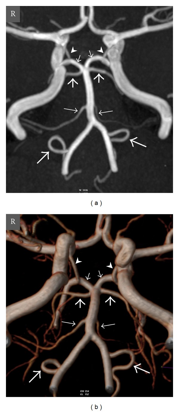
Figure 1.

Oblique coronal view maximum intensity projection (MIP) (a) and volume-rendered (VR) (b) time-of-flight (TOF) magnetic resonance (MR) angiography images show normal anatomy of the vertebrobasilar circulation in a 20-year-old woman. Thick long arrows = right and left posterior inferior cerebellar arteries (PICAs), thin long arrows = right and left anterior inferior cerebellar arteries (AICAs), thick short arrows = right and left superior cerebellar arteries (SCAs), thin short arrows = right and left posterior cerebral arteries (PCAs), arrowheads = right and left posterior communicating arteries (PCoAs), and R = right.
