Abstract
Transient transfection of small interfering RNA (siRNA) provides a powerful approach for studying cellular protein functions, particularly when the target protein can be re-expressed from an exogenous siRNA-resistant construct in order to rescue the knockdown phenotype, confirm siRNA target specificity and support mutational analyses. Rescue experiments often fail, however, when siRNA-resistant constructs are expressed at suboptimal levels. Here, we describe an ensemble of mammalian protein expression vectors with CMV promoters of differing strengths. We show, using CHMP2A rescue of HIV-1 budding, that these vectors can combine high transfection efficiencies with tunable protein expression levels to optimize the rescue of cellular phenotypes induced by siRNA transfection.
Keywords: siRNA, CMV promoter, mammalian protein expression, phenotypic rescue, HIV-1 budding, ESCRT, CHMP2
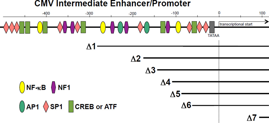
Small interfering RNAs are commonly employed, both individually and on a genome-wide scale, to degrade specific mRNAs and test the cellular requirements for their encoded proteins (1–7). The basic siRNA depletion experiment can be extended further using “rescue” experiments, in which the target protein is re-expressed from a transiently transfected vector that encodes an altered mRNA that is resistant to the siRNA silencing (8–10). This experiment is useful for confirming siRNA specificity because the exogenously expressed protein should rescue the loss-of-function phenotype. The experiment also enables genetic analyses in cultured mammalian cells because the functional effects of specific mutations can be tested. Phenotypic rescue experiments can fail, however, when the rescuing protein is expressed at such a high level that it dominantly inhibits the pathway of interest. This problem can often be alleviated by reducing the quantity of transfected expression vector, but this approach fails if the overall transfection efficiency is reduced. To address this problem, we have created an ensemble of seven mammalian expression vectors designed to allow more precise control of exogenous protein expression levels. These vectors have nested deletions that successively eliminate transcription factor binding sites within the human cytomegalovirus (CMV) intermediate early enhancer/promoter (summarized in Fig. 1 and Supplemental Table 1, and see Supplemental Fig. 1 for promoter DNA sequences and a summary of the design strategy). The deletions were made in the context of the mammalian expression vector pcDNA™3.1/myc-His(-)A that contained a custom-designed multiple cloning site (MCS) cassette. These vectors allow optimized expression of siRNA-resistant constructs, while maintaining the high transfection efficiencies necessary for potent phenotypic rescue.
Figure 1. Human Cytomegalovirus Major Immediate Early Enhancer/Promoter constructs used for attenuated gene expression.
The human CMV promoter structure is shown above, with the transcription start site at the +1 position and (putative) upstream binding sites for different transcription factors. Promoter deletion positions are shown below. Transcription factor binding elements were identified using the TESS analysis tool (http://www.cbil.upenn.edu/cgi-bin/tess/tess?RQ=WELCOME, (22)), with a consensus sequence cutoff of >12.0. Similar but slightly different variants of this binding site map were also generated by the program TFSEARCH (http://www.cbrc.jp/research/db/TFSEARCH.html, (23)) and have been published (24). Promoter deletions were introduced by cloning PCR fragments MluI-XbaI sites) with the designated deletions into a pcDNA™3.1/myc-His(-)A expression vector (Life Technologies) that carried a custom multicloning site between the XbaI and AflII sites. Deletion design and DNA sequences are provided in Supplemental Fig. 1, and construct numbers are provided in Supplemental Table 1.
HIV-1 and many other enveloped viruses recruit the cellular endosomal sorting complexes required for transport (ESCRT) pathway to facilitate the final membrane fission step of virus budding (11–14). As is true for many other cellular pathways, siRNA depletion/rescue experiments have contributed to our understanding of the role of the ESCRT pathway in HIV-1 budding (9,15). We have found, however, that it is often difficult to rescue virus budding to wild type levels following siRNA depletion because many ESCRT proteins, particularly those of the ESCRT-III family, can potently inhibit HIV-1 budding when overexpressed at elevated levels (16–20). The ESCRT-III/HIV-1 system therefore represents an attractive test case for examining the utility of our family of attenuated CMV expression vectors.
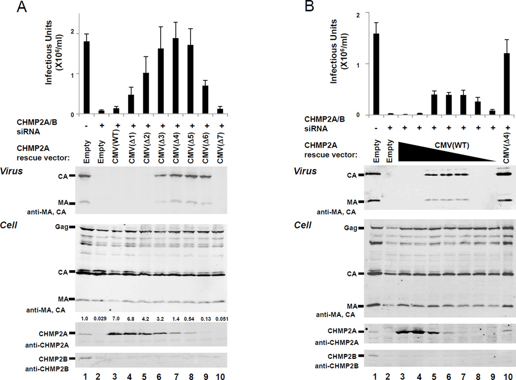
HIV-1 budding from cultured 293T cells can be potently inhibited by co-depletion of both members of the human CHMP2 family of ESCRT-III proteins (denoted CHMP2A and CHMP2B) (15). Hence, vector titers were dramatically reduced 48 h after co-transfection of a proviral HIV-1 vector together with siRNAs that targeted both CHMP2 proteins (Fig. 2A, top panel, 24±5-fold reduction, compare lanes 1 and 2). CHMP2 depletion also blocked virus release into the culture supernatants, as measured by immunoblotting for the virion-associated structural proteins, MA and CA (panel 2, compare lanes 1 and 2). Western blots of the 293T producer cells demonstrated that both CHMP2A and CHMP2B were depleted efficiently (panels 4 and 5, compare lanes 1 and 2) and that cellular levels of the structural HIV-1 Gag protein and its MA and CA cleavage products were not altered significantly by CHMP2 protein depletion (panel 3, compare lanes 1 and 2).
Figure 2. Rescue of HIV-1 budding from 293T cells that lack endogenous CHMP2 proteins by expression of human CHMP2A from attenuated CMV expression vectors.
(A) Differential rescue of HIV-1 budding by CHMP2A proteins expressed from the ensemble of different pCMV-CHMP2A expression vectors. HIV-1 vector infectivity titers (top panel) and western blots showing protein levels in culture supernatants (Virus, panel 2) or 293T cells (Cell, panels 3–5) co-transfected with: a proviral HIV-1 vector (500 ng of pCMV-dR8.2, 500 ng pLox-GFP, 250 ng pMD-G)(25) (all lanes), either 20 nM control siRNA duplex (CGUACGCGGAAUACUUCGAtt, where “tt” represents two overhanging deoxyribothymidines, lanes 1) or 10 nM each of siRNA duplexes against CHMP2A and CHMP2B (AGGCAGAGAUCAUGGAUAUtt and GGAACAGAAUCGAGAGUUAtt, lanes 2–10)(15), and 500 ng of either an empty vector control (lane 2) or the designated pCMV-CHMP2A vector expressing an siRNA-resistant CHMP2A construct (lanes 3–10). Integrated CHMP2A band intensities, normalized to the endogenous CHMP2A level, are provided over each lane in panel 4. 293T cells (2×10⁵ cells/well, 6-well plates, 2 ml volume) were seeded at t=0, transfected with siRNA (20 nM final total concentration, 7.5 µl Lipofectamine RNAiMAX; Life Technologies, Carlsbad, California) at t=24h, and co-transfected with siRNA, the designated pCMV-CHMP2A vector (500 ng), and the HIV-1 vector (20nM final total siRNA concentration, 500 ng pCMV-dR8.2, 500 ng pLox-GFP, 250 ng pMD-G, 10 µl Lipofectamine 2000; Life Technologies) at t=48h. The following silent mutations were introduced into the CHMP2A cDNA coding sequence to make the CHMP2A mRNA siRNA resistant: AGGCAGAGATCATGGATAT to AaGCtGAaATtATGGATAT (nucleotides 395–413). Cells and supernatant were collected and analyzed at t=96h. Released virions were pelleted through a 20% sucrose cushion at 15,000 × g and viral Gag-derived proteins were detected by western blotting using our rabbit anti-HIV-1 CA (UT415, 1:2000) and MA (UT556, 1:1000) antisera. Cells were lysed with buffer (50mM Tris-HCl pH 7.4, 150mM NaCl, 1% Triton-X100 and PMSF) for western blotting of intracellular proteins. Anti-CHMP2A and CHMP2B were detected with UT589 (our antibody) and Ab33174 (Abcam, Cambridge, MA) as described (26). Secondary antibodies were anti-mouse IgG or anti-rabbit IgG polyclonal conjugated to IRdye700 or IRdye800 (1:10000, Rockland Immunochemicals Inc., Gilbertsville, PA). Western blots were visualized using an Odyssey scanner (Li-Cor Biosciences, Lincoln, NB). For titer measurements, 293T cells were infected with viral supernatants and GFP-positive cells were quantified by flow cytometry (FL1 channel, FACScan, BD). Values show the average of three independent repetitions with standard errors.
(B) Rescue of HIV-1 budding by CHMP2A proteins expressed from different quantities of the wild type CMV expression vector, pCMV(WT)-CHMP2A. The figure and experiments are equivalent to panel (A) except that the following quantities of the siRNA-resistant CHMP2A rescue construct, pCMV(WT)-CHMP2A, were transfected: 500 ng (lane 3), 170 ng (lane 4), 56 ng (lane 5), 19 ng (lane 6), 6.2 ng (lane 7), 2.1 ng lane 8), and 0.69 ng (lane 9). In the experiments shown in lanes 4–9, total expression vector levels were adjusted to 500 ng with pCMV(WT) empty vector. The sample shown in Lane 10 was transfected with 500 ng of the pCMV(Δ4)-CHMP2A expression vector positive control).
To test for rescue of virus budding, 500 ng of each of the different siRNA-resistant pCMV-CHMP2A expression vectors were co-transfected together with the siRNA and proviral HIV-1 see Fig. 2A, caption). As expected, CHMP2A expression levels were highest for the construct that carried the wild type CMV promoter (denoted pCMV(WT)-CHMP2A), and decreased successively over two orders of magnitude as larger and larger promoter deletions were introduced (denoted pCMV(Δ1)-CHMP2A to pCMV(Δ7)-CHMP2A, panel 4, compare lanes 3–10). In contrast, the rescue of virus budding was biphasic: virion release and infectivity were low when CHMP2A levels were highest, increased when CHMP2A was expressed at intermediate levels, and then decreased again at the lowest CHMP2A expression levels (Panels 1 and 2, compare lanes 3–10). Levels of virion release and infectivity generally correlated well, but maximal infectivity occurred at slightly higher CHMP2A levels, perhaps because rapid virus release kinetics contribute more to viral infectivity than to total virion release as measured in the endpoint release assay. The pCMV(Δ4)-CHMP2A and pCMV(Δ5)-CHMP2A constructs expressed CHMP2A at levels that most closely approximated the normal level of the endogenous protein (panel 4, compare lanes 7 and 8 to lane 1). These two CHMP2A expression constructs also rescued virus release and infectivity best (panels 1 and 2). Importantly, the pCMV(Δ4)-CHMP2A construct rescued viral titers very efficiently, to 102±12% of untreated control levels. These data imply that: 1) CHMP2A alone can fully rescue HIV-1 budding, even in the absence of CHMP2B, 2) CHMP2A functions best when expressed at near-native levels, and 3) The attenuated pCMV(Δ4)-CHMP2A and pCMV(Δ5)-CHMP2A constructs can express near-native levels of CHMP2A under conditions where transfection efficiencies apparently remain high.
We next tested whether HIV-1 budding could be rescued to comparable levels simply by varying the quantity of pCMV(WT)-CHMP2A used in the transfection reaction. Three-fold dilutions over a range of 500-0.69 ng of pCMV(WT)-CHMP2A were tested for rescue of HIV-1 budding from cells that lacked endogenous CHMP2 proteins. CHMP2A expression levels correlated well with the quantity of pCMV(WT)-CHMP2A vector used (Fig. 2B, panel 4, lanes 3–9), and CHMP2A levels most closely approximated normal endogenous protein levels when 56 and 19 ng of pCMV(WT)-CHMP2A were used (compare lane 1 to lanes 6 and 7). Rescue of HIV-1 budding again followed a biphasic curve, with optimal rescue observed when CHMP2A was expressed at intermediate levels (170-19 ng pCMV(WT)-CHMP2A, lanes 5–7). In this case, however, HIV-1 titers never exceeded 26% of control levels, even when the bulk levels of exogenous CHMP2A approximated endogenous control levels (panel 1, compare lane 1 to lanes 6 and 7). In a parallel control experiment, HIV-1 release was again rescued to nearly wild type levels upon co-transfection of 500 ng of the pCMV(Δ4)-CHMP2A construct (lane 10). We therefore conclude that although optimizing pCMV(WT)-CHMP2A vector levels improved HIV-1 budding, overall rescue levels were never as high as could be achieved with the attenuated pCMV(Δ4)-CHMP2 expression construct.
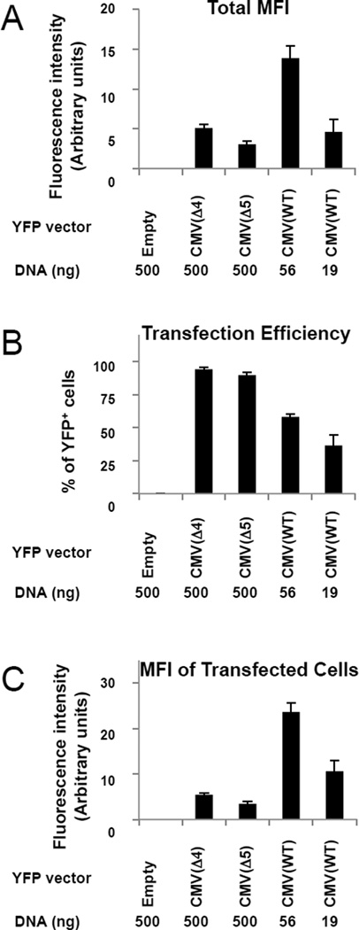
We hypothesized that the pCMV(Δ4)-CHMP2A and pCMV(Δ5)-CHMP2A vectors worked well in the rescue experiment because they could be used at concentrations that coupled high transfection efficiencies with restricted protein expression. To test this idea, we created pCMV(WT)-YFP, pCMV(Δ4)-YFP and pCMV(Δ5)-YFP expression vectors and used YFP fluorescence as a measure of protein expression in 293T cells. This approach allowed us to use flow cytometry to quantify transfection efficiencies and relative protein expression levels at the single-cell level. Titrations were again performed to determine the quantity of pCMV(WT)-YFP required to express YFP at levels comparable to those produced by transfections with 500 ng of pCMV(Δ4)-YFP or pCMV(Δ5)-YFP. This was achieved with 19 ng of pCMV(WT)-YFP, in reasonable agreement with the analogous CHMP2A titration experiments (Fig. 3A, compare total mean fluorescence levels for 500 ng of pCMV(Δ4)-YFP or pCMV(Δ5)-YFP DNA with 19 ng of pCMV(WT)-YFP). As shown in Fig. 3B, overall transfection efficiencies under these three conditions were: 94±1% for 500 ng of pCMV(Δ4)-YFP, 90± 2% for 500 ng of pCMV(Δ5)-YFP DNA and 36±8% for 19 ng pCMV(WT)-YFP (compare lanes 2, 3 and 5). Thus, overall transfection efficiencies dropped off significantly when the quantity of vector was reduced from 500 to 19 ng. We also quantified the mean fluorescence intensity (MFI) in the subsets of cells that were actually transfected in each reaction (i.e., now excluding cells in which YFP expression was undetectable). As shown in Fig. 3C, transfected cells in the 19 ng pCMV(WT)-YFP reaction had a mean fluorescence intensity of 11±2, whereas transfected cells in the 500 ng pCMV(Δ4)-YFP and pCMV(Δ5)-YFP reactions had mean fluorescence intensities of 5.4±0.4 and 3.4±0.6. These data demonstrate that although bulk YFP expression levels were comparable for the three conditions, this was achieved in different ways: the pCMV(Δ4)-YFP and pCMV(Δ5)-YFP vectors supported low-level YFP expression in nearly all of the cells, whereas the pCMV(WT)-YFP vector supported higher expression levels per cell, but in fewer than half of the cells. Thus, the attenuated vectors appear to work better in rescue experiments because, unlike the wild type pCMV(WT) vector, they can be used at sufficiently high concentrations to maintain high overall transfection efficiencies, yet they express low levels of the target protein in each cell. It is possible that varying HIV-1 vector levels could also affect the degree of rescue, but our experiments did not test this parameter.
Figure 3. Comparisons of transfection efficiencies and protein expression levels for the pCMV(WT)-YFP, pCMV(Δ4)-YFP, and pCMV(Δ5)-YFP vectors.
(A) Total mean fluorescence intensity of YFP (MFI, in arbitrary units) for all cells in each of the cultures following transfection with 500 ng empty pCMV(WT)(negative control, lane 1), 500 ng pCMV(Δ4)-YFP (lane 2), 500 ng pCMV(Δ5)-YFP (lane 3), 56 ng pCMV(WT)-YFP (lane 4) or 19 ng pCMV(WT)-YFP (lane 5). The YFP expression vectors were created by PCR amplification of the yfp gene and subcloned into the KpnI/XhoI sites of the custom multiple cloning site of the pcDNA™3.1/myc-His(-)A expression vector. 293T cells were seeded (t=0, 2×10⁵ cells/well, 6-well plates) and transfected (t=24h) with the designated pCMV-YFP constructs adjusted to 500 ng total DNA with pCMV(WT) empty vector where necessary), 10 µl Lipofectamine 2000). At t=72h, cells were trypsinized and analyzed by flow cytometry. YFP-positive cells were scored using a control-transfected sample to set the negative background level (BD CellQuest™ Pro software). YFP intensity was determined after subtracting control-transfected samples (FL1). Values here and in panels B and C show the average of five independent repetitions with standard errors.
(B) Percentages of cells with detectable YFP fluorescence in each of the cultures described in part (A).
(C) Mean YFP fluorescence intensity (arbitrary units) for the subsets of cells that were transfected (as judged by detectable YFP expression) in each of the cultures described in part (A).
In summary, we have created mammalian expression vectors that allow tunable expression of siRNA-resistant constructs, and demonstrated their utility in rescuing HIV-1 budding from cells that lacked endogenous CHMP2 proteins. We have also used this system successfully in other experiments, for example to achieve high-level rescue of retrovirus budding from cells depleted of endogenous ALIX and CHMP4 proteins (although the relative advantages of using the attenuated CMV vector system were somewhat less pronounced in these two cases, data not shown). The optimal CMV vector must, of course, be determined empirically for each new system because the correct choice will be influenced by differences in endogenous protein levels, protein expression efficiencies, and the degree to which the specific pathway and cell type can tolerate protein overexpression. Although we are not aware of previous studies that have employed the approach described here, related approaches such as the use of inducible promoters to optimize the expression of siRNA-resistant rescue constructs have been described (21). In principle, this is an elegant approach that can also be used to maximize phenotypic rescue, but it requires the creation of stable cell lines and is therefore less convenient than transient transfection, particularly when the functions of multiple mutant proteins are being screened. Hence, our system is likely to be most useful in cases where levels of the rescue protein must be tightly controlled and where the creation of stable cell lines is overly time consuming or problematic. Our vectors should also be useful in other applications where it is desirable to attenuate protein expression while maintaining high transfection levels.
Supplementary Material
Acknowledgments
This work was supported by National Institutes of Health grant AI051174 (W.I.S) and research fellowships from the Japanese Herpesvirus Infectious Forum (J.A.) and the Deutsche Forschungsgemeinschaft (J.V., VO 1836/1-1). This paper is subject to the NIH Public Access Policy.
Footnotes
Author Summary:
We have created a family of mammalian protein expression vectors with cytomegalovirus promoters of differing strengths and shown that these vectors can combine high transfection efficiencies with tunable protein expression levels to optimize the rescue of cellular phenotypes induced by siRNA transfection.
Competing interests
The authors declare no competing interests.
References
- 1.Elbashir SM, Harborth J, Lendeckel W, Yalcin A, Weber K, Tuschl T. Duplexes of 21-nucleotide RNAs mediate RNA interference in cultured mammalian cells. Nature. 2001;411:494–498. doi: 10.1038/35078107. [DOI] [PubMed] [Google Scholar]
- 2.Houzet L, Jeang KT. Genome-wide screening using RNA interference to study host factors in viral replication and pathogenesis. Exp Biol Med (Maywood) 2011;236:962–967. doi: 10.1258/ebm.2010.010272. [DOI] [PMC free article] [PubMed] [Google Scholar]
- 3.Sigoillot FD, King RW. Vigilance and validation: Keys to success in RNAi screening. ACS chemical biology. 2011;6:47–60. doi: 10.1021/cb100358f. [DOI] [PMC free article] [PubMed] [Google Scholar]
- 4.Falschlehner C, Steinbrink S, Erdmann G, Boutros M. High-throughput RNAi screening to dissect cellular pathways: a how-to guide. Biotechnol J. 2010;5:368–376. doi: 10.1002/biot.200900277. [DOI] [PubMed] [Google Scholar]
- 5.Martin SE, Caplen NJ. Applications of RNA interference in mammalian systems. Annu Rev Genomics Hum Genet. 2007;8:81–108. doi: 10.1146/annurev.genom.8.080706.092424. [DOI] [PubMed] [Google Scholar]
- 6.Pache L, Konig R, Chanda SK. Identifying HIV-1 host cell factors by genome-scale RNAi screening. Methods. 2011;53:3–12. doi: 10.1016/j.ymeth.2010.07.009. [DOI] [PubMed] [Google Scholar]
- 7.Sakurai K, Chomchan P, Rossi JJ. Silencing of gene expression in cultured cells using small interfering RNAs. Curr Protoc Cell Biol. 2010;Chapter 27(Unit 27):21–28. doi: 10.1002/0471143030.cb2701s47. 21. [DOI] [PubMed] [Google Scholar]
- 8.Lassus P, Rodriguez J, Lazebnik Y. Confirming specificity of RNAi in mammalian cells. Sci STKE. 2002;2002:l13. doi: 10.1126/stke.2002.147.pl13. [DOI] [PubMed] [Google Scholar]
- 9.Garrus JE, von Schwedler UK, Pornillos OW, Morham SG, Zavitz KH, Wang HE, Wettstein DA, Stray KM, et al. Tsg101 and the vacuolar protein sorting pathway are essential for HIV-1 budding. Cell. 2001;107:55–65. doi: 10.1016/s0092-8674(01)00506-2. [DOI] [PubMed] [Google Scholar]
- 10.Cullen BR. Enhancing and confirming the specificity of RNAi experiments. Nat Methods. 2006;3:677–681. doi: 10.1038/nmeth913. [DOI] [PubMed] [Google Scholar]
- 11.Henne WM, Buchkovich NJ, Emr SD. The ESCRT Pathway. Developmental cell. 2011;21:77–91. doi: 10.1016/j.devcel.2011.05.015. [DOI] [PubMed] [Google Scholar]
- 12.Martin-Serrano J, Neil SJ. Host factors involved in retroviral budding and release. Nature reviews. Microbiology. 2011;9:519–531. doi: 10.1038/nrmicro2596. [DOI] [PubMed] [Google Scholar]
- 13.Hurley JH, Hanson PI. Membrane budding and scission by the ESCRT machinery: it's all in the neck. Nat Rev Mol Cell Biol. 2010;11:556–566. doi: 10.1038/nrm2937. [DOI] [PMC free article] [PubMed] [Google Scholar]
- 14.Dordor A, Poudevigne E, Gottlinger H, Weissenhorn W. Essential and supporting host cell factors for HIV-1 budding. Future Microbiol. 2011;6:1159–1170. doi: 10.2217/fmb.11.100. [DOI] [PubMed] [Google Scholar]
- 15.Morita E, Sandrin V, McCullough J, Katsuyama A, Baci Hamilton I, Sundquist WI. ESCRT-III Protein Requirements for HIV-1 Budding. Cell Host Microbe. 2011;9:235–242. doi: 10.1016/j.chom.2011.02.004. [DOI] [PMC free article] [PubMed] [Google Scholar]
- 16.Howard TL, Stauffer DR, Degnin CR, Hollenberg SM. CHMP1 functions as a member of a newly defined family of vesicle trafficking proteins. J Cell Sci. 2001;114:2395–2404. doi: 10.1242/jcs.114.13.2395. [DOI] [PubMed] [Google Scholar]
- 17.von Schwedler UK, Stuchell M, Muller B, Ward DM, Chung HY, Morita E, Wang HE, Davis T, et al. The protein network of HIV-1 budding. Cell. 2003;114:701–713. doi: 10.1016/s0092-8674(03)00714-1. [DOI] [PubMed] [Google Scholar]
- 18.Martin-Serrano J, Yaravoy A, Perez-Caballero D, Bieniasz PD. Divergent retroviral late-budding domains recruit vacuolar protein sorting factors by using alternative adaptor proteins. Proc Natl Acad Sci USA. 2003;100:12414–12419. doi: 10.1073/pnas.2133846100. [DOI] [PMC free article] [PubMed] [Google Scholar]
- 19.Strack B, Calistri A, Craig S, Popova E, Gottlinger HG. AIP1/ALIX Is a Binding Partner for HIV-1 p6 and EIAV p9 Functioning in Virus Budding. Cell. 2003;114:689–699. doi: 10.1016/s0092-8674(03)00653-6. [DOI] [PubMed] [Google Scholar]
- 20.Zamborlini A, Usami Y, Radoshitzky SR, Popova E, Palu G, Gottlinger H. Release of autoinhibition converts ESCRT-III components into potent inhibitors of HIV-1 budding. Proc Natl Acad Sci USA. 2006;103:19140–19145. doi: 10.1073/pnas.0603788103. [DOI] [PMC free article] [PubMed] [Google Scholar]
- 21.Ma HT, Poon RY. Gene down-regulation with short hairpin RNAs and validation of specificity by inducible rescue in mammalian cells. Curr Protoc Cell Biol. 2010;Chapter 27(Unit 27):22. doi: 10.1002/0471143030.cb2702s49. [DOI] [PubMed] [Google Scholar]
- 22.Schug J. Using TESS to predict transcription factor binding sites in DNA sequence. In: Baxevanis AD, editor. Current Protocols in Bioinformatics. John Wiley & Sons, Ltd; 2009. [DOI] [PubMed] [Google Scholar]
- 23.Heinemeyer T, Wingender E, Reuter I, Hermjakob H, Kel AE, Kel OV, Ignatieva EV, Ananko EA, et al. Databases on transcriptional regulation: TRANSFAC, TRRD and COMPEL. Nucleic acids research. 1998;26:362–367. doi: 10.1093/nar/26.1.362. [DOI] [PMC free article] [PubMed] [Google Scholar]
- 24.Stinski MF, Isomura H. Role of the cytomegalovirus major immediate early enhancer in acute infection and reactivation from latency. Med Microbiol Immunol. 2008;197:223–231. doi: 10.1007/s00430-007-0069-7. [DOI] [PubMed] [Google Scholar]
- 25.Naldini L, Blomer U, Gage FH, Trono D, Verma IM. Efficient transfer, integration, and sustained long-term expression of the transgene in adult rat brains injected with a lentiviral vector. Proc Natl Acad Sci USA. 1996;93:11382–11388. doi: 10.1073/pnas.93.21.11382. [DOI] [PMC free article] [PubMed] [Google Scholar]
- 26.Morita E, Colf LA, Karren MA, Sandrin V, Rodesch CK, Sundquist WI. Human ESCRT-III and VPS4 proteins are required for centrosome and spindle maintenance. Proc Natl Acad Sci USA. 2010;107:12889–12894. doi: 10.1073/pnas.1005938107. [DOI] [PMC free article] [PubMed] [Google Scholar]
Associated Data
This section collects any data citations, data availability statements, or supplementary materials included in this article.


