

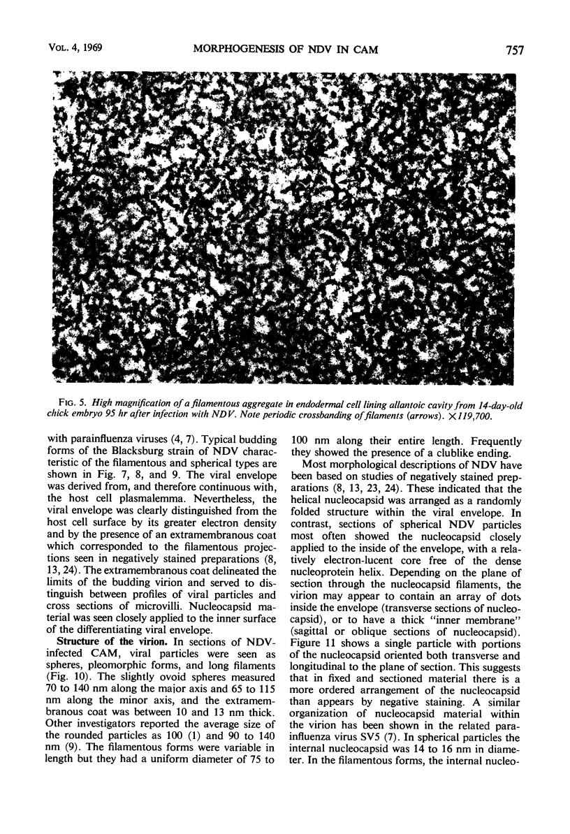
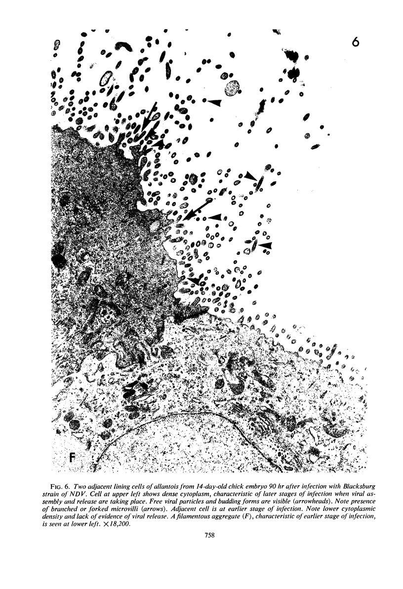
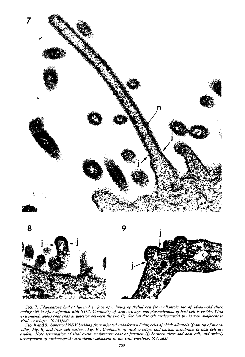
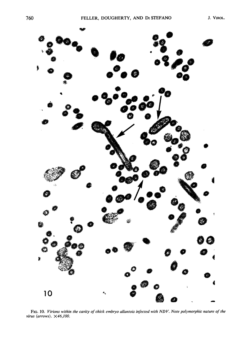
Abstract
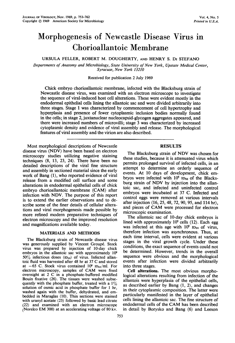
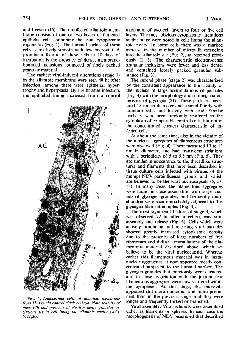
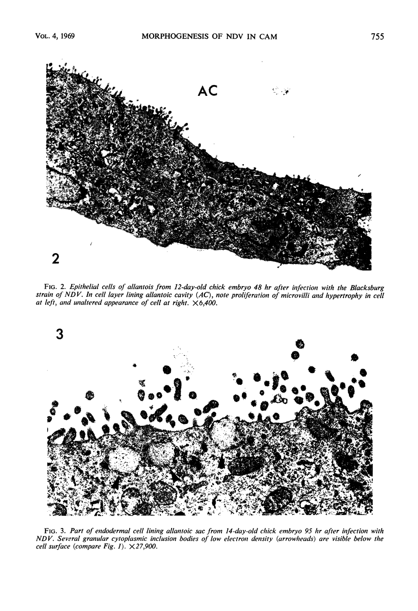
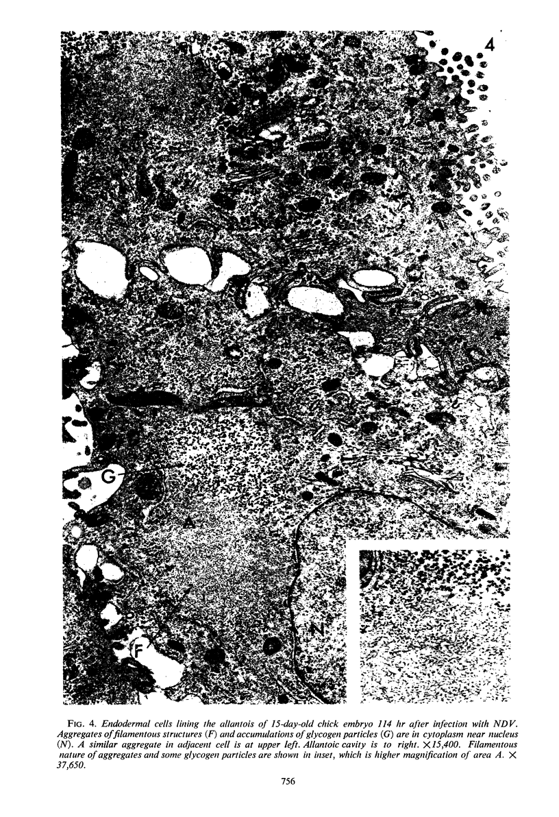
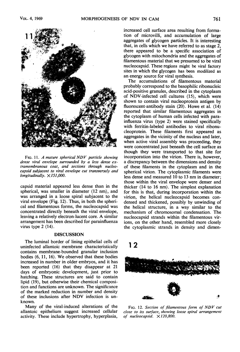
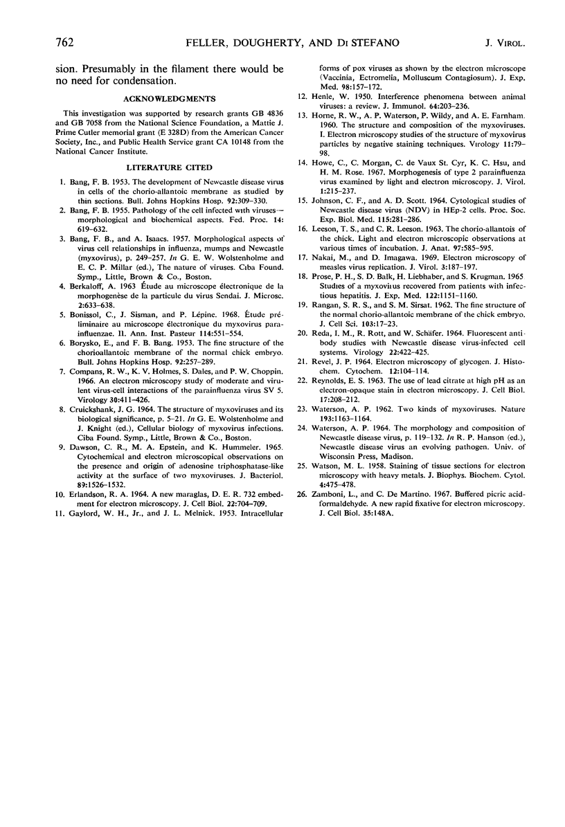
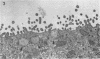
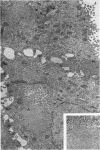
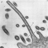
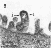
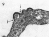
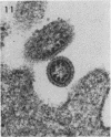
Chick embryo chorioallantoic membrane, infected with the Blacksburg strain of Newcastle disease virus, was examined with an electron microscope to investigate the sequence of viral-induced host cell alterations. These were evident mostly in the endodermal epithelial cells lining the allantoic sac and were divided arbitrarily into three stages. Stage 1 was characterized by commencement of cell hypertrophy and hyperplasia and presence of fewer cytoplasmic inclusion bodies normally found in the cells; in stage 2, juxtanuclear nucleocapsid-glycogen aggregates appeared, and there were increased numbers of microvilli; stage 3 was characterized by increased cytoplasmic density and evidence of viral assembly and release. The morphological features of viral assembly and the virion are also described.
Full text
PDF
Images in this article
Selected References
These references are in PubMed. This may not be the complete list of references from this article.
- BANG F. B. Pathology of the cell infected with viruses-morphological and biochemical aspects. Fed Proc. 1955 Jun;14(2):619–632. [PubMed] [Google Scholar]
- BANG F. B. The development of Newcastle disease virus in cells of the chorio-allantoic membrane as studied by thin section. Bull Johns Hopkins Hosp. 1953 Apr;92(4):309–329. [PubMed] [Google Scholar]
- BORYSKO E., BANG F. B. The fine structure of the chorio-allantoic membrane of the normal chick embryo. Bull Johns Hopkins Hosp. 1953 Apr;92(4):257–289. [PubMed] [Google Scholar]
- Bonissol C., Sisman J., Lépine P. Etude préliminaire au microscope électronique du Myxovirus parainfluenzae II. Ann Inst Pasteur (Paris) 1968 Apr;114(4):551–554. [PubMed] [Google Scholar]
- Compans R. W., Holmes K. V., Dales S., Choppin P. W. An electron microscopic study of moderate and virulent virus-cell interactions of the parainfluenza virus SV5. Virology. 1966 Nov;30(3):411–426. doi: 10.1016/0042-6822(66)90119-x. [DOI] [PubMed] [Google Scholar]
- DAWSON C. R., EPSTEIN M. A., HUMMELER K. CYTOCHEMICAL AND ELECTRON MICROSCOPICAL OBSERVATIONS ON THE PRESENCE AND ORIGIN OF ADENOSINE TRIPHOSPHATASE-LIKE ACTIVITY AT THE SURFACE OF TWO MYXOVIRUSES. J Bacteriol. 1965 Jun;89:1526–1532. doi: 10.1128/jb.89.6.1526-1532.1965. [DOI] [PMC free article] [PubMed] [Google Scholar]
- ERLANDSON R. A. A NEW MAGAGLAS, D.E.R.(R) 732, EMBEDMENT FOR ELECTRON MICROSCOPY. J Cell Biol. 1964 Sep;22:704–709. doi: 10.1083/jcb.22.3.704. [DOI] [PMC free article] [PubMed] [Google Scholar]
- GAYLORD W. H., Jr, MELNICK J. L. Intracellular forms of pox viruses as shown by the electron microscope (Vaccinia, Ectromelia, Molluscum Contagiosum). J Exp Med. 1953 Aug;98(2):157–172. doi: 10.1084/jem.98.2.157. [DOI] [PMC free article] [PubMed] [Google Scholar]
- HENLE W. Interference phenomena between animal viruses; a review. J Immunol. 1950 Mar;64(3):203–236. [PubMed] [Google Scholar]
- HORNE R. W., WATERSON A. P., WILDY P., FARNHAM A. E. The structure and composition of the myxoviruses. I. Electron microscope studies of the structure of myxovirus particles by negative staining techniques. Virology. 1960 May;11:79–98. doi: 10.1016/0042-6822(60)90056-8. [DOI] [PubMed] [Google Scholar]
- Howe C., Morgan C., de Vaux St Cyr C., Hsu K. C., Rose H. M. Morphogenesis of type 2 parainfluenza virus examined by light and electron microscopy. J Virol. 1967 Feb;1(1):215–237. doi: 10.1128/jvi.1.1.215-237.1967. [DOI] [PMC free article] [PubMed] [Google Scholar]
- JOHNSON C. F., SCOTT A. D. CYTOLOGICAL STUDIES OF NEWCASTLE DISEASE VIRUS (NDV) IN HEP-2 CELLS. Proc Soc Exp Biol Med. 1964 Feb;115:281–286. doi: 10.3181/00379727-115-28891. [DOI] [PubMed] [Google Scholar]
- LEESON T. S., LEESON C. R. THE CHORIO-ALLANTOIS OF THE CHICK. LIGHT AND ELECTRON MICROSCOPIC OBSERVATIONS AT VARIOUS TIMES OF INCUBATION. J Anat. 1963 Oct;97:585–595. [PMC free article] [PubMed] [Google Scholar]
- Nakai M., Imagawa D. T. Electron microscopy of measles virus replication. J Virol. 1969 Feb;3(2):187–197. doi: 10.1128/jvi.3.2.187-197.1969. [DOI] [PMC free article] [PubMed] [Google Scholar]
- Prose P. H., Balk S. D., Liebhaber H., Krugman S. Studies of a myxovirus recovered from patients with infectious hepatitis. II. Fine structure and electron microscopic demonstration of intracytoplasmic internal component and viral filament formation. J Exp Med. 1965 Dec 1;122(6):1151–1160. doi: 10.1084/jem.122.6.1151. [DOI] [PMC free article] [PubMed] [Google Scholar]
- REDA I. M., ROTT R., SCHAEFER W. FLUORESCENT ANTIBODY STUDIES WITH NDV-INFECTED CELL SYSTEMS. Virology. 1964 Mar;22:422–425. doi: 10.1016/0042-6822(64)90033-9. [DOI] [PubMed] [Google Scholar]
- REVEL J. P. ELECTRON MICROSCOPY OF GLYCOGEN. J Histochem Cytochem. 1964 Feb;12:104–114. doi: 10.1177/12.2.104. [DOI] [PubMed] [Google Scholar]
- REYNOLDS E. S. The use of lead citrate at high pH as an electron-opaque stain in electron microscopy. J Cell Biol. 1963 Apr;17:208–212. doi: 10.1083/jcb.17.1.208. [DOI] [PMC free article] [PubMed] [Google Scholar]
- WATERSON A. P. Two kinds of myxovirus. Nature. 1962 Mar 24;193:1163–1164. doi: 10.1038/1931163a0. [DOI] [PubMed] [Google Scholar]
- WATSON M. L. Staining of tissue sections for electron microscopy with heavy metals. J Biophys Biochem Cytol. 1958 Jul 25;4(4):475–478. doi: 10.1083/jcb.4.4.475. [DOI] [PMC free article] [PubMed] [Google Scholar]