Abstract
Paired box (PAX) genes are transcription factors that play important roles in embryonic development. Although the PAX gene family occurs in animals only, it is widely distributed. Among the vertebrates, its 9 genes appear to be the product of complete duplication of an original set of 4 genes, followed by an additional partial duplication. Although some studies of PAX genes have been conducted, no comprehensive survey of these genes across the entire taxonomic unit has yet been attempted. In this study, we conducted a detailed comparison of PAX sequences from 188 chordates, which revealed restricted variation. The absence of PAX4 and PAX8 among some species of reptiles and birds was notable; however, all 9 genes were present in all 74 mammalian genomes investigated. A search for signatures of selection indicated that all genes are subject to purifying selection, with a possible constraint relaxation in PAX4, PAX7, and PAX8. This result indicates asymmetric evolution of PAX family genes, which can be associated with the emergence of adaptive novelties in the chordate evolutionary trajectory.
Introduction
Paired box (PAX) genes are transcription factors that play key roles in several aspects of embryonic development and organogenesis [1]–[3]. Although the PAX family is specific to the animal lineage, the evolutionary history of these genes remains uncertain. A unique PAX gene (TriPaxB) has been isolated from Trichoplax adhaerens (Placozoa), the most morphologically simple species of all non-parasitic multicellular metazoan animals. The TriPaxB protein contains the characteristic DNA-binding domain of the PAX family, the paired domain (PD) of 128 amino acids, an octapeptide motif (OC), and a paired-type homeobox DNA-binding domain (HD) [1], [4].
Four PAX genes (PAX1/9, PAX2/5/8, PAX3/7, and PAX4/6) have been found in the basal chordates, amphioxus (e.g. Brachiostoma floridae) and tunicates (e.g. Ciona intestinalis) [5]–[7]. Phylogenetic analyses indicated that a single PAX gene of each subfamily was present in the ancestral chordate and gave rise to the amphioxus PAX. Afterwards, a plus round of whole genome duplications, gave origin to the multiple vertebrate PAX subfamily copies [4]–[8]. Ohno [9], suggested that the early vertebrate lineage underwent one (1 R) or more (≥2 R) whole genome duplications (WGDs). These processes were considered to provide additional possibilities for diversified evolution and/or speciation. Rapid and widespread evolutionary changes could lead to macroevolutionary emergent properties, since WGD products are able to evolve and reach a greater level of interaction and complexity than would otherwise be possible through cumulative single gene duplications [10]–[15]. The second round of whole genome duplication most likely occurred after the divergence of invertebrate chordate lineages from the ancestral vertebrate, although there is controversy about the exact branch at which the phenomenon occurred [11], [16]–[18].
After these 2 major duplication events occurred, probably 8 PAX genes emerged. Another partial duplication occurred subsequently, resulting in the 9 PAX genes currently found in mammals (subfamilies: (1) PAX1 and PAX9; (2) PAX2, PAX5, and PAX8; (3) PAX3 and PAX7; (4) PAX4 and PAX6 [1], [6]). An alternative scenario would be that more PAX genes would have arisen after 2 WGRD and then lost during the vertebrate evolution history [19], [20].
In vertebrates, as well as in other chordates, PAX genes are notably expressed during development. They are also known to play an important role in mature life stages, based on observations of organ/tissue-specific signals (Table S1; [21]). For instance, PAX3 and PAX7 proteins are found in adult cells of the vertebrate muscle tissue [22]. Analogously, in amphioxus, the PAX3/7 gene is most highly expressed in adult muscle [7]. These observations, along with other similar findings, indicate that a PAX-derived gene can maintain similar roles to those present in the putative ancestor. Nonetheless, various novel roles for PAX genes have also emerged during the evolutionary history of vertebrates: they were co-opted for new regulatory networks, diverged, and subsequently gained new functions [5], [7], [19]–[27].
Although the presence of PAX genes has been investigated in a variety of organisms [6], [19]–[23], [26], a broad survey of chordate PAX genes has yet to be conducted. Therefore, the aim of the present study was to address questions regarding the occurrence and evolution of the PAX family in chordates. We used publicly available sequences to evaluate: (1) the presence/absence of PAX genes in 188 organisms, and (2) the evolutionary rates and properties of PAX genes in chordates. Results of this analysis will contribute to a greater understanding of the mechanisms of change in complex gene families.
Results and Discussion
Search and Identification for Vertebrate PAX Genes
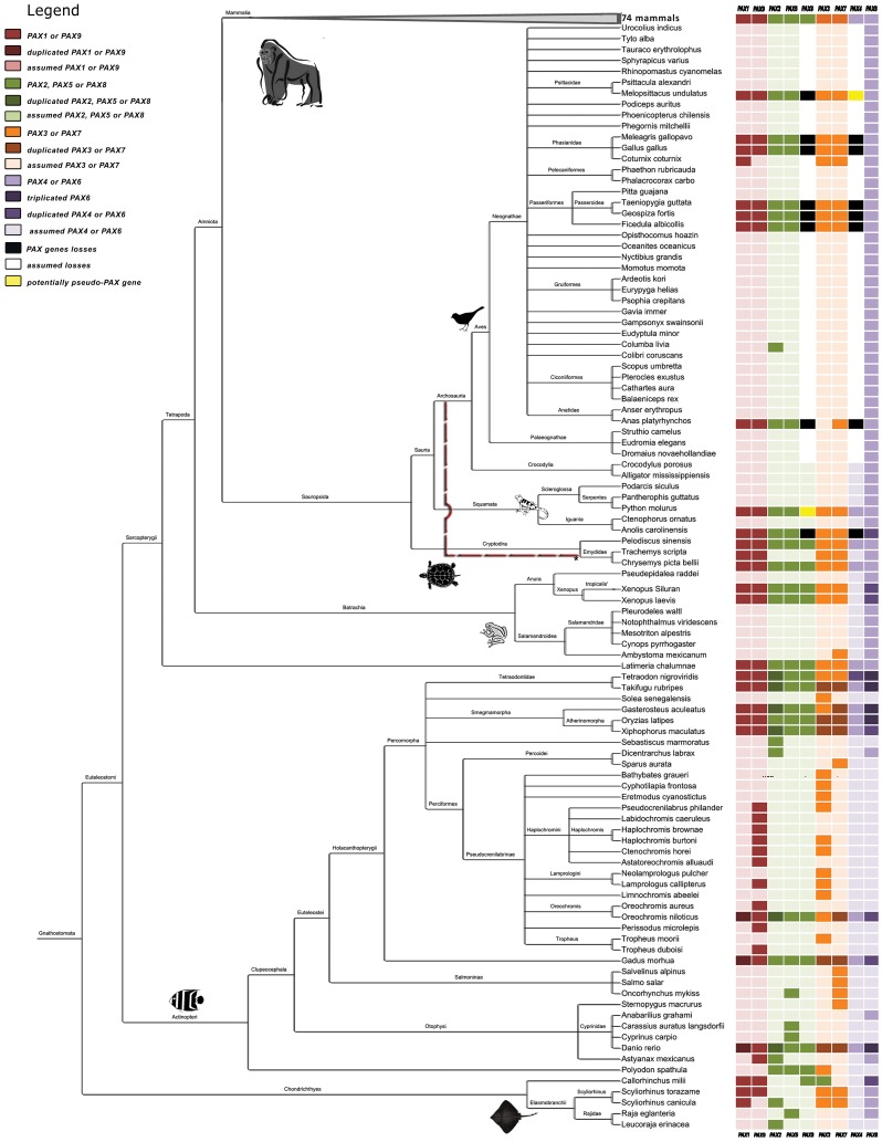
We characterized chordate PAX genes using sequences that were available in the Ensembl, UCSC, NCBI, and UniProt databases [28]–[31]. A total of 188 species were evaluated, including vertebrates (175 jawed and 4 jawless), urochordates (6 tunicates), and cephalochordates (3 amphioxus) [32]; see details in Tables S2 and S3 and Figure 1.
Figure 1. Phylogenetic tree for the 175 jawed vertebrate species considered in this study.
Sequences were obtained from the NCBI Taxonomy Browser and edited with Figtree and hitmap for the presence of PAX genes. The dashed red line indicates the relationship suggested by Crawford et al [46].
Basal Chordates
We retrieved 4 PAX genes in cephalochordates (Branchiostoma belcheri, B. floridae, and B. lanceolatum), which is the most basal chordate subphylum. In tunicates, the most basal animals belonging to the Olfactores clade, we recovered 5 PAX genes. Three of the genes (PAX1/9, PAX 3/7, and PAX4/6) could be considered equivalent to the ancestral vertebrate PAX types, while the others (PAX2/5/8a and PAX2/5/8b in Branchiostoma lanceolatum) were derived [5]–[7], [33].
Jawless Vertebrates
Jawless vertebrates were represented in our study by 1 hagfish species (Eptatretus burger) and 3 lamprey species (Lampetra fluviatilis, Lethenteron camtschaticum, and Petromyzon marinus). Together, they form a sister group of the gnathostome vertebrates, making them a good model to investigate ancestral vertebrate characteristics. The hidden Markov models (HMMER) search recovered 3 PAX genes for the hagfish and 4 for all lampreys. In Petromyzon marinus, we recovered 2 PAX genes (PAX1/9 and PAX1/9b), and found 2 segments of 158 bp and 144 bp, respectively, showing 88% of identity with the PAX3 and PAX7 genes. Interestingly, in vitro studies identified PAX1/9 and PAX1/9b in this species [33], as well as the PAX7, PAX2 [34], PAX6 [33]–[37], PAX3/PAX7 genes [36]. This in vitro information, which was confirmed by our genome data, suggests that Petromyzon marinus contains genes corresponding to the 4 ancestral PAX genes in addition to a second copy of the PAX1/9 and PAX3/7 type. This suggests that a duplication of the PAX1/9 and PAX3/7 genes occurred in the lamprey or jawless lineage, although the possibility of an ancient genome duplication event (before the split between jawless and jawed vertebrates) with subsequent lineage-specific modifications cannot be discarded [12]. The difference between the numbers of PAX genes found in the basal chordates, tunicates (4 or 5) and lampreys (6) and the basal jawed vertebrates (9) can be associated with the emergence of adaptive novelties at the tunicate/vertebrate and agnathan/gnathostome transitions.
Jawed Vertebrates
We found 6 PAX genes in the most basal taxon of this group, the Chondrichthyes (2 skates, 2 sharks, and 1 chimaera). The chimaera species (Callorhinchus milii; elephant shark), for which the draft genome is already available, contained all 6 genes (1, 9, 8, 3, and 2 copies of 6; Figure 1 and Table S3). PAX4 was not retrieved in any search (Genomes, HMMER protein, and BLAT/BLAST; Table S3). However, the absence of PAX4 should be interpreted with caution since the elephant shark genome has low coverage (1.4x) and the sequence databases are biased toward the most popular/known genes. The duplicate PAX6 (named PAX6.2) was recently discovered, and based on experimental work and in the conservation of coding and noncoding elements, the authors suggested that although an ancient duplication event occurred in a gnathostome ancestor, the additional copy was independently lost in mammals and birds [38].
All of the expected 9 PAX genes were found in 37 species of ray-finned bony fishes. Considering only the 8 ray-finned bony fishes for which complete genomes are available (class Actinopterygii; Table S3), additional duplicate or triplicate copies were found in 7 of the 9 PAX genes (exception: PAX5 and PAX8; Figure 1). This situation was probably a consequence of whole genome duplication [39], [40], which occurred in the early evolution of teleost fishes approximately 320–350 million years ago (3 RWGD hypothesis; [41]).
Latimeria chalumnae (coelacanth), a lobe-finned fish, presents all 9 PAX genes, suggesting that the ancestor that gave rise to the tetrapod lineage contained all members of the PAX family.
The frog species Xenopus tropicalis and Xenopus laevis also presented 9 PAX genes. However, we found duplicated copies of PAX2 and PAX6 in X. laevis and X. tropicalis, respectively. The presence of the additional PAX2 copy in X. laevis could be the result to the fact that this species experienced a recent and specific polyploidization event approximately 40 million years ago. Approximately 32–47% of duplicated genes were observed in its whole genome [15], [39], [42]. An alternative is that PAX2 could have been duplicated through local gene duplication. The duplication of PAX6 in X. tropicalis has been reported in a previous study (PAX6.2 [38]). For the other 6 species of amphibians, we only retrieved PAX6 and PAX7 sequences (6 hits and 1 hit, respectively). For all amphibian species studied here, it was not possible to localize PAX4, corroborating a recent paper that proposed that X. tropicalis lost PAX4 [43]. These data suggest that the absence of PAX4 could be a general characteristic of amphibian taxa.
The analysis of the entire PAX family in reptile and bird species (Sauropsida, Table S3 and Figure 1) showed a surprising finding: some branches appear to have lost PAX4 and PAX8. It was recently suggested [44] that PAX8 gene was lost after turtles split from other reptiles and birds, which most likely occurred ∼240 million years ago [45]–[47]. We found PAX8 in 2 of the 3 turtle species studied (Chrysemys picta bellii and Pelodiscus sinensis; Table S3). We also found 9 PAX genes, including a PAX8 segment, in a snake (Python molurus). The unresolved Sauropsida phylogeny (Figure 1; [48]) raises the question as to whether the loss of PAX4 and PAX8 is an ancestral event, or whether these losses occurred independently in distinct reptile and bird lineages.
Overall, the searches showed that all 9 PAX genes appear to be present in the 74 mammalian species studied (Figure 1). Although some exceptions to this general pattern were found, they are likely a consequence of the low coverage of the genome in question (e.g. Dipodomys ordii (kangaroo rat), which had only a 2× coverage), or due to bias toward the most popular genes, rather than a reflection of actual gene loss.
Shared Synteny and/or Conserved Neighborhood Analysis
We performed an analysis of shared synteny (genes in the same chromosome) and/or conserved neighborhood (genes side-by-side in the same order) for all 4 PAX subfamilies: (1) PAX1 and PAX9; (2) PAX2, PAX5, and PAX8; (3) PAX3 and PAX7; (4) PAX4 and PAX6 [7], [21]. The most conserved and similar blocks were those in which PAX1 and PAX9 were inserted. The others presented distinct levels of neighboring and conserved synteny.
This analysis was also used as additional evidence for the absence of PAX4 and PAX8, as well as for evidence of PAX6 duplication in some taxa, as described in the previous section.
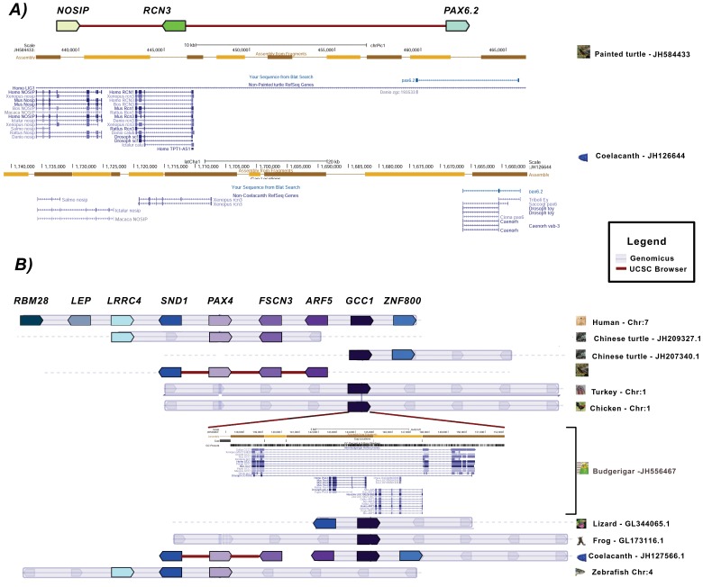
By using a similar approach, Ravi et al. [38] recently found that PAX6.2 was located in close proximity to the RCN3 and NOSIP genes in the elephant shark, lizard, zebrafish, and Xenopus. However, no PAX6-duplicated ortholog was found in the proximity of NOSIP-RCN3 or elsewhere in the genomes of birds or mammals. Our analysis confirmed the presence of PAX6.2 in RCN3-NOSIP in Xenopus and in a lizard species (Anolis carolinensis). Additionally, we found the PAX6.2 gene in this same region in the coelacanth and in the painted turtle (Chrysemys picta bellii; Figure 2A), but failed to find the PAX6.2-RCN3-NOSIP block in the genomes of another turtle species (Pelodiscus sinensis) or in mammals and birds. Consequently, our data support the proposal that the duplication that gave rise to the PAX6.2 gene must have occurred before the split between cartilaginous and bony fish, and that this duplication was followed by multiple independent PAX6.2 gene losses in distinct vertebrate lineages.
Figure 2. Synteny and neighborhood status for the PAX4 and PAX6 genes.
We found a possible fragment of the PAX4 gene in association with the ARF5 gene in the budgerigar (Melopsittacus undulatus) genome, which was in the intronic region of the GCC1 gene. In other vertebrate genomes, the GCC1-ARF5-FSCN3-PAX4 block formed a conserved neighborhood (Figure 2B). Although the PAX4 segment appeared to contain a complete paired domain, 3 independent approaches failed to predict a full functional protein.
The syntenic analysis of PAX8 showed that the block in which it is inserted in mammals and fish is relatively well conserved in birds and reptiles, whose some genomes lack PAX8, providing supporting evidence for its loss.
Based on the presence of putatively nonfunctional relics (PAX4 and PAX8 segments in Melopsittacus undulatus and Python molurus, respectively), along with the other findings presented above, we can suggest that the loss of PAX4 and/or PAX8 occurred multiple times in tetrapod lineages, which also appears to be the case for PAX6.2. This hypothesis is compatible with findings of previous studies [49]–[56].
Comparative Analysis of PAX Proteins
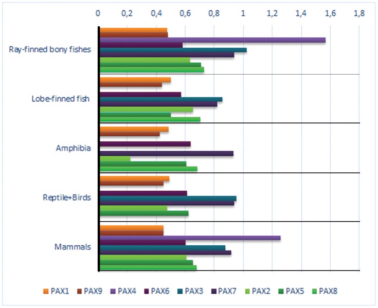
The PAX protein analysis was performed using the 53 species whose available sequences met our quality criteria (see Material and Methods). Pairwise amino acid distances were calculated between each PAX subfamily member and the PAX protein type of its probable outgroup (the urochordata Ciona intestinalis) (Table S4 and Figure 3). The distances of each protein from that of its probable outgroup were also compared among the 4 subfamilies ((1) PAX1 and PAX9; (2) PAX2, PAX5, and PAX8; (3) PAX3 and PAX7; (4) PAX4 and PAX6). For instance, the distance values of the human PAX1 and PAX9 to the PAX1/9 type from Ciona intestinalis were 0.447 and 0.455, respectively, which was not statistically significant (p = 0.706).
Figure 3. Pairwise PAX protein changes observed in different vertebrate taxa compared to that of the sea squirt.
Considering all comparisons (53 chordate species and 9 PAX proteins), some specific and general patterns emerged: the distance values between PAX1 and PAX9 paralogs to their putative PAX1/9 protein did not show significant differences (p = 0.069). In other words, the distance of PAX1 to the Ciona Intestinalis PAX1/9 is the same as the distance to its paralogous gene, PAX9. The same result was found when PAX3 and PAX7 were compared with PAX3/7 (p = 0.704).
On the other hand, the PAX4 protein was more dissimilar to its putative outgroup (PAX4/6) than it was to PAX6 (p = 0.001). Therefore, this suggests that PAX4 most likely emerged in 2 R together with PAX6. Possibly higher evolutionary rates (inferred by ω = dN/dS) would further confirm this result. A recent study indicated a possible loss of PAX4 expression from the brain in vertebrates, probably after 2 RWGD [43], may have led to relaxed constraints on gene conservation, as suggested by a higher rate of sequence divergence.
When PAX2, PAX5, and PAX8 were compared with their putative PAX 2/5/8 ancestral type, other suggestive patterns appeared. The distance between PAX8 and its ancestor was significantly different from those of the others (p = 0.001), whereas the PAX2 and PAX5 distances did not present a significant difference (p = 0.091).
These results could indicate that a round of complete vertebrate genome duplication most likely involved PAX2 and PAX5 ancestor, whereas PAX8 emerged through local gene duplication. This would support the idea that PAX8 is the most recent gene to appear by duplication in this family. An alternative scenario to evolution of the PAX2/5/8 subfamily is that after 1 RWGD, two copies of the subfamily genes emerged, one resembling PAX2/5 and the other PAX8. A second duplication event (2 WRGD), resulted in 4 copies of the PAX2/5/8 subfamily, followed by loss of one of the PAX8 duplicates. The result is the presence of PAX2, PAX5, and PAX8 genes in jawed vertebrates [19], [20]. The relaxation of selective pressure immediately after this last partial/total duplication would be expected, which could explain the higher variation observed in PAX8 relative to its outgroup (PAX2/5/8). The higher evolutionary rate (inferred by ω = dN/dS; see Material and Methods and next section) could be an alternative explanation; however, the 2 above mentioned possibilities, relative to PAX8, are not mutually exclusive. Redundancy in the expression of these genes likely played a central role in the loss and/or higher divergence rate of PAX8. In mammals, PAX8 is mainly expressed in the kidney, ear, and thyroid gland during development, whereas PAX2 is expressed not only in these organs and tissues, but also in others, such as the eye, pharyngeal arches, and brain [2], [3], [5], [7], [19], [20], [44]. Amphioxus (here considered as an outgroup) contains only PAX2/5/8 and shows pleiotropic expression in most organs and tissues, implying that PAX2, PAX5, and PAX8 have retained most of their ancestral expression patterns [19], [20], [44].
Selection on PAX Genes
Our molecular evolution analyses (Table 1) revealed ω values <1, indicating that purifying selection has acted on PAX over the majority of the evolutionary history of vertebrates. This result is consistent with the idea that developmental genes are under functional restriction in metazoa [56]. The clade model D [57] performed better in a likelihood ratio test (LTR; p<0.001; Table 1) when compared with the neutral M1a model. This result indicates that ω values obtained for the 4 PAX subfamilies can vary between branches, predicting distinct molecular evolutionary patterns.
Table 1. Branch-site clade model D values for ω (dN/dS ratio) estimated for 2 site classes.
| PAX subfamily | Clade Model D | M1a - Parameter estimates | P* (LRT) | ||
| Proportion (p) | Branch type 0 (ω) | Branch type 1 (ω) | M1a X Clade Model | ||
| PAX9 | PAX1 | PAX1/PAX9 | |||
| PAX1 and PAX9 | 0.87268 | 0.00186 | 0.00186 | p: 0.98611 0.01389 | <0.001 |
| 0.12732 | 0.08140 | 0.09185 | ω: 0.01023 1.00000 | ||
| PAX2/PAX5 | PAX8 | PAX2/PAX5/PAX8 | |||
| PAX2, PAX5, and PAX8 | 0.62462 | 0.01192 | 0.01192 | p: 0.99999 0.00001 | <0.001 |
| 0.37538 | 0.00027 | 0.00117 | ω: 0.00650 1.00000 | ||
| PAX3 | PAX7 | PAX3/PAX7 | |||
| PAX3 and PAX7 | 0.68577 | 0.01016 | 0.01016 | p: 0.99360 0.00640 | <0.001 |
| 0.31423 | 0.09976 | 0.18923 | ω: 0.03997 1.00000 | ||
| PAX4 | PAX6 | PAX4/PAX6 | |||
| PAX4 and PAX6 | 0.61493 | 0.02335 | 0.02335 | p: 0.83385 0.16615 | <0.001 |
| 0.38507 | 0.30208 | 0.01784 | ω: 0.06400 1.00000 | ||
Degrees of freedom: 2; LRT: 2Δl = 2(l 1− l 0);
Although all estimated ω values were less than 1, which suggested the action of negative selection, a possible relaxation of this selective constraint was revealed when the subfamilies were compared. In 38% of the sites, the PAX4 ω value was 16 times greater than that of the PAX6 ω value. Additionally, in 37% and 31% of the sites, the PAX8 and PAX7 ω values were approximately 2 and 4 times greater than the PAX2-PAX5 and PAX3 ω values, respectively (Table 1). These results suggest that PAX4, PAX8, and PAX7 have experienced relatively more modifications than the other PAX genes.
Gene Phylogeny Analysis
Bayesian Monte Carlo Markov Chain trees were built from 2 PAX subfamily data sets (PAX2, PAX5, and PAX8; PAX4 and PAX6), in which genes were lost in some lineages, and presented greater molecular evolutionary rates wherever they were not lost. Well-defined clusters were observed that separated the 3 and 2 genes of each subfamily, respectively (Figure S1 illustrates the PAX2, PAX5, and PAX8 tree). These results indicate again the conservative nature of purifying selection that has driven molecular evolution of the PAX gene. As expected, the PAX genes found in the tunicate Ciona intestinalis (PAX2/5/8 and PAX4/6) lead to basal branches in Figure S1 in the other subfamily (data not shown). In some cases, recovery of the class phylogeny of the species is apparent as clear mammal, fish, and bird clades can be observed in the PAX2 cluster. Similar topologies and statistical robustness were obtained using the maximum likelihood method (data not shown). The trees, however, do not provide additional evidence about the differences in evolutionary rates of genes observed within each subfamily.
Conclusion
Overall, purifying selection appears to be the main factor responsible for molecular evolution of the PAX family in chordate species. However, there are some indications of potential group-specific changes that are beyond this general pattern. There was a loss of PAX4 and PAX8 in lizards and birds. Accelerated evolutionary rates were suggested for the PAX4, PAX8 and PAX7 genes. The accumulation of variation (at least in some sites), due to an initial relaxation of purifying selection, may indicate the beginning of a process that enabled evolvability of the system.
Results of the present study revealed that some PAX genes experienced striking changes in the course of their evolutionary trajectory, which emphasizes the point that even developmental master genes might not follow universal patterns of molecular evolution. Functional retention and loss, subfunctionalization, as well as neofunctionalization can also be observed in developmental genes.
The asymmetric evolution of the PAX family genes observed here, as evidenced by uneven events of duplications and deletions are compatible with the emergence of adaptive novelties during chordate radiation.
Materials and Methods
Data Collection
Nucleotide and amino acid sequences for all available PAX genes in chordate species were obtained using Biomart (Ensembl v66–70 - http://www.ensembl.org/biomart/martview/; [58], [59]). The Protein Domains/Limit filter (InterPro (ID): IPR00152) was used as a parameter to identify the PAX genes or the paired box domains. A second approach was the inspection of one-to-one ortholog gene maps, which were also obtained from Biomart.
BLAST/BLAT searches in Ensembl (http://www.ensembl.org/Multi/blastview), UCSC databases (http://genome.ucsc.edu/), and in the NCBI Genebank (genomic BLAST http://www.ncbi.nlm.nih.gov/sutils/genom_table.cgi?organism=8496&database=8496) were also conducted in order to identify possible unannotated orthologs. The Pre!Ensembl database (http://pre.ensembl.org/index.html) was used to access the new draft released genomes. Finally, we applied hidden Markov models (HMMER) web service searching sequence databases for homologs of PAX amino acid sequences in the NR and Uniprot collections [60].
The genomic sequences of possible unannotated orthologs were verified using three programs that can predict open reading frames (ORF): BESTORF (http://linux1.softberry.com/berry.phtml?topic=bestorf&group=programs&subgroup=gfind), GeneWise (http://www.ebi.ac.uk/Tools/psa/genewise/) [61], and STAR ORF (http://star.mit.edu/orf/index.html).
The following procedures were also adopted: (a) for genes encoding multiple transcripts, the transcript with the longest genomic transcribed length was selected; (b) a high identity (up to 70%) with the paired domain was accepted as indicating a PAX family member; (c) possible gene losses were accepted only when they were observed in multiple species as well as in high coverage genome assemblies; (d) a subset of 53 species was selected for the evolutionary analyses, since their sequences had the best alignment, and they were optimized for analysis with a higher number of sites.
PAX Gene Family Synteny and Neighborhood Status
Mapping adjacent genes into PAX synteny regions was achieved with the Genomicus website v70.01 (http://www.dyogen.ens.fr/genomicus-70.01/cgi-bin/search.pl [62]). Additionally, we manually searched the Ensembl and UCSC genome browsers for the same purpose.
Variation in the PAX Family
The amino acid sequences were aligned using the MUSCLE algorithm [63] included in Mega (version 5.0) [64], which were verified with the GUIDANCE web service using the MAFFT algorithm [65]. Mega (version 5.0) software was employed to evaluate variability in the PAX groups using the pairwise distance of members of each subfamily from the gene of its probable outgroup, the tunicate (Ciona intestinalis). SPSS (version 16) software was used to calculate the statistical significance of differences between PAX1/9, PAX2/5/8, PAX3/7, and PAX4/6 paralogous sequences using the paired Student’s t-test.
Tests for Selection
Patterns of selection and rates of evolutionary changes in the PAX family were evaluated using standard tests [66]–[68]. We used the phylogeny-based maximum likelihood analysis of ω (dN/dS) as implemented in the CODEML program of the PAML 4.4 package to statistically test for positive selection and/or relaxation of functional constraints. The heterogeneity of evolutionary rates among paralogous groups was tested using the CODEML program in PAML4.4 clade models [57]. Branches on the phylogeny were divided into 2 clades a priori, and a likelihood ratio test (LRT) was used to evaluate divergences in selective pressures between them, as indicated by different ω ratios. We employed the clade model type D that assumes 2 site classes, which was compared with the neutral model M1a by an LRT with 2 degrees of freedom. This comparison was primarily used to detect positive selection, but our goal here was also to evaluate acceleration during the evolutionary history through direct inferences of dN/dS differences.
Empirical Bayes approaches, implemented in CODEML, were also used to infer which of the PAX sequences sites might have evolved under positive selection. To determine sites under selection, the naive-empirical Bayes (NEB) test was employed. The unrooted tree input file for PAML4.4 analyses was a phylogenetic tree provided by Ensembl, which was edited using PhyloWidget for the 53 species included in this study.
Gene Phylogeny
Data from PAX4 and PAX6, as well as from PAX2, PAX5, and PAX8 gene subfamilies were used to construct phylogenetic trees. The comparison was performed using a mixed Bayesian Monte Carlo Markov Chain sampler for phylogenetic reconstruction using protein alignments in PhyloBayes [69] on the web server Bioportal from the University of Oslo. Additionally, we built trees using the maximum likelihood method (Mega, version 5.0 [64]) using the same dataset.
Supporting Information
PAX2, PAX5 , and PAX8 gene subfamilies phylogeny based on the Bayesian Monte Markov Chain method.
(JPG)
Description and functions of PAX genes.
(DOCX)
Species considered in this study and font of hits.
(XLSX)
Presence of PAX genes in 188 Chordate species.
(XLSX)
Pairwise distances between the PAX subfamilies and the outgroup PAX sea squirt.
(DOCX)
Acknowledgments
We thank the Bioportal at the University of Oslo, where part of the phylogenetic analysis was conducted. We are grateful to reviewers and to Dr. Philippe Vernier and Dr. Marc Robison-Rechavi (Academic Editor) for valuable comments that greatly improved our manuscript.
Funding Statement
This research was financially supported by the Coordenação de Aperfeiçoamento de Pessoal de Nível Superior (CAPES), Conselho Nacional de Desenvolvimento Científico e Tecnológico (CNPq) and Fundação de Amparo à Pesquisa do Estado do Rio Grande do Sul, (FAPERGS), Programa de Apoio a Núcleos de Excelência (PRONEX). VRP-C received a doctoral fellowship from CNPq. The funders had no role in study design, data collection and analysis, decision to publish, or preparation of the manuscript.
References
- 1. Chi N, Epstein JA (2002) Getting your PAX straight: Pax proteins in development and disease. Trends Genet 1: 41–47. [DOI] [PubMed] [Google Scholar]
- 2. Lang D, Powell SK, Plummer RS, Young KP, Ruggeri BA (2007) PAX genes: roles in development, pathophysiology, and cancer. Biochem Pharmacol 73: 1–14. [DOI] [PubMed] [Google Scholar]
- 3. Dressler GR (2011) Patterning and early cell lineage decisions in the developing kidney: the role of PAX genes. Pediatr Nephrol 26: 1387–1394. [DOI] [PMC free article] [PubMed] [Google Scholar]
- 4. Hadrys T, DeSalle R, Sagasser S, Fischer N, Schierwater B (2005) The Trichoplax PAXB gene: a putative Proto-PaxA/B/C gene predating the origin of nerve and sensory cells. Mol Bio Evol 22: 1569–1578. [DOI] [PubMed] [Google Scholar]
- 5. Mazet F, Hutt JA, Millard J, Shimeld SA (2003) PAX gene expression in the developing central nervous system of Ciona intestinalis . Gene Expr Patt 3: 743–745. [DOI] [PubMed] [Google Scholar]
- 6. Vorobyov E, Horst J (2006) Getting the proto-PAX by the tail. J Mol Evol 63: 153–164. [DOI] [PubMed] [Google Scholar]
- 7. Chen L, Zhang Q, Wang W, Wang Y (2010) Spatiotemporal expression of PAX genes in amphioxus: insights into PAX-related organogenesis and evolution. Sci China Life Sci 53: 1031–1040. [DOI] [PubMed] [Google Scholar]
- 8. Holland L Z, Schubert M, Kozmik Z, Holland ND (1999) AmphiPAX3/7, an amphioxus paired box gene: insights into chordate myogenesis, neurogenesis, and the possible evolutionary precursor of definitive vertebrate neural crest. Evol Dev 1: 153–165. [DOI] [PubMed] [Google Scholar]
- 9.Ohno S (1970) Evolution by gene duplication. New York: Springer Verlag.
- 10. Dehal P, Boore JL (2005) Two rounds of whole genome duplication in the ancestral vertebrate. PLoS Biol 3: e314. [DOI] [PMC free article] [PubMed] [Google Scholar]
- 11. Putnam NH, Butts T, Ferrier DE, Furlong RF, Hellsten U, et al. (2008) The amphioxus genome and the evolution of the chordate karyotype. Nature 453: 1064–1071. [DOI] [PubMed] [Google Scholar]
- 12. Kuraku S, Meyer A, Kuratani S (2009) Timing of genome duplications relative to the origin of the vertebrates: did cyclostomes diverge before or after? Mol Biol Evol 26: 47–59. [DOI] [PubMed] [Google Scholar]
- 13. Kuraku S, Meyer A (2009) The evolution and maintenance of Hox gene clusters in vertebrates and the teleost-specific genome duplication. Intern J Developm Biol 53: 765–774. [DOI] [PubMed] [Google Scholar]
- 14. Van de Peer Y, Maere S, Meyer A (2009) The evolutionary significance of ancient genome duplications. Nature Rev Genet 10: 725–732. [DOI] [PubMed] [Google Scholar]
- 15.Cañestro C (2012) Two rounds of whole-genome duplication: evidence and impact on the evolution of vertebrate innovations. In Polyploidy and genome evolution (ed. MS Soltis, DE Soltis), 309–339. Berlin: Springer.
- 16. Holland PW, Garcia-Fernandez J (1996) Hox genes and chordate evolution. Dev Biol 173: 382–395. [DOI] [PubMed] [Google Scholar]
- 17. Venkatesh B, Kirkness EF, Loh YH, Halpern AL, Lee AP, Johnson J, et al. (2007) Survey sequencing and comparative analysis of the elephant shark (Callorhinchus milii) genome. PLoS Biol 5: e101. [DOI] [PMC free article] [PubMed] [Google Scholar]
- 18. Kuraku S (2013) Impact of asymmetric gene repertoire between cyclostomes and gnathostomes. Semin in Cell Developm Biol 24: 119–127. [DOI] [PubMed] [Google Scholar]
- 19. Bassham S, Cañestro C, Postlethwait JH (2008) Evolution of developmental roles of Pax2/5/8 paralogs after independent duplication in urochordate and vertebrate lineages. BMC Biology 6: e35. [DOI] [PMC free article] [PubMed] [Google Scholar]
- 20. Goode DK, Elgar G (2009) The PAX258 gene subfamily: a comparative perspective. Dev Dynam 238: 2951–2974. [DOI] [PubMed] [Google Scholar]
- 21. Suga H, Tschopp P, Graziussi DF, Stierwald M, Schmid V, et al. (2010) Flexibly deployed PAX genes in eye development at the early evolution of animals demonstrated by studies on a hydrozoan jellyfish. Proc Natl Acad Sci USA 107: 14263–14268. [DOI] [PMC free article] [PubMed] [Google Scholar]
- 22. Buckingham M, Frédéric R (2007) The role of PAX genes in the development of tissues and organs: PAX3 and PAX7 regulate muscle progenitor cell functions. Annu Rev Cell Bio 23: 645–673. [DOI] [PubMed] [Google Scholar]
- 23. Lynch M, Conery JS (2000) The evolutionary fate and consequences of duplicate genes. Science 290: 1151–1155. [DOI] [PubMed] [Google Scholar]
- 24. Lynch VJ, Wagner GP (2008) Resurrecting the role of transcription factor change in developmental evolution. Evolution 62: 2131–2154. [DOI] [PubMed] [Google Scholar]
- 25. Boyle AP, Song L, Lee BK, London D, Keefe D, et al. (2010) High-resolution genome-wide in vivo footprinting of diverse transcription factors in human cells. Genome Res 21: 456–464. [DOI] [PMC free article] [PubMed] [Google Scholar]
- 26. Balczarek KA, Lai ZC, Kumar S (1997) Evolution of functional diversification of the paired box (PAX) DNA-binding domains. Mol Biol Evol 14: 829–842. [DOI] [PubMed] [Google Scholar]
- 27. Catmull J, Hayward DC, McIntyre NE, Reece-Hoyes JS, Mastro R, et al. (1998) PAX-6 origins – implications from the structure of two coral PAX genes. Dev Genet Evol 208: 352–356. [DOI] [PubMed] [Google Scholar]
- 28. Flicek P, Ahmed I, Amode MR, Barrell D, Beal K, et al. (2013) Ensembl 2013. Nucleic Acids Res 41: D48–D55. [DOI] [PMC free article] [PubMed] [Google Scholar]
- 29. Meyer LR, Zweig AS, Hinrichs AS, Karolchik D, Kuhn RM, et al. (2013) The UCSC Genome Browser database: extensions and updates. Nucleic Acids Res 41: 64–69. [DOI] [PMC free article] [PubMed] [Google Scholar]
- 30. Park Y, Sheetlin SL, Ma N, Madden TL, Spouge JL (2012) New finite-size correction for local alignment score distributions. BMC Res Notes 5: e286. [DOI] [PMC free article] [PubMed] [Google Scholar]
- 31. The UniProt Consortium (2012) Reorganizing the protein space at the Universal Protein Resource (UniProt). Nucleic Acids Res 40: D71–D75. [DOI] [PMC free article] [PubMed] [Google Scholar]
- 32. Delsuc F, Brinkmann H, Chourrout D, Philippe H (2006) Tunicates and not cephalochordates are the closest living relatives of vertebrates. Nature 439: 965–968. [DOI] [PubMed] [Google Scholar]
- 33. Ogasawara M, Shigetani S, Hirano S, Satoh N, Kuratani S (2000) PAX1/PAX9-related genes in an agnathan vertebrate, Lampetra japonica: expression pattern of LjPAX9 implies sequential evolutionary events toward the gnathostome body plan. Dev Biol 223: 399–410. [DOI] [PubMed] [Google Scholar]
- 34. McCauley DW, Bronner-Fraser M (2002) Conservation of PAX gene expression in ectodermal placodes of the lamprey. Gene 287: 129–139. [DOI] [PubMed] [Google Scholar]
- 35. Derobert Y, Baratte B, Lepage M, Mazan S (2002) PAX6 expression patterns in Lampetra fluviatilis and Scyliorhinus canicula embryos suggest highly conserved roles in the early regionalization of the vertebrate brain. Brain Res Bull 57: 277–280. [DOI] [PubMed] [Google Scholar]
- 36. Osorio J, Mazan S, Rétaux S (2005) Organisation of the lamprey (Lampetra fluviatilis) embryonic brain: insights from LIM-homeodomain, PAX and hedgehog genes. Dev Biol 288: 100–112. [DOI] [PubMed] [Google Scholar]
- 37. Murakami Y, Ogasawara M, Sugahara F, Hirano S, Satoh N, et al. (2001) Identification and expression of the lamprey Pax6 gene: evolutionary origin of the segmented brain of vertebrates. Development 128: 3521–3531. [DOI] [PubMed] [Google Scholar]
- 38. Ravi V, Bhatia S, Gautier P, Loosli F, Tay BH, et al. (2013) Sequencing of Pax6 loci from the elephant shark reveals a family of Pax6 genes in vertebrate genomes, forged by ancient duplications and divergences. PLoS Genet 9: e1003177. [DOI] [PMC free article] [PubMed] [Google Scholar]
- 39. Taylor JS, Raes J (2004) Duplication and divergence: the evolution of new genes and old. Annu Rev Genet 38: 615–643. [DOI] [PubMed] [Google Scholar]
- 40. Laisney JA, Braasch I, Walter RB, Meierjohann S, Schartl M (2010) Lineage-specific co-evolution of the Egf receptor/ligand signaling system. BMC Evol Biol 10: e27. [DOI] [PMC free article] [PubMed] [Google Scholar]
- 41. Braasch I, Volff JN, Schartl M (2008) The evolution of teleost pigmentation and the fish-specific genome duplication. J Fish Biol 73: 1891–1918. [Google Scholar]
- 42. Semon M, Wolfe KH (2008) Preferential subfunctionalization of slow-evolving genes after allopolyploidization in Xenopus laevis. Proc. Natl Acad Sci USA 105: 8333–8338. [DOI] [PMC free article] [PubMed] [Google Scholar]
- 43. Manousaki T, Feiner N, Begemann G, Meyer A, Kuraku S (2011) Co-orthology of Pax4 and Pax6 to the fly eyeless gene: molecular phylogenetic, comparative genomic, and embryological analyses. Evol Dev 13: 448–459. [DOI] [PubMed] [Google Scholar]
- 44. Freter S, Muta Y, O’Neill P, Vassilev VS, Kuraku S, et al. (2012) PAX2 modulates proliferation during specification of the optic and epibranchial placodes. Dev Dynam 241: 1716–1728. [DOI] [PubMed] [Google Scholar]
- 45. Benton MJ, Donoghue PCJ (2007) Paleontological evidence to date the tree of life. Mol Biol Evol 24: 26–53. [DOI] [PubMed] [Google Scholar]
- 46. Modesto SP, Anderson JS (2004) The phylogenetic definition of Reptilia. Syst Biol 53: 815–821. [DOI] [PubMed] [Google Scholar]
- 47. Kumar S, Hedges SB (2011) TimeTree2: species divergence times on the iPhone. Bioinformatics 27: 2023–2024. [DOI] [PMC free article] [PubMed] [Google Scholar]
- 48. Crawford NG, Faircloth BC, McCormack JE, Brumfield RT, Winker K, et al. (2012) More than 1000 ultraconserved elements provide evidence that turtles are the sister group of archosaurs. Biol Lett 8: 783–786. [DOI] [PMC free article] [PubMed] [Google Scholar]
- 49. Hughes AL, Friedman R (2008) Genome size reduction in the chicken has involved massive loss of ancestral protein-coding genes. Mol Biol Evol 25: 2681–2688. [DOI] [PMC free article] [PubMed] [Google Scholar]
- 50. He J, Irwin DM, Chen R, Zhang YP (2010) Stepwise loss of motilin and its specific receptor genes in rodents. J Mol Endocrinol 44: 37–44. [DOI] [PubMed] [Google Scholar]
- 51. Quesada V, Velasco G, Puente XS, Warren WC, López-Otín C (2010) Comparative genomic analysis of the zebra finch degradome provides new insights into evolution of proteases in birds and mammals. BMC Genomics 11: e220. [DOI] [PMC free article] [PubMed] [Google Scholar]
- 52. Zhang G, Cowled C, Shi Z, Huang Z, Bishop-Lilly KA, et al. (2013) Comparative analysis of bat genomes provides insight into the evolution of flight and immunity. Science 339: 456–460. [DOI] [PMC free article] [PubMed] [Google Scholar]
- 53. Kuraku S, Kuratani S (2011) Genome-wide detection of gene extinction in early mammalian evolution. Genome Biol Evol 3: 1449–1462. [DOI] [PMC free article] [PubMed] [Google Scholar]
- 54.Yamamoto K, Mirabeau O, Bureau C, Blin M, Michon-Coudouel S, et al.. (2012) Evolution of dopamine receptor genes of the D1 class in vertebrates. Mol Biol Evol (published online ahead of press). [DOI] [PMC free article] [PubMed]
- 55. Hughes CR, Miles S, Walbroehl JM (2008) Support for the minimal essential MHC hypothesis: a parrot with a single, highly polymorphic MHC class II B gene. Immunogenetics 60: 219–231. [DOI] [PubMed] [Google Scholar]
- 56. Bates MD, Wells JM, Venkatesh B (2005) Comparative genomics of the Hlx homeobox gene and protein: conservation of structure and expression from fish to mammals. Gene 352: 45–56. [DOI] [PubMed] [Google Scholar]
- 57. Bielawski JP, Yang Z (2004) A maximum likelihood method for detecting functional divergence at individual codon sites, with application to gene family evolution. J Mol Evol 59: 121–132. [DOI] [PubMed] [Google Scholar]
- 58. Haider S, Ballester B, Smedley D, Zhang J, Rice P, et al. (2009) BioMart Central Portal – unified access to biological data. Nucleic Acids Res 37: 23–27. [DOI] [PMC free article] [PubMed] [Google Scholar]
- 59. Durinck S, Huber W (2011) The biomaRt user’s guide. Database 1: 1–22. [Google Scholar]
- 60.Finn RD, Clements J, Eddy SR (2011) HMMER web server: interactive sequence similarity searching. Nucleic Acids Res 39(Web Server Issue): W29–W37. [DOI] [PMC free article] [PubMed]
- 61. Birney E, Clamp M, Durbin R (2004) GeneWise and genomewise. Genome Res 14: 988–995. [DOI] [PMC free article] [PubMed] [Google Scholar]
- 62. Louis A, Muffato M, Crollius HR (2012) Genomicus: five genome browsers for comparative genomics in eukaryota. Nucleic Acids Res 38: 23–28. [DOI] [PMC free article] [PubMed] [Google Scholar]
- 63. Edgar RC (2004) MUSCLE: multiple sequence alignment with high accuracy and high throughput. Nucleic Acids Res 32: 1792–1797. [DOI] [PMC free article] [PubMed] [Google Scholar]
- 64. Tamura K, Peterson D, Peterson N, Stecher G, Nei M, et al. (2011) MEGA5: Molecular Evolutionary Genetics Analysis using maximum likelihood, evolutionary distance, and maximum parsimony methods. Mol Bio Evol 28: 2731–2739. [DOI] [PMC free article] [PubMed] [Google Scholar]
- 65.Penn O, Privman E, Ashkenazy H, Landan G, Graur D, et al.. (2010) GUIDANCE: a web server for assessing alignment confidence scores. Nucleic Acids Res 38(Web Server Issue): W23–W28. [DOI] [PMC free article] [PubMed]
- 66. Nielsen R, Yang Z (1998) Likelihood models for detecting positively selected amino acid sites and applications to the HIV-1 envelope gene. Genetics 148: 929–936. [DOI] [PMC free article] [PubMed] [Google Scholar]
- 67. Yang Z, Bielawski J (2000) Statistical methods for detecting molecular adaptation. Trends Ecol Evol 15: 496–503. [DOI] [PMC free article] [PubMed] [Google Scholar]
- 68. Yang Z, Nielsen R (2008) Mutation-selection models of codon substitution and their use to estimate selective strengths on codon usage. Mol Bio Evol 25: 568–579. [DOI] [PubMed] [Google Scholar]
- 69. Lartillot N, Lepage T, Blanquart S (2009) PhyloBayes 3: a Bayesian software package for phylogenetic reconstruction and molecular dating. Bioinformatics 25: 2286–2288. [DOI] [PubMed] [Google Scholar]
Associated Data
This section collects any data citations, data availability statements, or supplementary materials included in this article.
Supplementary Materials
PAX2, PAX5 , and PAX8 gene subfamilies phylogeny based on the Bayesian Monte Markov Chain method.
(JPG)
Description and functions of PAX genes.
(DOCX)
Species considered in this study and font of hits.
(XLSX)
Presence of PAX genes in 188 Chordate species.
(XLSX)
Pairwise distances between the PAX subfamilies and the outgroup PAX sea squirt.
(DOCX)


