Abstract
Lymphoid papillary hyperplasia is a rare abnormality of the tonsils with a predilection for affecting young Asian girls. Herein, we report a 31-year-old Chinese woman presented as right lateral recurrent tonsillar hypertrophy with odynophagia and dysphagia over the past 5 years, worsening over a period of for half a year. Clinically, this lesion was similar to papillomatosis or lymphoid polyposis. However, histopathologic study showed a distinctive form of lymphoid hyperplasia with considerable distinct finger-like projections composed of many phyllodes which contained remarkable follicular lymphoid hyperplasia. This is the only Chinese case of lymphoid papillary hyperplasia of the palatine tonsils that has been reported in the most recent English literature so far. The importance of recognizing this disorder rests in the fact that in spite of the clinical features suggestive of both a benign and a malignant tumor, however, the process is a benign tumor-like proliferation, probably non-neoplastic, could easily be cured by tonsillectomy.
Keywords: Palatine tonsils, lymphoid hyperplasia, papilloma, lymphoid polyposis
Introduction
Most neoplasms occur in the palatine tonsils are malignant, benign tumors or tumor-like lesions are less common. Squamous papillomas account for the majority of the benign disorders. The palatine tonsils may be the center of some acute or chronic inflammations. Chronic tonsillitis represents the most frequent lesion within inflammatory pathology of the tonsil organ. Under these circumstances, they became hypertrophied, resulting in a lymphoid proliferation. Lymphoid papillary hyperplasia is a rare abnormality of the tonsils described most frequently in the Japanese populations [1], sporadically in the western literature [2,3]. However, this abnormality has not been well documented among the Chinese population. Herein, we report a Chinese case of this rare condition.
Case report
A 31-year-old Chinese woman presented to the ENT department of our hospital with a complaint of right lateral recurrent tonsillar hypertrophy, concomitant with odynophagia and dysphagia over the past 5 years which were progressively aggravated for half a year. The woman was absent of other symptoms such as fever, cough and expectoration, hemoptysis or dyspnea. The patient’s left lateral tonsil was innocent. Her past medical history was unremarkable, and her pedigree members denied any family medical history neither. Physical examination showed a right lateral papilloma-like lesion arising in the mesopharynx, posterior to the soft palate. The clinical impression was suggestive of considering a neoplastic lesion such mucosal papilloma, or even a malignant lymphoma as indicated by the surgeons, although no other clinical or any laboratory abnormalities were found with the woman. Surgical tonsillectomy was performed, with specimens fixed in neutral buffered formalin and embedded in paraffin for routine histologic examination. The postoperative period of the woman were uneventful, there has been no evidence of recurrence of disease within follow-up for 12 months since the patient’s discharge.
Pathological findings
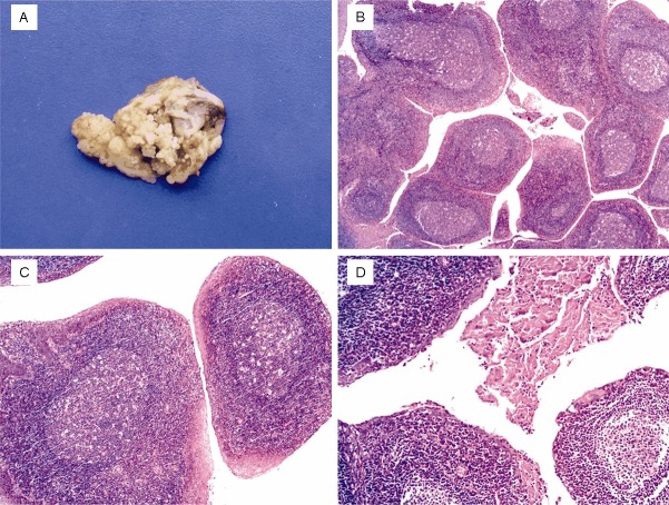
Macroscopically, the materials resected from the right lateral palatine tonsil measured, on average, 3.5x2.5x2cm, with pearl-albus and light brown coloration, and a friable granular surface with numerous tiny papilliform configuration (Figure 1A), which were grossly similar to papillomatosis occurred in the upper respiratory tract, or lymphoid polyposis of the gastrointestinal tract. There was no hemorrhage, necrosis, or infiltration present grossly. Histopathological examination of the surgical materials disclosed a branching condition with considerable distinct finger-like projections composed of many phyllodes which contained remarkable follicular lymphoid hyperplasia covered by stratified squamous epithelium on the surface (Figure 1B). The papillary lymphoid hyperplasia microscopically revealed that the rich lymphoid tissue with intense follicular hyperplasia with excessive increase of the germinal center and decrease of the follicular cortex (Figure 1C), scattered within the fibrous septa around the lymphoid nodules were a small number of plasma cells and mononuclear cells, which sometimes penetrated into the surface mucosa. There were no confluent solid sheets areas composed of monomorphic hyperplasia within the lymphoid tissues. The covering squamous mucosa showed mild hyperplasia with occasionally parakeratosis and hyperkeratosis (Figure 1D). As carefully examining the tissues, no evidence of malignancy was detected. The histopathologic appearance was indicated of a diagnosis of lymphoid papillary hyperplasia of the palatine tonsil.
Figure 1.
A: Grossly, the tonsil was occupied by numerous tiny papilliform projections similar to epithelial papillomatosis or lymphoid polyposis on the surface. B: Depicting distinct finger-like projections composed of many phyllodes which contained remarkable follicular lymphoid hyperplasia (H&Ex25). C: The core of the papillary configuration revealed the rich lymphoid tissue with excessive increase of the germinal center and decrease of the follicular cortex (H&Ex100). D: Squamous surface epithelium of the papillae displayed mild hyperplasia with parakeratosis and hyperkeratosis (H&Ex50).
Discussion
Lymphoid papillary hyperplasia is one of the rare abnormalities of the palatine tonsils, with a clinical impression of both a benign and a malignant tumor [1-3], such oral squamous papilloma and lymphoid polyposis, both of which could produce severe pharyngeal obstruction, as preoperatively indicated by the clinicians in the present case. Whereas based on the microscopical examination, this lesion, which revealed that the rich lymphoid tissue with intense follicular hyperplasia with excessive increase of the germinal center, without monomorphic lymphoid hyperplasia presented, and the covering mucosa showed only mild hyperplasia without atypia, could be readily identified as a benign, tumor-like lesion and easily be distinguished from other neoplastic lesions. Another rare nonneoplastic lesion, which was recently described by Kardon et al [4] as a hamartomatous proliferation, termed tonsillar lymphangiomatous polyps, can both clinically and macroscopically mimic lymphoid papillary hyperplasia, however, tonsillar lymphangiomatous polyps histologically show a characteristic submucosal proliferation of endothelial-lined lymph-vascular channels amid a fibrous, lymphoid, or adipose stroma, lacking the prominent lymphoid follicles hyperplasia characteristic of lymphoid papillary hyperplasia.
This condition has been long known as early as in 1896, when Roberts reported an interesting case of lymphoid papillary hyperplasia that was clinically described as resembling a papilloma of the tonsil [3]. From then on, lymphoid papillary hyperplasia was most commonly reported in Asian populations [1]. Since the first report of papillary hypertrophy of the tonsils by Ogino and Matsui in 1924, approximate three dozens of this disease has been documented in Japanese literature [3]. Notwithstanding, the current case, to the best knowledge of us, represents the only case of lymphoid papillary hyperplasia reported in recent years in a Chinese patient in the English literature. In addition, there have only been documented four examples of this disease in non-Asian individual currently [3].
The age and sex distribution of previously reported cases revealed females to be slightly more commonly affected than males, with an age range of 2 to 54 years [3], but the most recently reported case showed somewhat female predominance with predilection for young girls [1-3], it is not surprising, given that the greatest degree of lymphoid hyperplasia is found during childhood, and in children, symptoms of obstruction that are related to hyperplasia of pharyngeal tonsils are common because of the small size of the nasopharynx in this age group [3]. However, lymphoid hyperplasia may also affect adults, probably due to a local dysfunction of the epithelial structures [5]. Most reported lymphoid papillary hyperplasia presented with involving of bilateral tonsils, however the current patient presented with only right lateral palatine tonsil was affected, sparing the opposite one.
The etiology of lymphoid papillary hyperplasia remains an enigma [1-3]. Some of the suggested causative factors, as previously reviewed from the old archives by Dias et al [3] in 2003, include repeated inflammatory stimulation, hormonal influence, neoplasia, and congenital deformity with autosomal dominant inheritance, the last interesting thing was first documented in 1980 by Enomoto et al [2], who described a lesion named papillary hypertrophy of the tonsils, affecting a female young children with a complaint of obstructive feeling in the throat; surprisingly this disease was found in 13 members of her family pedigree, genetical analysis showed that this lesion was transmitted by an autosomal dominant gene. However, for the current case, the patient denied any family medical history of hypertrophy of the amygdale detected. It was impossible to determine whether any other evidence of abnormal hormonal stimuli potentially contributory to this lesion in the current case. Otherwise, repeated inflammatory stimulation may be a more potential factor contributing to the development of this disease [3]. As it has been established that chronic tonsillitis, after repeated antigenic stimulation, a lymphoid follicles hyperplasia took place, proportionally to the tonsils hypertrophic degree [3]. Lymphoid papillary hyperplasia may be the result of the excessively persistentantigenic stimulation on the tonsils. T-lymphocyte-mediated immune response may play a critical role in this process [3]. However, with which the exact regulatory mechanism is not entirely certain.
In conclusion, lymphoid papillary hyperplasia is a rare abnormality of the tonsils with a predilection for affecting young Asian girls, of which disease the pathogenesis is not well-known. Herein, we report a Chinese case of this rare condition, as far as we known, this case is the only Chinese patient of lymphoid papillary hyperplasia reported in the most recent English literature. The significance of recognizing this abnormality lies in its clinical appearance, given the fact that in spite of the clinical characteristics evoking a diagnosis of epithelial papillomas or even a malignance, the process is benign, probably non-neoplastic, and could readily be differentiated from other neoplastic lesions by histologic examination and easily be cured by tonsillectomy.
Disclosure of conflict of interest
The authors have disclosed that they have no significant relationships with, or financial interest in, any commercial companies pertaining to this article.
References
- 1.Enomoto T, Enomoto T, Matsui K, Tabata T. Papillary hypertrophy of the palatine tonsils. Ann Otol Rhinol Laryngol. 1980;89:132–4. doi: 10.1177/000348948008900208. [DOI] [PubMed] [Google Scholar]
- 2.Carrillo-Farga J, Abbud-Neme F, Deutsch E. Lymphoid papillary hyperplasia of the palatine tonsils. Am J Surg Pathol. 1983;7:579–82. doi: 10.1097/00000478-198309000-00008. [DOI] [PubMed] [Google Scholar]
- 3.Dias EP, Alfaro SE, De Piro SC. Lymphoid papillary hyperplasia: report of a case. Oral Surg Oral Med Oral Pathol Oral Radiol Endod. 2003;95:77–9. doi: 10.1067/moe.2003.48. [DOI] [PubMed] [Google Scholar]
- 4.Kardon DE, Wenig BM, Heffner DK, Thompson LD. Tonsillar lymphangiomatous polyps: a clinicopathologic series of 26 cases. Mod Pathol. 2000;13:1128–33. doi: 10.1038/modpathol.3880208. [DOI] [PubMed] [Google Scholar]
- 5.Mogoantă CA, Ioniţă E, Pirici D, Mitroi M, Anghelina F, Ciolofan S, Pătru E. Chronic tonsillitis: histological and immunohistochemical aspects. Rom J Morphol Embryol. 2008;49:381–6. [PubMed] [Google Scholar]


