

Abstract
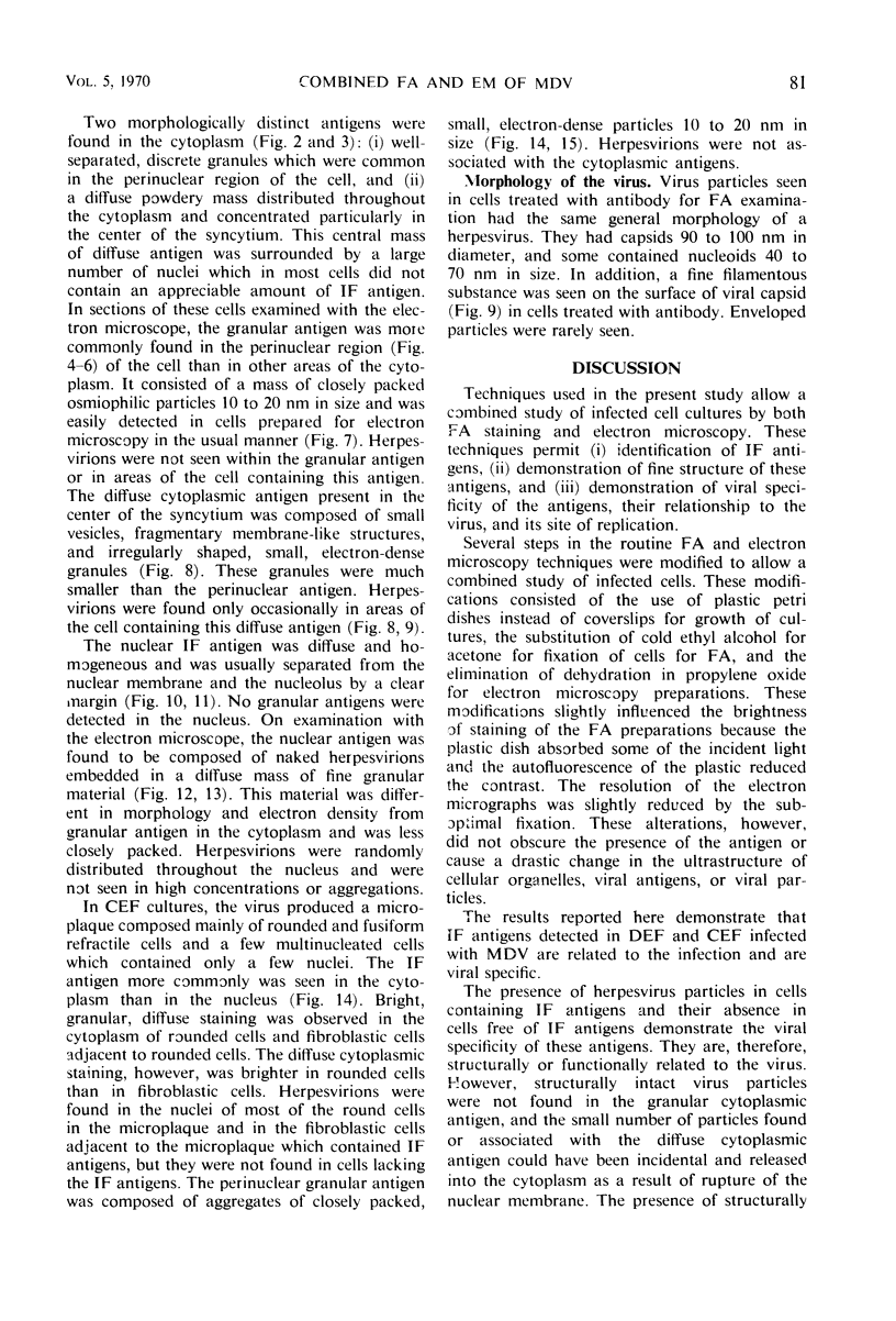
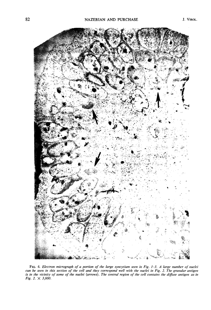
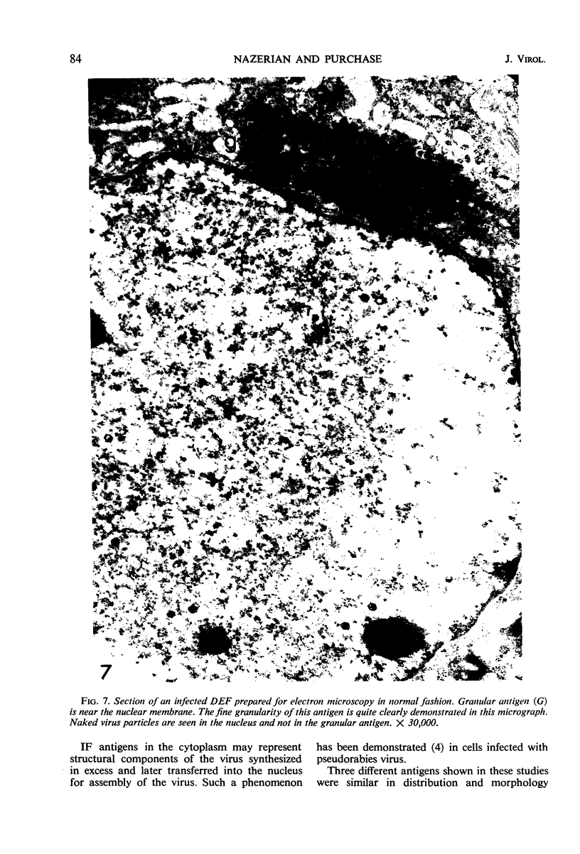
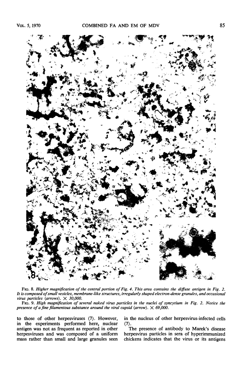
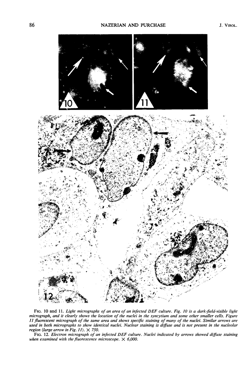
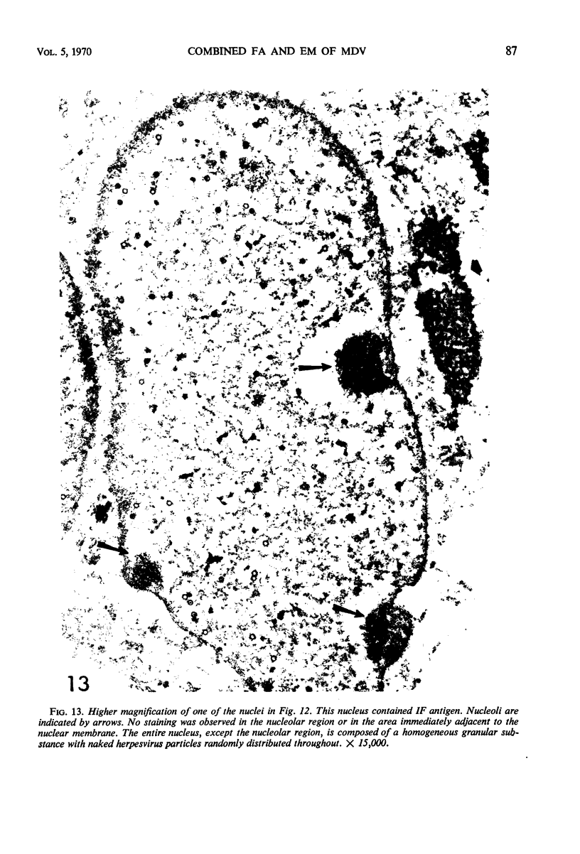
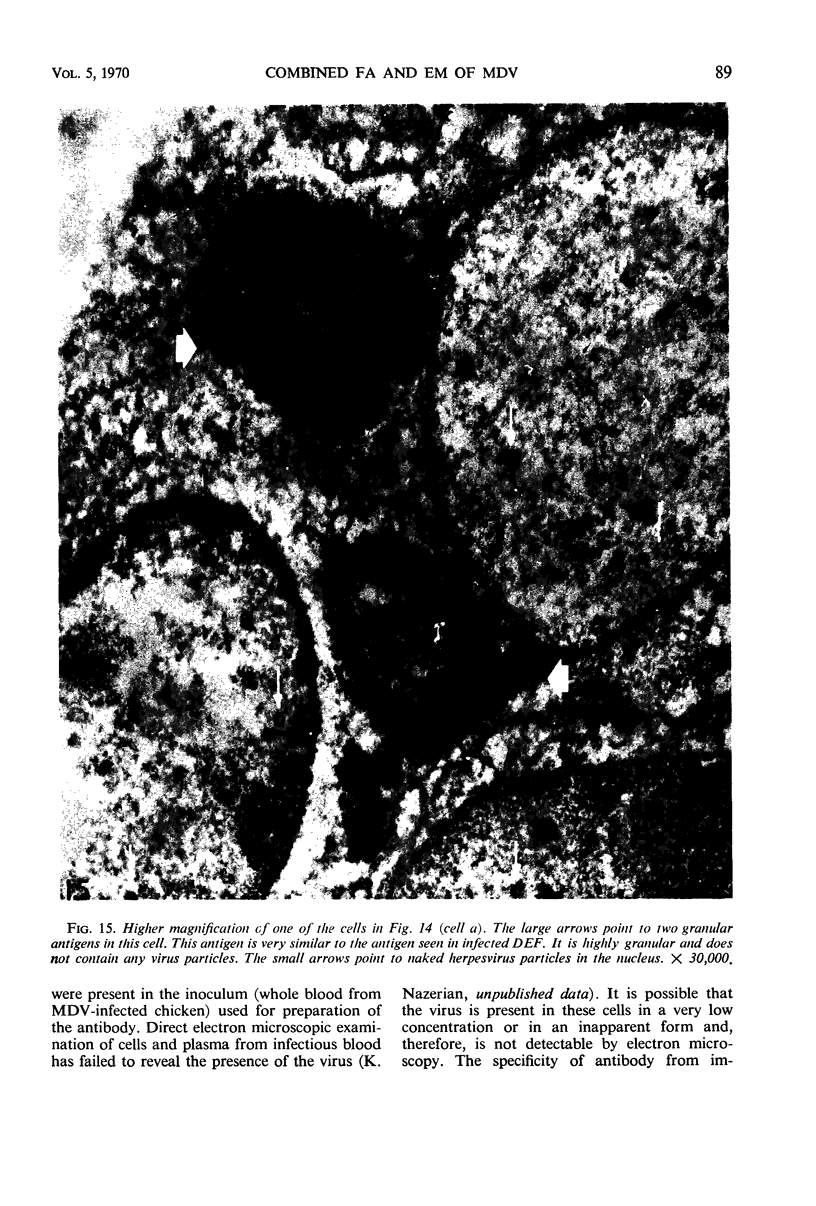
Duck embryo fibroblast (DEF) and chicken embryo fibroblast (CEF) cultures infected with Marek's disease virus were studied by combined fluorescent antibody and electron microscopy techniques. In both DEF and CEF cultures, cells containing immunofluorescent (IF) antigen also contained herpesvirus particles; conversely, cells lacking this antigen lacked herpesvirus particles. Two morphologically distinct IF antigens were detected in the cytoplasm. (i) A granular antigen in the perinuclear region was brightly stained with the conjugated antibody. This antigen was composed of a granular mass of osmiophilic material and did not contain virions. (ii) A diffuse antigen, present throughout the cytoplasm of infected cells, was less brightly stained. The area of the cell with the highest concentration of this antigen contained small vesicles, folded membranes, and fine electron-dense granules. Naked virions were occasionally seen in these areas. A diffuse nuclear IF antigen was occasionally seen in infected cells. This antigen was often separated from the nuclear membrane and the nucleolus by a clear margin. The intranuclear IF antigen was composed of a fine granular aggregate and naked herpesvirus particles which were randomly distributed throughout the nucleus. Viral capsids in antibody-treated cells were coated with fine filamentous material.
Full text
PDF


Images in this article

Selected References
These references are in PubMed. This may not be the complete list of references from this article.
- Churchill A. E., Biggs P. M. Agent of Marek's disease in tissue culture. Nature. 1967 Jul 29;215(5100):528–530. doi: 10.1038/215528a0. [DOI] [PubMed] [Google Scholar]
- Epstein M. A., Achong B. G. Specific immunofluorescence test for the herpes-type EB virus of Burkitt lymphoblasts, authenticated by electron microscopy. J Natl Cancer Inst. 1968 Mar;40(3):593–607. [PubMed] [Google Scholar]
- Fujiwara S., Kaplan A. S. Site of protein synthesis in cells infected with pseudorabies virus. Virology. 1967 May;32(1):60–68. doi: 10.1016/0042-6822(67)90252-8. [DOI] [PubMed] [Google Scholar]
- Nazerian K., Solomon J. J., Witter R. L., Burmester B. R. Studies on the etiology of Marek's disease. II. Finding of a herpesvirus in cell culture. Proc Soc Exp Biol Med. 1968 Jan;127(1):177–182. doi: 10.3181/00379727-127-32650. [DOI] [PubMed] [Google Scholar]
- Purchase H. G. Immunofluorescence in the study of Marek's disease. I. Detection of antigen in cell culture and an antigenic comparison of eight isolates. J Virol. 1969 Jun;3(6):557–565. doi: 10.1128/jvi.3.6.557-565.1969. [DOI] [PMC free article] [PubMed] [Google Scholar]
- Roizman B., Spring S. B., Roane P. R., Jr Cellular compartmentalization of herpesvirus antigens during viral replication. J Virol. 1967 Feb;1(1):181–192. doi: 10.1128/jvi.1.1.181-192.1967. [DOI] [PMC free article] [PubMed] [Google Scholar]
- Solomon J. J., Witter R. L., Nazerian K., Burmester B. R. Studies on the etiology of Marek's disease. I. Propagation of the agent in cell culture. Proc Soc Exp Biol Med. 1968 Jan;127(1):173–177. doi: 10.3181/00379727-127-32649. [DOI] [PubMed] [Google Scholar]