Abstract
Background:
Cone beam computed tomography (CBCT) is an alternative to a computed tomography (CT) scan, which is appropriate for a wide range of craniomaxillofacial indications. The long-term use of metallic materials in dentistry means that artifacts caused by metallic restorations in the oral cavity should be taken into account when utilizing CBCT and CT scanners. The aim of this study was to quantitatively compare the beam hardening artifacts produced by dental implants between CBCT and a 64-Slice CT scanner.
Materials and Methods:
In this descriptive study, an implant drilling model similar to the human mandible was used in the present study. The implants (Dentis) were placed in the canine, premolar and molar areas. Three series of scans were provided from the implant areas using Somatom Sensation 64-slice and NewTom VGi (CBCT) CT scanners. Identical images were evaluated by three radiologists. The artifacts in each image were determined based on pre-determined criteria. Kruskal-Wallis test was used to compare mean values; Mann-Whitney U test was used for two-by-two comparisons when there was a statistical significance (P < 0.05).
Results:
The images of the two scanners had similar resolutions in axial sections (P = 0.299). In coronal sections, there were significant differences in the resolutions of the images produced by the two scanners (P < 0.001), with a higher resolution in the images produced by NewTom VGi scanner. On the whole, there were significant differences between the resolutions of the images produced by the two CT scanners (P < 0.001), with higher resolution in the images produced by NewTom VGi scanner in comparison to those of Somatom Sensation.
Conclusion:
Given the high quality of the images produced by NewTom VGi and the lower costs in comparison to CT, the use of the images of this scanner in dental procedures is recommended, especially in patients with extensive restorations, multiple prostheses and previous implants.
Keywords: Beam hardening artifacts, cone beam computed tomography, dental implants
INTRODUCTION
Computed tomography (CT) is an important imaging technique for the diagnosis of soft- and hard-tissue lesions in the oral cavity and the head and neck region. However, the use of CT in dental procedures is limited due to its high costs, large size of the equipment and high radiation doses. Therefore, in recent years cone beam computed tomography (CBCT) has become a very important alternative diagnostic tool. It appears CBCT has a high potential in the diagnosis and treatment planning, especially in implant treatment, by providing three-dimensional images.[1,2,3,4]
If a mental is present in the area to be scanned, the images are prone to production of artifacts. Artifacts are the main cause of a decrease in image quality. In some, the artifacts render the image useless.[4] Some of these artifacts are produced due to a phenomenon, referred to as beam hardening. When the X-ray beam travels through an object, the low-energy photons are absorbed more than the high-energy photons; the phenomenon is referred to as beam hardening. This phenomenon is produced by objects with a high density, including metallic restorations, dental implants, etc.[4,5,6,7] Since CT and CBCT use back-projection beams to produce three-dimensional images and image production principles are the same, these artifacts can be seen in the images of both imaging systems.[4,5]
Although, there are many techniques to reduce the number of these artifacts in CT technique,[8,9,10,11,12,13,14] only a limited number of techniques have been introduced to counteract these artifacts in the CBCT technique.[15,16] In the clinic, decreasing the field of view, changing the position of patient head or separating dental arches in order to avoid scanning the areas susceptible to beam hardening have been recommended.[5] It appears the type of the machine, too, is effective in producing artifacts, although, only a limited number of studies have been carried out in this respect.[2,3,4]
Exposure conditions can have a great role in producing artifacts by influencing the energy of the photons; in this context, some studies have recommended imaging techniques with high kVp to decrease hardening of the beams.[1,4] Other factors that can have a role in beam hardening included the amount of rotation of the machine, the configuration of the X-ray beam and the type of the algorithm used for data processing.[17,18,19]
A large number of studies have evaluated metallic artifacts in CT[20,21,22,23,24] and CBCT;[1,4,6,15] however, the majority of these studies have been qualitative studies. Only a limited number of studies have quantitatively compared CT and CBCT machines,[1,17] necessitating further studies. The aim of the present study was to compare artifacts as a result of beam hardening during scanning procedures of dental implants using Somatom Sensation 64-slice CT and NewTom VGi cone beam CT scanners.
MATERIALS AND METHODS
Study design
In this descriptive study, a dry human skull was used. Since the aim of the present study was to evaluate artifacts produced by beam hardening in dental implants without interference of any other materials, an implant drilling model (Nissian, Kyoto, Japan), which is completely similar to a human mandible, was used instead of human mandible [Figure 1]. This model has a spongy structure like human mandible, except that it does not produce any images during scanning under different conditions and has no effect on beam hardening.[17]
Figure 1.
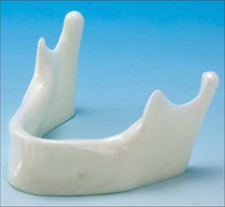
The implant drilling model used in the present study
Dentis implant system (Dentis, Daegu, Korea) was used to evaluate artifacts. Two implants were placed in the canine area, two in the second premolar area and two in the second molar area [Figure 2]. The implants measured 12 mm in length and 4.3 mm in diameter. On the whole, 3 series of scans (canine, premolar and molar) were provided using NewTom VGi Cone Beam CT (QR SRL Company, Verona, Italy) and Somatom Sensation 64-slice CT (Siemens, Germany) scanners. Gutta-percha was used as a marker to determine axial and coronal section locations. In each implant site identical sections were selected from axial and coronal sections.
Figure 2.
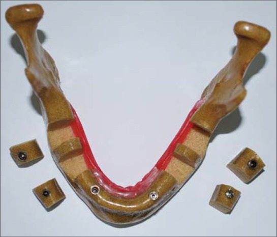
The implants placed in the canine, premolar and molar areas
CBCT scans
The examination with NewTom VGi was performed at 4.71 mA and 110 kVp, with a scan time of 3.6 s. Spatial resolution of primary image data reconstruction was 0.127 mm × 0.127 mm in the axial plane with 1-mm slice thickness. Primary and secondary reconstructions were performed with NNT viewer software version 2.21 (Quantitative Radiology, Verona, Italy). The sagittal images were based on multi-planner reconstruction.
64-slice CT scans
Scans were performed with a Somatom Sensation 64-slice CT at 120 Kvp and 104 mAs and a rotation time of 0.33 s. The matrix size was 512 × 512 pixels and the field of view was 174 mm × 174 mm, resulting in a pixel size of 0.39 mm. The slice thickness was 1 mm. Image analysis of the axial and coronal cross-sections was performed with eFilm Worksation Software version 2.1 (eFilm Medical Inc., Toronto, Canada). The sagittal image was based on multi-planner reconstruction.
Image quality assessment
Two equal sets of images from the cone beam and the 64-slice CT scanner were evaluated by three independent observers, who were oral and maxillofacial radiologists each with more than 4 years of experience in the analysis of CBCT and CT scans. Images as demonstrated in Figure 3, which includes axial and coronal displays, were presented to the observers.
Figure 3.
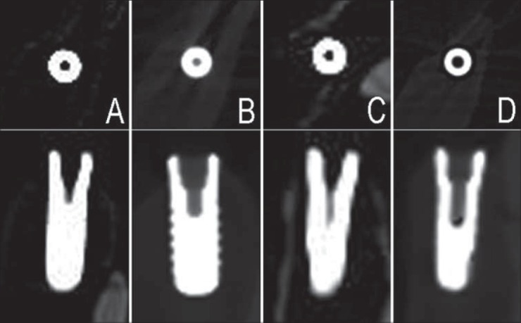
Somatom Sensation 64-Slice computed tomography images (Axial and Coronal; Molar region: A-Canine region:C), NewTom VGI CBCT images (Axial and Coronal ; Molar region: B-Canine region:D)
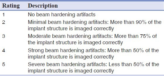
The images were evaluated on a 17-inch monitor (cathode ray tube) of a desk-top computer. The evaluation was carried out in a windowless room under mild lighting conditions. A standardized rating was used for this evaluation [Table 1].[17]
Table 1.
The image quality assessment evaluation rating
Statistical analysis
Kruskal-Wallis test was used to compare the means; Mann-Whitney U test was used for two-by-two comparisons when there were statistically significant differences, using SPSS 16 statistical software. Statistical significance was defined at P < 0.05.
RESULTS
Three series of scans (canine, premolar and molar) were provided by two scanners in the present study. In the axial cross-sections the means of image resolutions were 4.43 and 4.32 with NewTom VGi and Somatom Sensation scanners, respectively, with no statistically significant differences in the image resolutions between the two scanners (P = 0.299) [Table 2]. In the coronal cross-sections the means of image resolutions were 4.55 and 3.92 with NewTom VGi and Somatom Sensation scanners, respectively. Mann-Whitney U test revealed statistically significant differences in the resolutions between the two scanners (P < 0.0001), with higher resolution in the images provided by NewTom VGi scanners [Table 2].
Table 2.
The resolutions of the images in the axial, coronal and both sections of NewTom VGi (N) and Somatom Sensation 64-slice computed tomography (S) scanners*
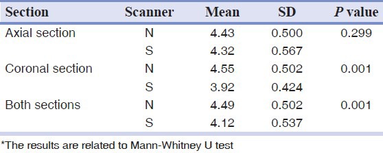
The overall image resolution means in the two cross-sections were 4.49 and 4.12 in the NewTom VGI and Somatom Sensation scanners, respectively, with significant differences between the two scanners (P < 0.0001); NewTom VGi scanner images had higher resolution [Table 2].
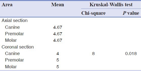
In addition, Kruskal-Wallis test was used to compare image resolution between the canine, premolar and molar areas, the axial and coronal cross-sections in the two scanners. With the NewTom VGi scanners in the axial cross-sections, image resolutions were similar in all the three canine, premolar and molar areas. In the coronal cross-sections, image resolutions in the three areas revealed statistically significant differences, with significantly less image resolution in the canine area compared to the other two areas [Table 3].
Table 3.
Comparison of image resolutions between the three canine, premolar and molar scans in the axial and coronal sections of NewTom VGI cone beam computed tomography scanner
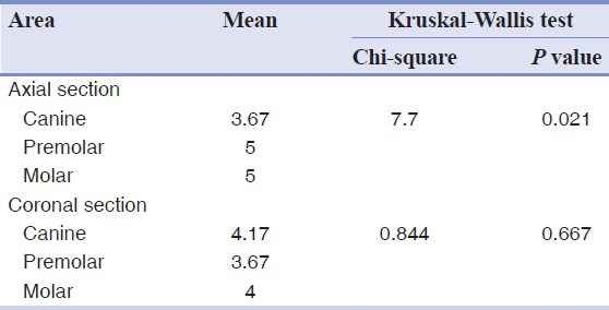
With the Somatom Sensation scanner, in the axial sections image resolutions exhibited statistically significant differences between the three areas under study, with the canine area exhibiting significantly less image resolution compared to the other two areas. There were no significant differences in image resolutions between the three areas under study in the coronal cross-sections [Table 4].
Table 4.
Comparison of image resolutions between the three canine, premolar and molar scans in the axial and coronal sections of Somatom Sensation 64-slice computed tomography scanner
DISCUSSION
At present three-dimensional imaging systems (CT and CBCT) are highly valuable in the diagnosis and treatment planning for dental and medical procedures in the head and neck region. Therefore, it is necessary to request the best-quality images for treatment planning.[5,7] The long-term use of metallic restorations in dentistry necessitates attention to the artifacts produced by these restorations in the oral cavity when three-dimensional imaging systems are used. The issue becomes more important when the patient has extensive prostheses, amalgam restorations or implants in the oral cavity. The artifacts produced by metallic objects are the result of beam hardening phenomenon which takes place in all the CT and CBCT imaging systems.[4,5,6,7] Evaluation of these artifacts and comparison of various imaging systems in this respect are necessary because in some cases these artifacts are so severe that they decrease the image quality or they even distort the images.[5,6,7]
Schulz et al.[18] evaluated the image qualities of NewTom 900 and Siemens Siremobil scanners in a dry skull and reported no artifacts as a result of beam hardening, which was attributed to the fact that no metallic structures were used in the study. In the present study, different amounts of metallic artifacts were observed due to the use of titanium implants in both scanners under study.
Chindasombatjareon et al.[1] evaluated the metallic artifacts produced by dental metals with the use of Light Speed QX/I (Multi Slice Computed Tomography [MDCT]) and Alpha Vega 3030 scanners (Cardiac Computed Tomography [CCT]). Cubes of aluminum, titanium, chromium-cobalt and Type IV gold alloy were scanned by the two scanners and the images were evaluated and compared by Image J software. The results showed fewer artifacts with CBCT scanners compared to MDCT scanners under identical conditions. In addition, increase in kVp in both scanners resulted in a decrease in artifacts. However, an increase in tube electric current had no effect on artifacts. An increase in kVp resulted in a decrease in beam hardening by influencing the energy of the photons. The results of the present study revealed a very high-resolution of the images produced by NewTom VGi and Somatom Sensation, which might be attributed to the high kVp in these two scanners. In addition, the amount of artifacts with NewTom VGi was less than that with Somatom Sensation, consistent with the results of the above-mentioned study. The dental implants used in the present study, similar to titanium blocks in the above-mentioned study, produced severe metallic artifacts.
Schulze et al.[4] evaluated the artifacts produced by dental implants with the use of Accuitomo and 3D Exam CBCT scanners by studying the geometric and physical parameters effective on data collected from scans for the reconstruction of three-dimensional images. The results showed a great amount of artifacts produced by titanium implants under standard conditions. Furthermore, the results showed that scanning under high kVp conditions reduces the amount of artifacts. In the present study, NewTom VGi and Somatom Sensation scanners produced less artifacts due to high kVp.
Draenert et al.[17] compared the artifacts of dental implants with the use of NewTom 9000 (CBCT) and Philips M × 8000 (4-row MDCT). The axial and coronal images of implants in the canine and molar areas of the maxilla were compared in a model of skull made from saw bone material. The results showed much less artifacts with MDCT in comparison to CBCT, with the image of the implant in MDCT correctly produced in all the axial and coronal cross-sections in comparison to the main implant. Only 16% of the implants in MDCT had artifacts while the images of CBCT had no artifacts in less than 25% of cases. In addition, the results showed that there were more artifacts in the canine area compared to the molar area.
In the present study NewTom VGi scanner was used, which is more advanced and newer with a higher kVp in comparison with the scanner used in a study by Draenert.[17] Fewer artifacts with NewTom VGi in comparison with NewTom 9000 might be attributed to higher kVp. Somatom Sensation exhibited some artifacts, which is consistent with the results reported by Draenert. The quality of the images produced by MDCT scanner in the present study were lower than those of CBCT, contrary to the results reported by Draenert,[17] which might be attributed to higher spatial resolution of CBCT compared to CT and the higher kVp of the CBCT scanner used in the present study.
In the present study, there were fewer artifacts with NewTom VGi in the axial sections and with Somatom Sensation in the coronal sections in the canine area compared to the molar and premolar areas, consistent with the results of the above-mentioned study, which might be attributed to the position of the canine because in the canine area the teeth are located in a more circumferential manner. In addition, Draenert[17] recommended another study with kVps higher than 90 with the CBCT scanner in order to reduce the amount of artifacts. In the present study, scanning with a kVp higher than 110 with the NewTom VGi scanner significantly decreased the amount of artifacts, confirming the accuracy of findings reported by Draenert.[17]
It is recommended that the effect of different exposure conditions (kVp and mAs) on the amount of artifacts be evaluated in future studies. In addition, it is recommended that different kinds of CBCT scanners be evaluated.
CONCLUSION
Generally NewTom VGi scanner produced images with higher resolutions. However, the resolution of the images produced by Somatom Sensation 64-slice CT scanner in axial sections was similar to that of NewTom VGi.
Given the higher resolution of the images produced by NewTom VGi scanner and its lower doses and costs compared to CT scanner, it appears the images produced by this scanner have high diagnostic values, the importance of which is highlighted when this scanner is used to produce images from patients with extensive restorations, multiple prostheses or previous implant treatments.
Footnotes
Source of Support: Tabriz University of Medical Science
Conflict of Interest: None declared
REFERENCES
- 1.Chindasombatjareon J, Kakimoto N, Murakami S, Maeda Y, Furukawa S. Quantitative analysis of metalic artifacts caused by dental metals: Comparison of cone-beam and multi-detector row CT scanners. Oral Radiol. 2011;27:114–20. [Google Scholar]
- 2.Ludlow JB, Davies-Ludlow LE, Brooks SL, Howerton WB. Dosimetry of 3 CBCT devices for oral and maxillofacial radiology: CB Mercuray, NewTom 3G and i-CAT. Dentomaxillofac Radiol. 2006;35:219–26. doi: 10.1259/dmfr/14340323. [DOI] [PubMed] [Google Scholar]
- 3.Ludlow JB, Ivanovic M. Comparative dosimetry of dental CBCT devices and 64-slice CT for oral and maxillofacial radiology. Oral Surg Oral Med Oral Pathol Oral Radiol Endod. 2008;106:106–14. doi: 10.1016/j.tripleo.2008.03.018. [DOI] [PubMed] [Google Scholar]
- 4.Schulze RK, Berndt D, d’Hoedt B. On cone-beam computed tomography artifacts induced by titanium implants. Clin Oral Implants Res. 2010;21:100–7. doi: 10.1111/j.1600-0501.2009.01817.x. [DOI] [PubMed] [Google Scholar]
- 5.White SC, Pharoah MJ. Oral Radiology: Principles and Interpretation. 6th ed. St. Louis: Mosby; 2009. Cone- beam computed tomography; pp. 235–7. [Google Scholar]
- 6.Schulze R, Heil U, Gross D, Bruellmann DD, Dranischnikow E, Schwanecke U, et al. Artefacts in CBCT: A review. Dentomaxillofac Radiol. 2011;40:265–73. doi: 10.1259/dmfr/30642039. [DOI] [PMC free article] [PubMed] [Google Scholar]
- 7.Zoller JE, Neugebauer J. Cone-beam Volumetric Imaging in Dental, Oral and Maxillofacial Medicine: Fundamentals, Diagnostics and Treatment Planning. 1th ed. Landshut: Quintessence Publishing; 2008. Image quality: Requirements and influencing factors; pp. 27–35. [Google Scholar]
- 8.Zhao S, Robertson DD, Wang G, Whiting B, Bae KT. X-ray CT metal artifact reduction using wavelets: An application for imaging total hip prostheses. IEEE Trans Med Imaging. 2000;19:1238–47. doi: 10.1109/42.897816. [DOI] [PubMed] [Google Scholar]
- 9.Wang G, Snyder DL, O’sullivan JA, Vannier MW. Iterative deblurring for CT metal artifact reduction. IEEE Trans Med Imaging. 1996;15:657–64. doi: 10.1109/42.538943. [DOI] [PubMed] [Google Scholar]
- 10.Kalender WA, Hebel R, Ebersberger J. Reduction of CT artifacts caused by metallic implants. Radiology. 1987;164:576–7. doi: 10.1148/radiology.164.2.3602406. [DOI] [PubMed] [Google Scholar]
- 11.Robertson DD, Weiss PJ, Fishman EK, Magid D, Walker PS. Evaluation of CT techniques for reducing artifacts in the presence of metallic orthopedic implants. J Comput Assist Tomogr. 1988;12:236–41. doi: 10.1097/00004728-198803000-00012. [DOI] [PubMed] [Google Scholar]
- 12.van der Schaaf I, van Leeuwen M, Vlassenbroek A, Velthuis B. Minimizing clip artifacts in multi CT angiography of clipped patients. AJNR Am J Neuroradiol. 2006;27:60–6. [PMC free article] [PubMed] [Google Scholar]
- 13.Moon SG, Hong SH, Choi JY, Jun WS, Kang HG, Kim HS, et al. Metal artifact reduction by the alteration of technical factors in multidetector computed tomography: A 3-dimensional quantitative assessment. J Comput Assist Tomogr. 2008;32:630–3. doi: 10.1097/RCT.0b013e3181568b27. [DOI] [PubMed] [Google Scholar]
- 14.Lee IS, Kim HJ, Choi BK, Jeong YJ, Lee TH, Moon TY, et al. A pragmatic protocol for reduction in the metal artifact and radiation dose in multislice computed tomography of the spine: Cadaveric evaluation after cervical pedicle screw placement. J Comput Assist Tomogr. 2007;31:635–41. doi: 10.1097/01.rct.0000250117.18080.d8. [DOI] [PubMed] [Google Scholar]
- 15.Wang G, Vannier MW, Cheng PC. Iterative X-ray cone-beam tomography for metal artifact reduction and local region reconstruction. Microsc Microanal. 1999;5:58–65. doi: 10.1017/S1431927699000057. [DOI] [PubMed] [Google Scholar]
- 16.Zhang Y, Zhang L, Zhu XR, Lee AK, Chambers M, Dong L. Reducing metal artifacts in cone–beam CT images by preprocessing projection data. Int J Radiat Oncol Biol Phys. 2007;67:924–32. doi: 10.1016/j.ijrobp.2006.09.045. [DOI] [PubMed] [Google Scholar]
- 17.Draenert FG, Coppenrath E, Herzog P, Müller S, Mueller-Lisse UG. Beam hardening artefacts occur in dental implant scans with the NewTom cone beam CT but not with the dental 4-row multidetector CT. Dentomaxillofac Radiol. 2007;36:198–203. doi: 10.1259/dmfr/32579161. [DOI] [PubMed] [Google Scholar]
- 18.Schulze D, Heiland M, Blake F, Rother U, Schmelzle R. Evaluation of quality of reformatted images from two cone-beam computed tomographic systems. J Craniomaxillofac Surg. 2005;33:19–23. doi: 10.1016/j.jcms.2004.07.004. [DOI] [PubMed] [Google Scholar]
- 19.Hunter A, McDavid D. Analyzing the beam hardening artifact in the Planmeca Promax. Oral Surg Oral Med Oral Pathol Oral Radiol Endod. 2009;107:28–9. [Google Scholar]
- 20.Sanders MA, Hoyjberg C, Chu CB, Leggitt VL, Kim JS. Common orthodontic appliances cause artifacts that degrade the diagnostic quality of CBCT images. J Calif Dent Assoc. 2007;35:850–7. [PubMed] [Google Scholar]
- 21.Sullivan PK, Smith JF, Rozzelle AA. Cranio-orbital reconstruction: Safety and image quality of metallic implants on CT and MRI scanning. Plast Reconstr Surg. 1994;94:589–96. [PubMed] [Google Scholar]
- 22.Dalal T, Kalra MK, Rizzo SM, Schmidt B, Suess C, Flohr T, et al. Metallic prosthesis: Technique to avoid increase in CT radiation dose with automatic tube current modulation in a phantom and patients. Radiology. 2005;236:671–5. doi: 10.1148/radiol.2362041565. [DOI] [PubMed] [Google Scholar]
- 23.Barrett JF, Keat N. Artifacts in CT: Recognition and avoidance. Radiographics. 2004;24:1679–91. doi: 10.1148/rg.246045065. [DOI] [PubMed] [Google Scholar]
- 24.Fiala TG, Novelline RA, Yaremchuk MJ. Comparison of CT imaging artifacts from craniomaxillofacial internal fixation devices. Plast Reconstr Surg. 1993;92:1227–32. [PubMed] [Google Scholar]


