

Abstract
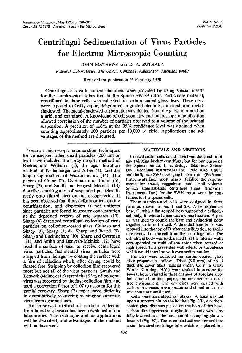
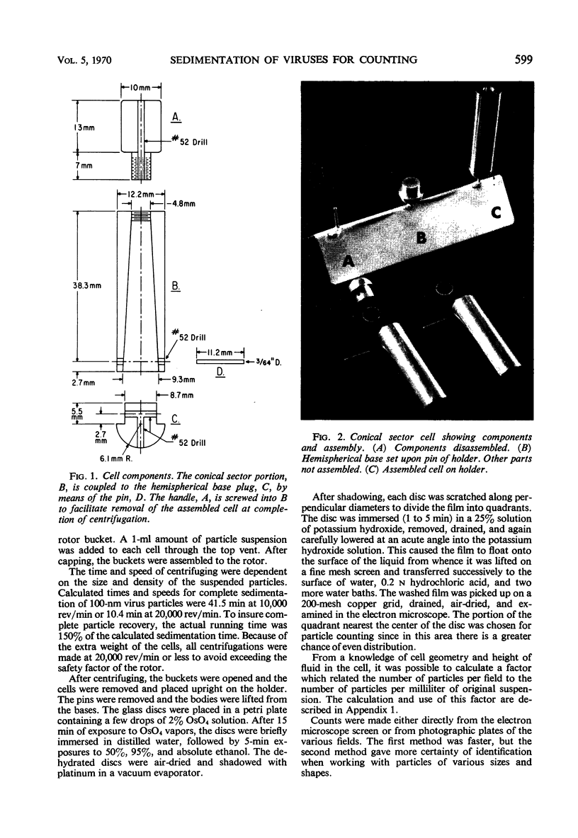
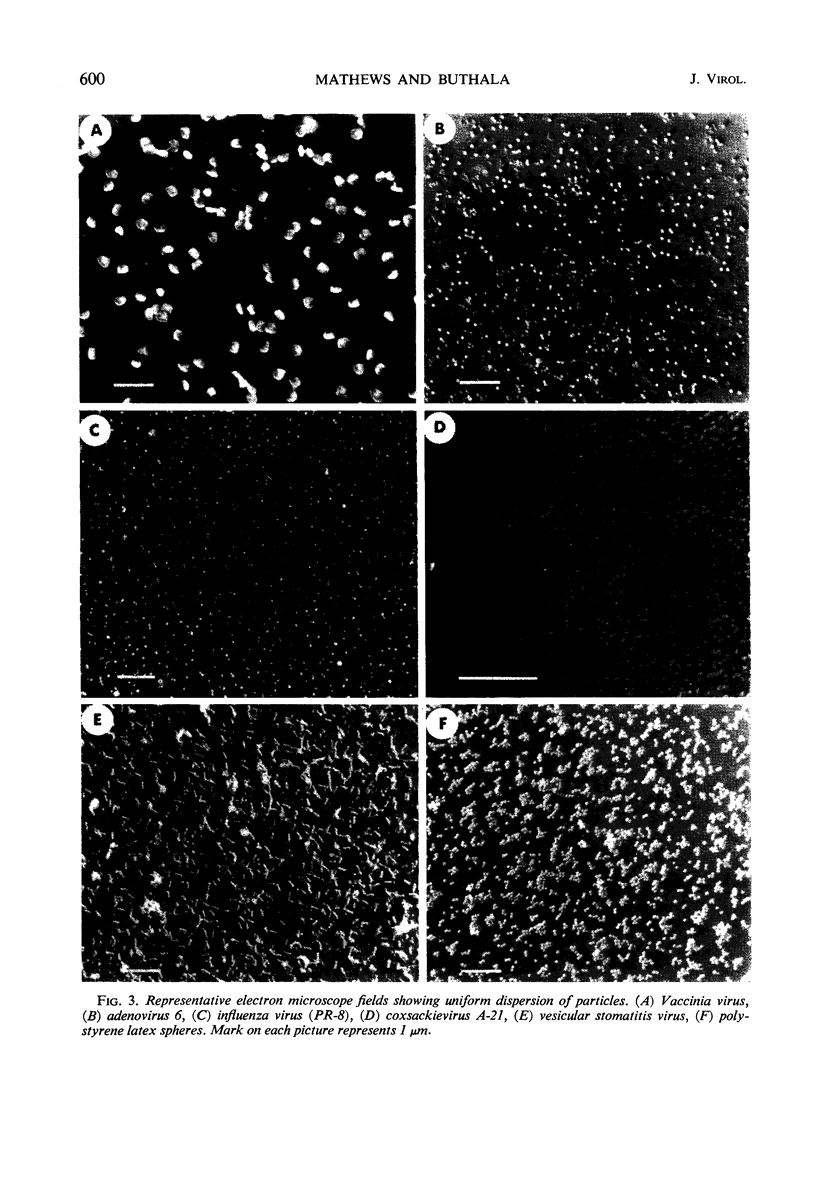
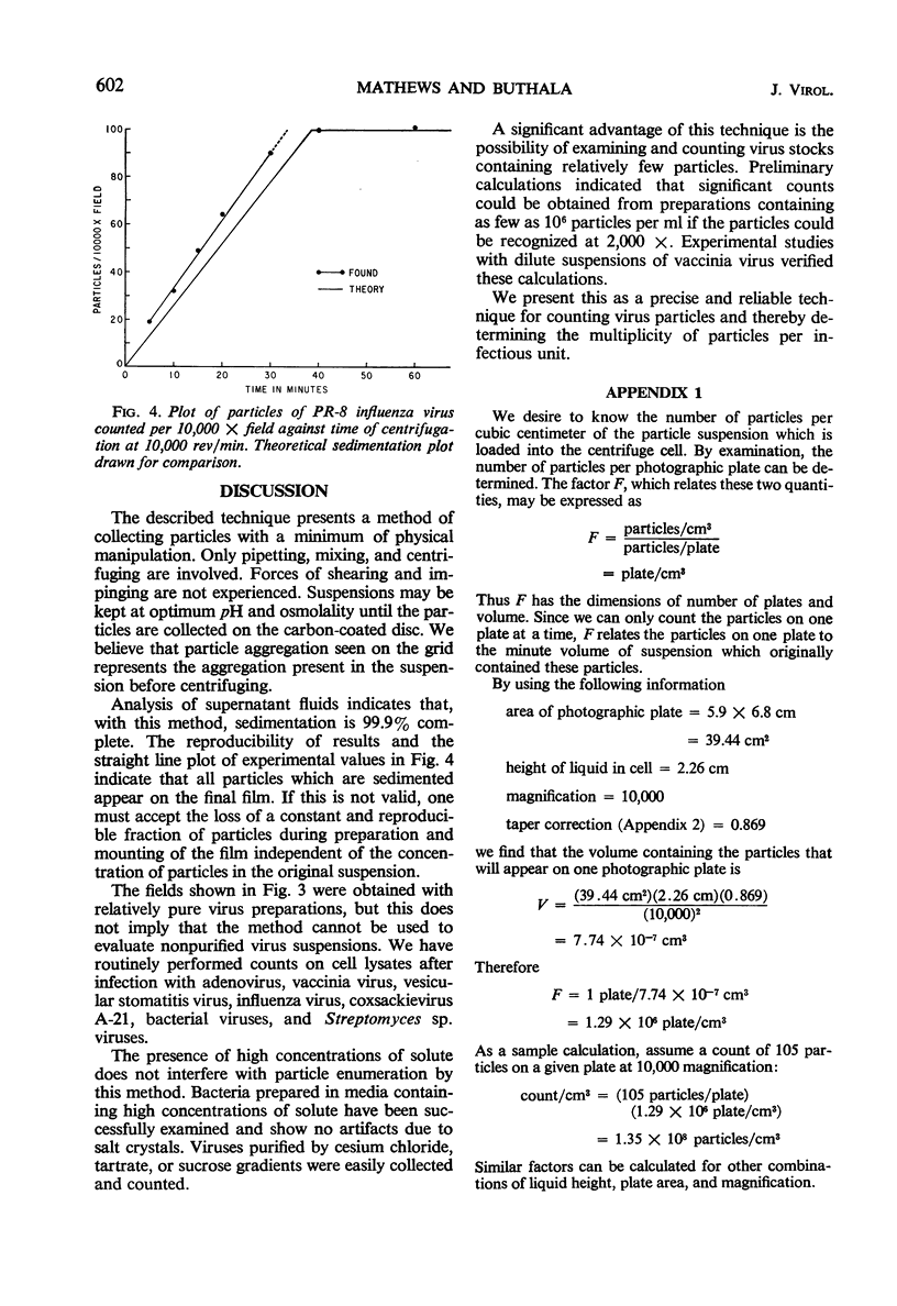
Centrifuge cells with conical chambers were provided by using special inserts for the stainless-steel tubes that fit the Spinco SW-39 rotor. Particulate material, centrifuged in these cells, was collected on carbon-coated glass discs. These discs were exposed to OsO4 vapor, dehydrated in graded alcohols, air-dried, and metal-shadowed. The metal-shadowed carbon film was floated from the glass, mounted on a grid, and examined. A knowledge of cell geometry and microscope magnification allowed correlation of the number of particles observed to a volume of the original suspension. A precision of ±6% at the 95% confidence level was attained when counting approximately 100 particles per 10,000 × field. Applications and advantages of the method are discussed.
Full text
PDF
Images in this article
Selected References
These references are in PubMed. This may not be the complete list of references from this article.
- GALASSO G. J., SHARP D. G. COMPLETELY DISPERSED SUSPENSIONS OF VACCINIA VIRUS PARTICLES. Proc Soc Exp Biol Med. 1964 Oct;117:101–105. doi: 10.3181/00379727-117-29506. [DOI] [PubMed] [Google Scholar]
- KELLENBERGER E., ARBER W. Electron microscopical studies of phage multiplication. I. A method for quantitative analysis of particle suspensions. Virology. 1957 Apr;3(2):245–255. doi: 10.1016/0042-6822(57)90091-0. [DOI] [PubMed] [Google Scholar]
- OVERMAN J. R., TAMM I. Equivalence between vaccinia particles counted by electron microscopy and infectious units of the virus. Proc Soc Exp Biol Med. 1956 Aug-Sep;92(4):806–810. doi: 10.3181/00379727-92-22621. [DOI] [PubMed] [Google Scholar]
- SHARP D. G., BEARD J. W. Counts of virus particles by sedimentation on agar and electron micrography. Proc Soc Exp Biol Med. 1952 Oct;81(1):75–79. doi: 10.3181/00379727-81-19782. [DOI] [PubMed] [Google Scholar]
- SHARP D. G., BUCKINGHAM M. J. Electron microscopic measure of virus particle dispersion in suspension. Biochim Biophys Acta. 1956 Jan;19(1):13–21. doi: 10.1016/0006-3002(56)90381-x. [DOI] [PubMed] [Google Scholar]
- SHARP D. G., OVERMAN J. R. Enumeration of vaccinia virus particles in crude extracts of infected tissues by electron microscopy. Proc Soc Exp Biol Med. 1958 Nov;99(2):409–413. doi: 10.3181/00379727-99-24366. [DOI] [PubMed] [Google Scholar]
- SMITH K. O., MELNICK J. L. Electron microscopic counting of virus particles by sedimentation on aluminized grids. J Immunol. 1962 Aug;89:279–284. [PubMed] [Google Scholar]
- WATSON D. H., RUSSELL W. C., WILDY P. Electron microscopic particle counts on herpes virus using the phosphotungstate negative staining technique. Virology. 1963 Mar;19:250–260. doi: 10.1016/0042-6822(63)90062-x. [DOI] [PubMed] [Google Scholar]