Abstract
Central nervous system (CNS) tuberculosis may manifest as meningitis, meningoencephalitis, tuberculoma, tubercular abscess, stroke due to tuberculous vasculitis and tuberculous encephalopathy. Occasionally, tubercular meningitis (TBM) can predispose to cerebral venous sinus thrombosis (CVST). We report a young man, who developed CVST as a complication of TBM. Worsening of pre-existing headache, impairment of consciousness and seizures should raise suspicion of CVST in any patient with CNS infection. Early diagnosis and appropriate clinical management are important for good outcome.
Background
Central nervous system tuberculosis (CNS TB) is a major cause of morbidity and mortality in the developing countries. Clinical spectrum of CNS TB includes meningitis, meningoencephalitis, tuberculoma, tubercular abscess, stroke due to vasculitis and cerebral venous sinus thrombosis (CVST).1 We report a young man with tubercular meningitis (TBM), who developed CVST as a complication. Early diagnosis and appropriate clinical management was associated with good outcome in this patient.
Case presentation
A 26-year-old man was admitted in our department with a history of low-grade fever for 4 months. The fever was accompanied with decreased appetite and significant unintentional weight loss. He also reported of headache for past 1 month, which localised around the nape of the neck and occipital region and later became generalised, continuous and severe in intensity. It would get aggravated on neck movements, on lying down and was associated with multiple episodes of projectile vomiting which were relieved partially with medications. He had binocular diplopia for distant objects for past 15 days. His clinical condition progressively deteriorated over past 7 days and he became drowsy, irritable and could not identify his relatives. He also had two episodes of generalised tonic-clonic seizures 2 days previously. On examination, pulse rate was 90/min with a blood pressure 130/80 mm Hg. The patient was drowsy, with localising pain, but no verbal output (Glasgow Coma Score E1M5V1). Examination of cranial nerves revealed bilateral papilloedema, small size and sluggishly reacting pupils and bilateral sixth cranial nerve palsy. Deep tendon reflexes were normal with plantar flexor response. The patient had neck stiffness with positive Kernig’s sign. Other systemic examination was normal.
Investigations
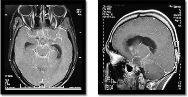
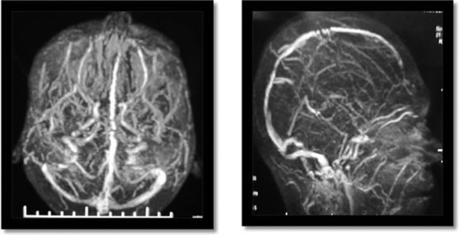
The patient's haemoglobin level was 12.2 g%, total leucocyte count was 8900/mm3 and erythrocyte sedimentation rate was 29 mm at the end of 1 h. Liver and renal function tests were within normal limits. Cerebrospinal fluid (CSF) was colourless 40 cells/mm3 (90% lymphocytes), proteins were 90 mg% and glucose was 17.4 mg% (corresponding plasma glucose 161.3 mg%). acid-fast bacilli stain, Gram stain and India ink preparation of CSF were negative. PCR for tuberculosis of CSF was positive. Contrast-enhanced CT of the cranium showed non-communicating hydrocephalus with leptomeningeal enhancement. MRI brain (figure 1) with contrast also showed non-communicating hydrocephalus with abnormal leptomeningeal enhancement. MR venogram (MRV) (figure 2) of brain was performed which showed focal segmental irregular narrowing in the mid part of superior sagittal sinus along with inferior sagittal sinus narrowing with increased collaterals suggestive of CVST. The patient's thrombophilia profile was performed, which showed normal antithrombin III (AT-III), protein C, protein S and factor V levels. Antinuclear antibodies and antiphospholipid antibodies were not detected.
Figure 1.
Axial and sagital T1-weighted MRI brain with contrast images showing hydrocephalus with leptomeningeal enhancement.
Figure 2.
MR venography showing superior and inferior sagittal sinus thrombosis with increased collaterals.
Differential diagnosis
This patient presented with features of raised intracranial pressure, later on developed seizures and altered mentation. Hence the following differential diagnosis was considered:
- Infections along with hydrocephalus
- Chronic meningoencephalitis like tuberculosis, cryptococcus, etc
- Tuberculomata
- Bacterial, tuberculous or fungal abscess
Space occupying lesions: primary CNS neoplasm, lymphoma, metastasis, abscess.
- Cerebral venous thrombosis
- Idiopathic
- Secondary to infections, malignancy, dehydration, trauma, dural fistula.
- Coagulation defects like protein C, protein S deficiency, factor V Leiden mutation, antithrombin deficiency, prothrombin gene mutation.
Treatment
The patient was started on once daily antitubercular treatment with rifampicin (10 mg/kg), isoniazid (5 mg/kg), pyrazinamide (25 mg/kg) and intramuscular streptomycin (15 mg/kg); with intravenous dexamethasone 0.4 mg/kg/day which was tapered over 8 weeks. Antiepileptic therapy was started with intravenous levetiracetam 20 mg/kg followed by oral levetiracetam 500 mg twice daily. Anticoagulation was achieved with subcutaneous enoxaparin 1 mg/kg twice daily followed by oral warfarin 4 mg/day with target international normalised ratio between 2 and 3. Symptomatic treatment to reduce intracranial pressure with intravenous mannitol (2 g/kg) was given for 5 days and oral acetazolamide 250 mg four times a day for about 3 weeks.
Outcome and follow-up
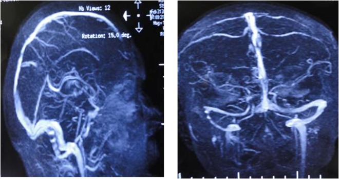
After 7 days of hospitalisation, the patient developed acute onset right hemiparesis. After this initial deterioration, he improved remarkably. There were no episodes of seizures and diplopia. Residual weakness in the right half of the body was noted on follow-up at 2 months. Follow-up MRV (figure 3) was performed after 3 months which showed resolution of cortical venous thrombosis.
Figure 3.
MR venography showing resolution of superior and inferior sagittal sinus.
Discussion
Clinical manifestations of CVST are highly variable, including headache, transient visual obscurations (due to papilloedema), diplopia, focal neurological deficits like hemiparesis, dysphasia, cranial nerve deficits, seizures and altered sensorium. Increasing number of cases are now being diagnosed due to better non-invasive diagnostic tools like MRV (sensitivity 84–100%).1–3
Superior sagittal sinus thrombosis usually presents with headache and papilloedema. Headache is mostly continuous, throbbing or constrictive in nature but episodic headaches are also seen with cerebral venous thrombosis (CVT).2 4 Occlusion of cortical veins cause localised oedema and venous infarction, whereas occlusion of major venous sinuses results in impaired CSF flow, raising the intracranial pressure.5
Vascular endothelial damage, stasis and hypercoagulability act synergistically to cause vascular thrombosis. Endothelial damage to the vessel may occur due to infection or inflammatory response with leucocyte recruitment. This further promotes venous stasis and proinflammatory cytokines like tumour necrosis factor (TNF)-α, interleukin (IL) 1β induce thrombin generation and decrease thrombomodulin expression.2 6 7 Defects in coagulation cascade seem to be the most common and important cause of CVST. Factor V Leiden mutation is the most common inherited thrombophilia worldwide and is responsible for about 20% cases of CVST.7–9 A recent study of CVST in India found that protein C deficiency in men and protein S deficiency in women were most commonly associated coagulation defects.10 Vascular endothelial damage leading to CVST is relatively unusual.2 It commonly results from infections (bacterial, tuberculosis, fungal), tumours, trauma and inflammatory conditions, including Behcets, sarcoidosis, ulcerative colitis and systemic lupus erythematosis. With early use of antibiotics, sepsis (local or systemic) induced CVST has become relatively uncommon.2 5
Despite high prevalence of tuberculosis in India, its association with CVST has been scarcely reported. The postulated mechanism of tuberculosis causing thrombosis is (1) endothelial injury due to inflammatory response, (2) sluggish venous flow, (3) increase platelet aggregation and release of procoagulant factors.11 Mycobacterium tuberculosis-infected microglia mount robust immune response and release several cytokines and chemokines like TNF-α, IL-6, IL-1β, CCL2, CCL5 and CXCL10.1 TNF-α and IL-1β have been found to have additive effect in procoagulant activity on human endothelial cells and may play important role in thrombosis.12 TNF α-promotes platelet aggregation and thrombosis especially in patients who are TNF receptor 1 (TNFR1) deficient.13 However, in our patient, we were not able to estimate the cytokine levels and TNFR1 receptor levels due to lack of availability of these tests. Rarely acute or recurrent infarction can occur due to paradoxical reactions14 There is a dearth of larger studies to establish causal relationship between CVST and TBM, hence it is difficult to conclude that this is a chance association or causally related.
MRI using combination of T1-weighted spin echo (T1SE), T2SE and MRV is considered the best diagnostic tool for diagnosis of CVT. Inability to differentiate between hypoplasia and thrombosis, flow artefacts and lack of hyperintense signal in acute thrombosis are few limitations of MRV. Newer MR techniques like T2*susceptibility-weighted imaging sequence can be useful in diagnosing CVT, especially in the acute phase.15
There are no randomised trials for treatment protocols in CVST secondary to infection. However, in the presence of transient risk factor, recommended treatment of CVST is by anticoagulation with heparin (conventional or low molecular weight) followed by oral warfarin for 3 months. In our patient, a similar treatment protocol was followed and resolution of thrombosis was demonstrated in follow-up MRV, which was performed 3 months later.
Learning points.
Cerebral venous sinus thrombosis (CVST) is an unusual complication of tubercular meningitis (TBM).
Clinical features of CVST could be difficult to identify in presence of TBM.
In patients with TBM, a high index of suspicion is needed to consider a possibility of CVST, which is further confirmed on MRV.
Antitubercular treatment and early institution of therapeutic anticoagulation are vital in the management of TBM associated CVST.
Footnotes
Contributors: RV made the observation and rest of the authors helped in preparing the manuscript.
Competing interests: None.
Patient consent: Obtained.
Provenance and peer review: Not commissioned; externally peer reviewed.
References
- 1.Rock RB, Olin M, Baker CA, et al. Central nervous system tuberculosis: pathogenesis and clinical aspects. Clin Microbiol Rev 2008;2013:243–61 [DOI] [PMC free article] [PubMed] [Google Scholar]
- 2.van Gjin J. Cerebral venous thrombosis: pathogenesis, presentation and prognosis. J R Soc Med 2000;2013:230–3 [DOI] [PMC free article] [PubMed] [Google Scholar]
- 3.Meckel S, Reisinger C, Bremerich J, et al. Cerebral venous thrombosis: diagnostic accuracy of combined, dynamic and static, contrast-enhanced 4D MR venography. AJNR Am J Neuroradiol 2010;2013:527–35 [DOI] [PMC free article] [PubMed] [Google Scholar]
- 4.Cumurciuc R, Crassard I, Sarov M, et al. Headache as the only neurological sign of cerebral venous thrombosis: a series of 17 cases. J Neurol Neurosurg Psychiatry 2005;2013:1084–7 [DOI] [PMC free article] [PubMed] [Google Scholar]
- 5.Stam J. Thrombosis of the cerebral veins and sinuses. N Engl J Med 2005;2013:1791–8 [DOI] [PubMed] [Google Scholar]
- 6.Wolberg AS, Aleman MM, Leiderman K, et al. Procoagulant activity in hemostasis and thrombosis: Virchow's triad revisited. Anesth Analg 2012;2013:275–85 [DOI] [PMC free article] [PubMed] [Google Scholar]
- 7.Zuber M, Toulon P, Marnet L, et al. Factor V Leiden mutation in cerebral venous thrombosis. Stroke 1996;2013:1721–3 [DOI] [PubMed] [Google Scholar]
- 8.Martinelli I, Sacchi E, Landi G, et al. High risk of cerebral vein thrombosis in carriers of a prothrombin-gene mutation and in users of oral contraceptives. N Engl J Med 1998;2013:1793–7 [DOI] [PubMed] [Google Scholar]
- 9.Tufano A, Guida A, Coppola A, et al. Risk factors and recurrent thrombotic episodes in patients with cerebral venous thrombosis. Blood Transfus. Published Online First: 6 Feb 2013. doi:10.2450/2013.0196-12 [DOI] [PMC free article] [PubMed] [Google Scholar]
- 10.Pai N, Ghosh K, Shetty S. Hereditary thrombophilia in cerebral venous thrombosis: a study from India. Blood Coagul Fibrinolysis 2013;24:540–3 [DOI] [PubMed] [Google Scholar]
- 11.FiorotJúnior JA, Felício AC, Fukujima MM, et al. Tuberculosis: an uncommon cause of cerebral venous thrombosis? Arq Neuropsiquiatr 2005;2013:852–4 [DOI] [PubMed] [Google Scholar]
- 12.Bevilacqua MP, Pober JS, Majeau GR, Jr, et al. Recombinant tumor necrosis factor induces procoagulant activity in cultured human vascular endothelium: characterization and comparison with the actions of interleukin 1. Proc Natl Acad Sci USA 1986;2013:4533–7 [DOI] [PMC free article] [PubMed] [Google Scholar]
- 13.Pircher J, Merkle M, Wörnle M, et al. Prothrombotic effects of tumor necrosis factor alpha in vivo are amplified by the absence of TNF-alpha receptor subtype 1 and require TNFalpha receptor subtype 2. Arthritis Res Ther 2012;2013:R225. [DOI] [PMC free article] [PubMed] [Google Scholar]
- 14.Morioka H, Matsumoto S, Kojima E, et al. Paradoxical infarct in tuberculous meningitis: a case report. Intern Med 2012;2013:949–51 [DOI] [PubMed] [Google Scholar]
- 15.Idbaih A, Boukobza M, Crassard I, et al. MRI of clot in cerebral venous thrombosis: high diagnostic value of susceptibility-weighted images. Stroke 2006;2013:991–5 [DOI] [PubMed] [Google Scholar]


