Abstract
The aim of this study was to evaluate sagittal thoracic and lumbar spinal curvatures and pelvic tilt in elite and master cyclists when standing on the floor, and sitting on a bicycle at three different handlebar-hand positions. A total of 60 elite male cyclists (mean age: 22.95 ± 3.38 years) and 60 master male cyclists (mean age: 34.27 ± 3.05 years) were evaluated. The Spinal Mouse system was used to measure sagittal thoracic and lumbar curvature in standing on the floor and sitting positions on the bicycle at three different handlebar-hand positions (high, medium, and low). The mean values for thoracic and lumbar curvatures and pelvic tilt in the standing position on the floor were 48.17 ± 8.05°, -27.32 ± 7.23°, and 13.65 ± 5.54°, respectively, for elite cyclists and 47.02 ± 9.24°, -25.30 ± 6.29°, and 11.25 ± 5.17° for master cyclists. A high frequency of thoracic hyperkyphosis in the standing position was observed (58.3% in elite cyclists and 53.3% in master cyclists), whereas predominately neutral values were found in the lumbar spine (88.3% and 76.7% in elite and master cyclists, respectively). When sitting on the bicycle, the thoracic curve was at a lower angle in the three handlebar-hand positions with respect to the standing position on the floor in both groups (p < 0.01). The lumbar curve adopted a kyphotic posture. In conclusion, cyclists present a high percentage of thoracic hyperkyphotic postures in standing positions on the floor. However, thoracic hyperkyphosis is not directly related to positions adopted on the bicycle.
Key points.
This study evaluated thoracic and lumbar spinal curvatures and pelvic tilt in elite and master cyclists while standing and sitting on the bicycle.
Elite and master cyclists showed a high frequency of thoracic hyperkyphosis and neutral lumbar lordosis in standing.
Cyclists adopted a significantly lower thoracic kyphosis on the bicycle at the three handlebar positions analysed (upper, middle and lower handlebars) than in standing posture. The lumbar spine showed a kyphotic posture.
The high percentage of standing thoracic hyperkyphosis in both groups of cyclists may be related to factors other than the specific posture adopted while cycling. Lumbar kyphosis on the bicycle may not affect the sagittal configuration of the lumbar spine in standing.
Keywords: Cycling, sagittal, curvature, sport
Introduction
Systematic sport training may generate specific spinal adaptations depending on the postures adopted during training. Several studies have analysed the influence of systematic sport training on sagittal spinal curvatures (López-Miñarro and Alacid, 2010; López-Miñarro et al., 2009; 2010; Rajabi et al., 2008). These studies found a relationship between postures during training and adaptations in the spinal curves, since in sports where trunk flexion postures are predominant have been related to a trend toward increased thoracic kyphosis (López-Miñarro and Alacid, 2010; López-Miñarro et al., 2009; 2010; Rajabi et al., 2008). Most studies have focused on the evaluation of spinal curvatures in the standing, sitting, and flexion positions, but not on a sport’s specific training positions. These studies did not evaluate pelvic positions, which have a direct influence on the lumbar curve (Levine and Whittle, 1996). In addition, only a few studies have analysed different sport categories within a specific sport.
In recent years, professional and recreational cycling has increased. Cycling is characterised by a sitting position on a bicycle, with the trunk flexed to reach the handlebars of the bicycle. Several studies have evaluated the sagittal spinal curvatures of cyclists. Rajabi et al., 2000 found greater standing thoracic kyphosis in cyclists than in sedentary individuals; however, this study did not analyse the cyclist’s position on the bicycle. Usabiaga et al., 1997 showed that cyclists modified the lumbar lordosis curve to kyphosis when they were seated on a bicycle. McEvoy et al., 2007 found that cyclists had higher anterior pelvic tilt in comparison with sedentary subjects when sitting on the floor with extended knees. However, no studies have analysed the thoracic and lumbar curvatures and pelvic posture in elite and master cyclists on their bicycles at different handlebar- hand positions.
Changes in posture with age have been described in previous studies. Some studies report age-related changes on sagittal spinal curvatures in standing and when trunk flexion movements are performed (Gelb et al., 1995; Lin and Liao, 2011; Maejima et al., 2004; Vedantam et al., 1998). Schwab et al., 2006 found age-related changes in spinal curvatures with an increasing anterior global inclination and a significantly greater thoracic hyperkyphosis with advancing age. For this reason, analysis of spinal posture in several age-groups is necessary.
A prolonged sitting position with the trunk flexed for an extended period increases intervertebral stress (Beach et al., 2005) as well as thoracic and lumbar intradiscal pressure (Nachemson, 1976; Polga et al., 2004; Sato et al., 1999; Wilke et al., 1999). Moreover, this posture has been associated with viscoelastic deformation of lumbar tissues (Solomonow et al., 2003). Alterations in spinal curvatures may potentially influence the development of lower back pain (Harrison et al., 2005; Smith et al., 2008), which is a common overuse injury in cyclists (Asplund et al., 2005; Clarsen et al., 2010; Marsden et al., 2010; Salai et al., 1999). Furthermore, other studies have found an association between spinal curvatures and ventilatory responses. Slumped sitting has been related to decreased lung capacity, expiratory flow, tidal volume and breathing frequency (Landers et al., 2003; Lin et al., 2006).
Due to cyclists’ maintenance of a sitting position with slight trunk flexion for several hours per day, and age differences between cyclists’ categories may influence on the sagittal spinal curvatures, the aims of this study were as follows: 1) to evaluate the thoracic and lumbar spinal curvatures and pelvic tilt while standing and sitting on a bicycle in elite and master cyclists; 2) to compare the spinal posture and pelvic tilt between different handlebar-hand positions; and 3) to determine the frequency of thoracic hyperkyphosis and lumbar hypolordosis during standing in elite and master cyclists.
Methods
Participants
A total of 120 male cyclists (60 elite cyclists and 60 master cyclists) participated in this study. Sample characteristics are shown in Table 1.
Table 1.
Descriptive characteristics of elite and master cyclists. Data are means (SD).
| Elite (n=60) | Master (n=60) | |
|---|---|---|
| Age (years) | 22.95 (3.38) | 34.27 (3.05) *** |
| Stretch stature (m) | 1.77 (.06) | 1.75 (.05) * |
| Body mass (kg) | 71.61 (9.66) | 77.12 (8.52) ** |
| BMI (kg·m-2) | 22.62 (2.54) | 25.04 (2.48) *** |
| Training (years) | 7.13 (4.16) | 7.28 (6.54) |
| Training (days/week) | 5.55 (1.46) | 3.23 (1.35) *** |
| Training (hours/day) | 3.23 (.69) | 3.06 (1.28) |
*, ** and *** indicate p <0.05, 0.01 and 0.001, respectively, with respect to elite cyclists.
The inclusion criteria were 1) daily training on bicycles of between two and four hours, 2) training between three and six days per week, and 3) at least four years of training experience. The exclusion criteria were 1) a history of spinal pain in the three months prior to the study, 2) a history of spinal surgery, or 3) a medically diagnosed spinal disorder. All participants were instructed to avoid physical activity 24 hours prior to the study.
Procedures
This study was approved by the Ethics and Research Committee of the University of Almería. All participants were informed of the procedures and signed an informed consent prior to the measurements.
Sagittal spinal curvatures and pelvic tilt were measured in the standing position on the floor and while sitting on a bicycle with different handlebar-hand posi-tions using a Spinal Mouse system (Idiag, Fehraltdorf, Switzerland). The Spinal Mouse is an electronic computer-aided measuring device, which measures sagittal spinal range of motion and intersegmental angles in a non-invasive way, a so-called surface-based technique. The device is connected radiographically via an analog-digital converter to a standard PC. For global spinal angles, the Spinal Mouse is a valid and reliable device (Guermazi et al., 2006; Mannion et al., 2004; Post and Leferink, 2004).
In the current study, each subject was evaluated by the same examiner in a single session. Prior to taking measurements, the main researcher determined the spinous process of C7 (starting point) and the top of the anal crease (end point) by palpation and marked the skin surface with a pencil. The Spinal Mouse was guided along the midline of the spine (or slightly paravertebrally, particularly in thin individuals with prominent processus spinous) starting at the processus spinous of C7 and finishing at the top of the anal crease (approximately S3). For each testing position, the position of the thoracic (T1-2 to T11-12) and lumbar (T12-L1 to the sacrum) spine and the position of the sacrum and the hips (difference between the sacral angle and the vertical) were recorded. Negative values corresponded to lumbar lordosis (posterior concavity). With respect to the pelvic position, a value of 0° represented the vertical position. Thus, a greater angle reflected an anterior pelvic tilt, and a lower angle (negative values) reflected a posterior pelvic tilt.
Standing
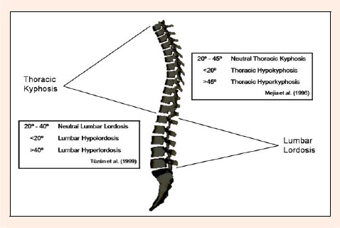
The cyclists wearing a culotte and barefoot assumed a straight position standing on the floor with the eyes and ears in line with the horizontal, arms relaxed at the side of the body, knees close to individual full extension, and feet shoulder-width apart. The values proposed by Mejia et al., 1996 and Tüzün et al., 1999 were used to classify the posture in categories for thoracic kyphosis and lumbar lordosis, respectively (Figure 1).
Figure 1.

Angle references for thoracic kyphosis and lumbar lordosis
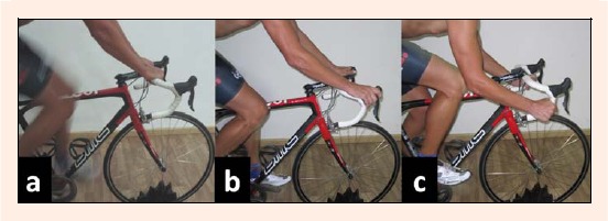
Sitting on the bicycle: Spinal curvatures and pelvic tilt were measured in a random order at three different handlebar-hand positions: high, medium, and low (Figure 2). Cyclists wore their own culotte and cycling shoes. They used their own bicycle. The subjects sat and pedalled for five minutes at a cadence of 90 revolutions per minute (measured by a cadence meter) in a cycling trainer (CycleOps PowerBeamTM, USA). The cycle resistance was controlled with Borg’s 6-20 points RPE scale. Each cyclist pedalled at “moderate intensity ”(12-13 points). After five minutes the cyclists was asked to stop pedalling, to maintain both pedals parallel to the floor. At this moment the tester measured the spinal angles and pelvic tilt. There was a 30-second of rest period after each hand position measurement.
Figure 2.

Handlebar-hand positions: upper handlebar (a), middle handlebar (b) and lower handlebar (c).
Statistical analysis
Intra-tester reliability of thoracic and lumbar curvatures and pelvic tilt was calculated in a previous pilot study. Twenty subjects who did not participated in the final sample were measured three times by the same tester in standing position on the floor, sitting on a stool, and prone lying in a single session. Intra-class correlation coefficients (ICC) with 95% confidence intervals (CI) were calculated. An ICCs upper or equal to 0.98 (95% CI: 0.98 - 0.99) were obtained for thoracic kyphosis, lumbar lordosis and pelvic tilt in all postures evaluated.
Means and standard deviations were calculated for all variables. The hypotheses of normality and homogeneity of variance were analysed using the Kolmogorov-Smirnov test and Levene’s test, respectively. A two-way ANOVA (group and posture) with repeated measurements for the second factor was used to compare spinal curvatures and pelvic tilt. The significance of the repeated multivariate measurements was confirmed by Wilk’s lambda, Pillai’s trace, Hotelling trace, and Roy’s tests, all of which obtained similar results. If a significant p-value was obtained for the main effect of the ANOVA, a post hoc comparison was conducted using Bonferroni correction for multiple comparisons, which adjusted the significance criterion to a value of 0.0125 (0.05/4). Between-group effect sizes (Cohen’s d) were calculated by using a pooled standard deviation. An effect size greater than 0.8 was considered large, around 0.5 was moderate, and less than 0.2 was small (Cohen, 1988). The data were analysed using SPSS (version 15.0), and the significance level was set at p < 0.05.
Results
The means and standard deviations of the measured postures in the elite and master cyclists are shown in Table 2. No significant differences between elite and master cyclists were found in thoracic kyphosis and lumbar lordosis while standing on the floor. The elite cyclists showed lower thoracic kyphosis in the three handlebar positions compared to the master cyclists, but they were only significantly different in the upper handlebar-hand position (p < 0.05). The lumbar curve changed from lordosis in standing to kyphosis on the bicycle. The elite cyclists showed greater lumbar flexion than master cyclists (Table 2). For all of the postures evaluated, the elite cyclists showed greater pelvic tilt than the master cyclists (p < 0.05).
Table 2.
Mean values (SD) of the thoracic, lumbar spine and pelvic tilt (°) in the four postures measured in elite and master cyclists.
| Elite | Master | Mean difference (95% CI) | p- value | Effect size | ||
|---|---|---|---|---|---|---|
| Standing | Thoracic spine | 48.2 (8.1) | 47.0 (9.2) | 1.15° (-1.98 to 4.28) | .469 | .13 |
| Lumbar spine | -27.3 (7.2) | -25.3 (6.3) | -2.01° (-4.46 to .43) | .106 | .29 | |
| Pelvic tilt | 13.7 (5.5) | 11.3 (5.2) | 2.40° (.46 to 4.33) | .016 | .44 | |
| Upper handlebar | Thoracic spine | 35.0 (10.8) | 39.7 (8.5) | -4.61° (-8.12 to -1.11) | .010 | .46 |
| Lumbar spine | 25.3 (7.6) | 22.9 (7.5) | 2.40° (-.32 to -5.12) | .084 | .31 | |
| Pelvic tilt | 24.6 (5.9) | 21.7 (6.2) | 2.93° (.74 to 5.12) | .009 | .47 | |
| Middle handlebar | Thoracic spine | 35.3 (10.8) | 37.5 (10.5) | -2.18° (-6.03 to 1.66) | .264 | .20 |
| Lumbar spine | 26.0 (7.8) | 24.2 (7.5) | 1.85° (-.91 to 4.61) | .188 | .31 | |
| Pelvic tilt | 29.0 (6.3) | 26.5 (6.5) | 2.45° (0.13 to 4.76) | .038 | .37 | |
| Lower handlebar | Thoracic spine | 37.18 (9.79) | 40.6 (10.0) | -3.36° (-6.94 to .21) | .065 | .33 |
| Lumbar spine | 28.5 (7.8) | 25.2 (7.3) | 3.26° (.53 to 5.99) | .200 | .42 | |
| Pelvic tilt | 36.3 (6.3) | 33.4 (6.5) | 2.95° (0.63 to 5.26) | .013 | .45 |
The ANOVA analysis showed significant differences for thoracic and lumbar angle and pelvic tilt in the different evaluated postures (p < 0.05). The post hoc analysis with Bonferroni adjustment showed a significantly lower thoracic kyphosis in all handlebar positions than in the standing position, and identical results in the significance values for both groups were found (p < 0. 0125). However, small effect sizes between both postures were found in elite cyclists (d around 0. 1) and master cyclists (d < 0.1).
The lumbar angle changed from lumbar lordosis in standing to lumbar kyphosis when sitting on the bicycle. The effect size was large in both groups (d = 0.99 in elite and master cyclists). A greater intervertebral flexion in the lower handlebar- hand position compared to the upper and middle handlebar-hand positions was found but a small effect size between these positions was found.
The pelvic tilt was significantly more flexed when sitting on the bicycle in the lower handlebar-hand position than in the other postures, although the effect sizes were small (d between 0.1 and 0.2 in elite cyclists and 0.1 and 0.3 in master cyclists) (Tables 3 and 4).
Table 3.
Pairwise comparisons (effect size) between positions for thoracic and lumbar curves and pelvic tilt in elite cyclists.
| Upper handlebar | Middle handlebar | Lower handlebar | ||
|---|---|---|---|---|
| Thoracic spine | Standing | * (.14) | * (.14) | * (.12) |
| Upper handlebar | - | NS (.00) | NS (.02) | |
| Middle handlebar | - | - | NS (.01) | |
| Lumbar spine | Standing | * (.95) | * (.93) | * (.98) |
| Upper handlebar | - | NS (.01) | * (.05) | |
| Middle handlebar | - | - | * (.04) | |
| Pelvic tilt | Standing | * (.33) | * (.43) | * (.64) |
| Upper handlebar | - | * (.11) | * (.31) | |
| Middle handlebar | - | - | * (.18) |
* p < 0.0125; NS: no significant.
Table 4.
Pairwise comparisons (effect size) between positions for thoracic and lumbar curves and pelvic tilt in master cyclists.
| Upper handlebar | Middle handlebar | Lower handlebar | ||
|---|---|---|---|---|
| Thoracic spine | Standing | * (.09) | * (.09) | * (.06) |
| Upper handlebar | - | NS (.02) | NS (.01) | |
| Middle handlebar | - | - | NS (.02) | |
| Lumbar spine | Standing | * (.99) | * (.99) | *(.99) |
| Upper handlebar | - | NS (.02) | * (.04) | |
| Middle handlebar | - | - | * (.01) | |
| Pelvic tilt | Standing | * (.31) | * (.44) | * (.63) |
| Upper handlebar | - | * (.11) | * (.28) | |
| Middle handlebar | - | - | * (.16) |
* p < 0.0125; NS: no significant.
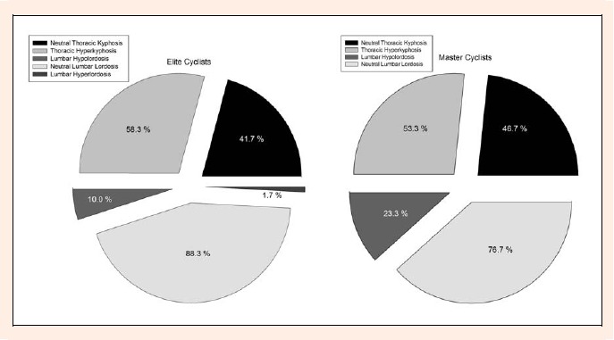
The frequencies of each thoracic and lumbar category in standing on the floor are presented in Figure 3. A higher percentage of hyperkyphotic postures in standing on the floor were found in the elite cyclists. However, master cyclists had a higher frequency of lumbar hypolordosis.
Figure 3.

Percentage of participants in each category of thoracic and lumbar curves while relaxed standing.
Discussion
A prolonged sitting position on a bicycle for an extended period may result adaptations in sagittal spinal angles. The main objective of this study was to evaluate the thoracic and lumbar spinal curvatures and pelvic tilt while standing on the floor and sitting on a bicycle in elite and master cyclists. A discussion of the standing on the floor and sitting on the bicycle positions is outlined below.
Group comparison
A high training volume while trunk is flexed, which characterises the position on the bicycle, might generate specific spinal adaptations in standing, such as higher thoracic kyphosis or reduced lumbar lordosis. When cyclist groups were classified by thoracic angle values, proposed by Mejia et al., 1996, a high percentage of thoracic hyperkyphosis in standing position on the floor in both groups (58.3% in elite and 53.3% in master cyclists) was found. However, a greater percentage of cyclists had neutral angles in standing (88.3% and 76.7% in elite and master cyclists, respectively). Recently, similar results were found for young kayakers. López-Miñarro et al., 2010 observed that kayakers adopt a lumbar flexed posture when sitting in the kayak, while the standing lumbar curve presented a high percentage (87.5%) of neutral postures.
Training volume appears to be an important variable in spinal adaptations. Wojtys et al., 2000 found a proportional increase of thoracic kyphosis in relation to training time per year (hours/year) when comparing different sports. However, lumbar lordosis did not change until training time exceeded 400 hours/year. Alricsson and Werner, 2006 observed increased thoracic kyphosis after a period of 5 years of intensive cross-country skiing. Ogurkowska (2007) reported morphological changes of intervertebral discs of the lumbosacral spine of competitive rowers who had practised competitive rowing for 8 to 20 years. In the current study no significant differences were found between elite and master cyclists. A possible explanation could be the similar training period (around 7 years) which may be insufficient to generate adaptation in the lumbar curve. However, elite cyclists trained more days per week and showed a slightly greater thoracic curve than master cyclists (48.17 ± 8.05° and 47.02 ± 9.24° in elite and master cyclists, respectively).
The pelvis is considered the base of the spine, and its anteroposterior orientation affects the sagittal curves of the spine (Levine and Whittle, 1996). In the current study elite cyclists reported a significantly greater pelvic tilt and lumbar lordosis when standing on the floor than master cyclists. This finding is in agreement with Barrey et al., 2007 and Schwab et al., 2009, who found a greater lumbar lordosis in subjects with a high anterior pelvic tilt. In contrast, subjects with reduced pelvic tilt showed a lumbar hypolordotic spine. These researchers explained their findings according to the principal function of the pelvis, related to postural balance (Barrey et al., 2007; Schwab et al., 2006). In our study, both groups of cyclists showed a greater anterior pelvic tilt at the three handlebar-hand positions than when standing. Elite cyclists showed a significantly higher anterior pelvic tilt than master cyclists in all handlebar-hand positions analysed. These findings may be explained by several factors. First, each cyclist used his own bicycle, and the handlebar on the bicycles of elite cyclists was lower, in order to reach an efficient aerodynamic posture. Moreover, elite cyclists may have a greater capacity for anterior pelvic tilt as an adaptation to the greater total training volume on the bicycle. McEvoy et al., 2007 reported that elite cyclists had a significantly higher anterior pelvic tilt than sedentary subjects while sitting with knees extended. These researchers explained this as a specific adaptation to the bending trunk in more aerodynamic positions.
The differences in pelvic position could be related to hamstring flexibility. Greater hamstring flexibility has been associated with increased lumbar flexion and pelvic tilt (Gajdosik et al., 1992; 1994; López-Miñarro and Alacid, 2010). However, the flexed position of the knee, around 20° in cyclists when the pedal is situated in the lowest position (De Vey Mestdagh, 1998), reduces tension in hamstring muscles and limits its effect on the pelvis and sagittal spinal curvatures (López-Miñarro and Alacid, 2010).
Sitting on the bicycle
When cyclists were evaluated sitting on their own bicycles, the percentage of thoracic hyperkyphosis was significantly lower than in the standing position. Several studies have observed a trend towards increased thoracic kyphosis in sports where specific technical movements involve maintained or cyclic trunk flexion, such as wrestling, canoeing, and cross-country skiing (Alricsson and Werner, 2006; López-Miñarro and Alacid, 2010; López-Miñarro et al., 2010; Rajabi et al., 2008), but these studies do not evaluate sports’ specific techniques postures.
Measurement of specific spinal postures is necessary in sports. In the present study we found a significant decrease of thoracic kyphosis on the bicycle (main differences: 13.14°, 12.84°, 10.99°, and 7.37°, 9.50°, 6.47° for elite and master cyclists for upper, middle and lower handlebars, respectively), although a small effect size was detected in both cyclist categories. This straighter posture, may be related to hand support on the handlebar, which leads to scapular retropulsion and thoracic intervertebral extension. These postural changes lead to a more neutral spinal posture. For this reason, the high percentage of standing thoracic hyperkyphosis in both groups of cyclists may be related to other factors, such as inadequate postural habits or thoracic muscle fatigue, rather than to the specific posture adopted while cycling. Rajabi et al., 2000 found a significantly higher thoracic kyphosis while relaxed standing position on the floor in elite cyclists than in sedentary individuals. They justified the increased thoracic kyphosis in cyclists due to a prolonged seated position on a bicycle with the trunk flexed. However, this study did not measure the specific posture on the bicycle and they did not reporter any further arguments. More investigation is needed in this line.
Increased spinal angles in sports where trunk flexion postures are predominant might be due to loss of disc height, which would tend to reduce the length of the anterior column of the spine (Rajabi et al., 2008; Wojtys et al., 2000). Greater intervertebral flexion has been associated with significantly higher intervertebral disc pressure, multisegmental spinal loads and spinal shear forces in relation to training volume (Briggs et al., 2007; Polga et al., 2004; Sato et al., 1999).
The lumbar angle while sitting on the bicycle was characterised by kyphotic posture in the three handlebar-hands position analysed. Lord et al., 1997 reported that lumbar lordosis while standing was nearly 50% greater on average than sitting lumbar lordosis. Lumbar kyphosis is associated with hand support on the handlebar, which is usually at a lower height than the seat. For this reason, De Vey Mestdagh, 1998 defined cyclists’ posture as not natural. In our study, a greater lumbar flexion and pelvic tilt was found with a lower handlebar. This posture is associated with a more aerodynamic posture. These results are in agreement with Usabiaga et al., 1997, who found a change from lumbar lordosis when standing to lumbar kyphosis when seated on the bicycle.
Sitting on the bicycle with a greater anterior pelvic tilt could help to maintain a neutral lumbar curve (De Vey Mestdagh, 1998). Salai et al., 1999 reported in cyclists with lumbar pain that an increase in the seat inclination between 10° and 15° was related to greater anterior pelvic tilt and trunk flexion and to a significant decrease in lumbar pain. However, this posture produces lumbar discomfort due to higher myoelectric activation in the lumbar multifidus muscle when the position is maintained for an extended period (O’Sullivan et al., 2006).
Previous studies have found increases in spinal load (Keller et al., 2005), disc pressure (Nachemsom, 1976; Polga et al., 2004; Sato et al., 1999; Wilke et al., 1999), creep deformation in lumbar tissues (Caldwell and Peters, 2009; Solomonow et al., 2003) and low back pain (Harrison et al., 2005; Smith et al., 2008) when static or cyclic spinal flexions are performed. This may be why low back pain is the most common overuse injury in cyclists (Asplund et al., 2005; Clarsen et al., 2010; Marsden et al., 2010; Salai et al., 1999).
Limitations and future directions
This study has several limitations. The first is the influence of personal and environment factors in spinal angles. The current study was based on a cross-sectional comparison between two age-groups. Longitudinal studies are necessary in ascertaining the long-term effects of cycling training in spinal posture.
A second limitation was that cyclists had to stop pedalling before spinal evaluation on the bicycle, since the vibrations and movements could affect spinal values. Another limitation was that we did not analyse the saddle inclination. Salai et al., 1999 found changes in lumbar spine and pelvic tilt when saddle inclination was modified. However, in the current study the cyclists used their own bicycles, maintaining their usual set-up equipment for keeping the usual parameters they maintain during training and competition.
Conclusion
The thoracic hyperkyphotic posture in standing in elite and master cyclists may be related to factors other than the posture adopted on the bicycle. Cyclists adopted a more neutral thoracic posture when sitting on the bicycle in all handlebar-hand positions. The lumbar flexed posture and high anterior pelvic tilt when sitting on the bicycle do not influence the sagittal configuration of the lumbar spine in standing.
Biographies
José M. Muyor
Employment
Professor of Physical Education Teaching. Faculty of Education. University of Almería, Spain.
Degree
MSc, PhD.
Research interests
Spine posture and hamstring flexibility.
E-mail: josemuyor@ual.es
Pedro A. López-Miñarro
Employment
Professor of Physical Activity and Health at the Research Department of Physical Education, Faculty of Education, University of Murcia, Spain.
Degree
PhD.
Research interests
Hamstring flexibility and spine posture.
E-mail: palopez@um.es
Fernando Alacid
Employment
Professor of Canoeing and Kayaking. Faculty of Sport Sciences, University of Murcia, Spain.
Degree
MSc, PhD.
Research interests
Kinanthropmetry and kinematics.
E-mail: fernando.alacid@um.es
References
- Alricsson M, Werner S. (2006) Young élite cross-country skiers and low back pain. A 5-year study. Physical Therapy in Sport, 7, 181-184 [DOI] [PubMed] [Google Scholar]
- Asplund C., Webb C., Barkdull T. (2005) Neck and back pain in bicycling. Current Sports Medicine Reports 4, 271-274 [DOI] [PubMed] [Google Scholar]
- Barrey C., Jund J., Noseda O., Roussouly P. (2007) Sagittal balance of the pelvis-spine complex and lumbar degenerative diseases. A comparative study about 85 cases. European Spine Journal 9, 1459-1467 [DOI] [PMC free article] [PubMed] [Google Scholar]
- Beach T., Parkinson R., Stothart P., Callaghan J. (2005) Effects of prolonged sitting on the passive flexion stiffness of the in vivo lumbar spine. The Spine Journal 5, 145-154 [DOI] [PubMed] [Google Scholar]
- Briggs A., Van Dieën J., Wrigley T., Greig A., Phillips B., Lo S., Bennell K. (2007) Thoracic kyphosis affects spinal loads and trunk muscle force. Physical Therapy 87, 595-607 [DOI] [PubMed] [Google Scholar]
- Caldwell B., Peters D. (2009) Seasonal variation in physiological fitness of a semiprofessional soccer team. Journal of Strength and Conditioning Research 23, 1370-1377 [DOI] [PubMed] [Google Scholar]
- Clarsen B., Krosshaug T., Bahr R. (2010) Overuse injuries in professional road cyclists. The American Journal of Sports Medicine 38, 2494-2501 [DOI] [PubMed] [Google Scholar]
- Cohen J. (1988). Statistical power analysis for the behavioral science. 2nd edition Hillsdale, NJ: Lawrence Erlbaum Associates [Google Scholar]
- De Vey Mestdagh K. (1998) Personal perspective: in search of an optimum cycling posture. Applied Ergonomics 29, 325-334 [PubMed] [Google Scholar]
- Gajdosik R., Albert C., Mitman J. (1994) Influence of hamstring length on the standing position and flexion range of motion of the pelvic angle, lumbar angle, and thoracic angle. Journal of Orthopaedic and Sports Physical Therapy 20, 213-219 [DOI] [PubMed] [Google Scholar]
- Gajdosik R., Hatcher C., Whitsell S. (1992) Influence of short hamstring muscles on the pelvis and lumbar spine in standing and during the toe-touch test. Clinical Biomechanics 7, 38-42 [DOI] [PubMed] [Google Scholar]
- Guermazi M., Ghroubi S., Kassis M., Jaziri O., Keskes H., Kessomtini W., Hammouda I.B., Elleuch M.H. (2006) Validity and reliability of Spinal Mouse® to assess lumbar flexion. Annales de Réadaptation et de Médecine Physique, 49, 172-177 [DOI] [PubMed] [Google Scholar]
- Gelb D.E., Lenke L.G., Bridwell K. H., Blanke K., McEnery K.W. (1995) An analysis of sagittal spinal alignment in 100 asymptomatic middle and older aged volunteers. Spine 15, 1351-1358 [PubMed] [Google Scholar]
- Harrison D.E., Colloca C.J., Harrison D.D., Janik T.J., Haas J.W., Keller T.S. (2005) Anterior thoracic posture increases thoracolumbar disc loading. European Spine Journal 14, 234-242 [DOI] [PMC free article] [PubMed] [Google Scholar]
- Keller T.S., Colloca C.J., Harrison D.E., Harrison D.D., Janik T.J. (2005) Influence of spine morphology on intervertebral disc load and stresses in asymptomatic adults: implications for the ideal spine. Spine 5, 297-300 [DOI] [PubMed] [Google Scholar]
- Landers M., Barker G., Vallentine S., Mc Whorter J.W, Peel C.A. (2003) A comparison of tidal volume, brething frequency, and minute ventilation between two sitting posutres in healthy adults. Physioterapy. Theory and Practice 19, 109-119 [Google Scholar]
- Levine D., Whittle M.W. (1996) The effects of pelvic movement on lumbar lordosis in the standing position. Journal of Orthopaedic and Sports Physical Therapy, 24, 130-135 [DOI] [PubMed] [Google Scholar]
- López-Miñarro P.A., Alacid F. (2010) Influence of hamstring muscle extensibility on spinal curvatures in young athletes. Science & Sports 25, 88-93 [Google Scholar]
- López-Miñarro P.A., Alacid F., Muyor J.M. (2009) Comparación del morfotipo raquídeo y extensibilidad isquiosural entre piragüistas y corredores. Revista Internacional de Medicina y Ciencias de la Actividad Física y del Deporte 9, 379-392 (in Spanish) [Google Scholar]
- López-Miñarro P.A., Muyor J.M., Alacid F. (2010) Sagittal spinal curvatures and pelvic tilt in elite young kayakers. Medicina dello Sport 63, 509-519 [Google Scholar]
- Lord M.J., Small J.M., Dinsay JM., Watkins RG. (1997) Lumbar lordosis: effects of sitting and standing. Spine 22, 2571-2574 [DOI] [PubMed] [Google Scholar]
- Lin S.I, Liao C.F. (2011) Age-related changes in the performance of forward reach. Gait & Posture, 33, 18-22 [DOI] [PubMed] [Google Scholar]
- Lin F., Parthasarathy S., Taylor S.J., Pacci D., Hendrix R.W, Makhsous M. (2006) Effect of different sitting postures on lung capacity, expiratory flow, and lumbar lordosis. Archives of Physical Medicine and Rehabilitation, 87, 504-509 [DOI] [PubMed] [Google Scholar]
- Maejima H., Takeishi K., Sunahori H., Yamawaki A., Nakajima K, Yoshimura O. (2004) The relationship between postural deformation and standing balance in elderly person. Journal of the Japanese Physical Therapy Association, 7, 7-14 [DOI] [PMC free article] [PubMed] [Google Scholar]
- Mannion A.F., Knecht K., Balaban G., Dvorak J., Grob D. (2004). A new skin-surface device for measuring the curvature and global and segmental ranges of motion of the spine: reliability of measurements and comparison with data reviewed from the literature. European Spine Journal, 13, 122-136 [DOI] [PMC free article] [PubMed] [Google Scholar]
- Marsden M., Schwellnus M. (2010) Lower back pain in cyclists: A review of epidemiology, pathomechanics and risk factors. International SportMed Journal 11, 216-225 [Google Scholar]
- McEvoy M., Wilkie K., Williams M. (2007) Anterior pelvic tilt in élite cyclist- A comparative matched pairs study. Physical Therapy in Sport 8, 22-29 [Google Scholar]
- Mejia E.A., Hennrikus W.L., Schwend R.M., Emans J.B. (1996) A prospective evaluation of idiopathic left thoracic scoliosis with MRI. Journal of Pediatric Orthopedics 16, 354-358 [DOI] [PubMed] [Google Scholar]
- Nachemson A. (1976) The load on lumbar disks in different positions of the body. Clinical Orthopaedic 45, 107-112 [PubMed] [Google Scholar]
- Ogurkowska M.B. (2007) Pathological change of intervertebral disc of the lumbosacral spine of competitive rowers. Biology of Sport 24, 375-388 [Google Scholar]
- O’Sullivan P.B, Dankaerts W., Burnett A.F., Farrell G.T, Jefford E., Naylor C.S., O’Sullivan K.J. (2006) Effect of different upright sitting postures on spinal-pelvic curvature and trunk muscle activation in a pain-free population. Spine 31, E707-12 [DOI] [PubMed] [Google Scholar]
- Polga D., Beaubien B., Kallemeier P., Schellhas K., Lew W., Buttermann G., Wood K. (2004) Measurement of in vivo intradiscal pressure in healthy thoracic intervertebral discs. Spine 29, 1320-1324 [DOI] [PubMed] [Google Scholar]
- Post R.B., Leferink V.J. (2004) Spinal mobility: sagittal range of motion measured with the SpinalMouse, a new non-invasive device. Archives of Orthopaedic and Trauma Surgery, 124, 187-192 [DOI] [PubMed] [Google Scholar]
- Rajabi R., Doherty P., Goodarzi M., Hemayattalab R. (2008) Comparison of thoracic kyphosis in two groups of élite Greco-Roman and free style wrestlers and a group of non-athletic subjects. British Journal of Sports Medicine 42, 229-232 [DOI] [PubMed] [Google Scholar]
- Rajabi R., Freemont A., Doherty P. (2000) The investigation of cycling position on thoracic spine. A novel method of measuring thoracic kyphosis in the standing position. Archives of Physiology and Biochemistry 1, 142 [Google Scholar]
- Salai M., Brosh T., Blankstein A., Oran A., Chechik A. (1999) Effect of changing the saddle angle on the incidence of low back pain in recreational bicyclists. British Journal of Sports Medicine 33, 398-400 [DOI] [PMC free article] [PubMed] [Google Scholar]
- Sato K., Kikuchi S., Yonezawa T. (1999) In vivo intradiscal pressure measurement in healthy individuals and in patients with ongoing back problems. Spine 24, 2468-2474 [DOI] [PubMed] [Google Scholar]
- Schwab F., Lafage V., Boyce R., Skalli W., Farcy J.P. (2006) Gravity line analysis in adults volunteers. Age-related correlation with spinal parameters, pelvic parameters and foot position. Spine 31, E959-E967 [DOI] [PubMed] [Google Scholar]
- Schwab F., Lafage V., Patel A., Farcy J.P. (2009) Sagittal plane considerations and the pelvis in the adult patient. Spine 31, 1828-1833 [DOI] [PubMed] [Google Scholar]
- Smith A., O’Sullivan P., Straker L. (2008) Classification of sagittal thoraco-lumbo-pelvic alignment of the adolescent spine in standing and its relationship to low back pain. Spine 33, 2101-2107 [DOI] [PubMed] [Google Scholar]
- Solomonow M., Zhou B., Baratta R.V., Burger E. (2003) Biomechanics and electromyography of a cumulative lumbar disorder: response to static flexion. Clinical Biomechanics 18, 883-889 [DOI] [PubMed] [Google Scholar]
- Tüzün C., Yorulmaz I., Cindas A., Vatan S. (1999) Low back pain and posture. Clinical Rheumatology 18, 308-312 [DOI] [PubMed] [Google Scholar]
- Usabiaga J., Crespo R., Iza I., Aramendi J., Terrados N., Poza J. (1997) Adaptation of the lumbar spine to different positions in bicycle racing. Spine 22, 1965-1969 [DOI] [PubMed] [Google Scholar]
- Vedantam R., Lenke L.G., Keeney J.A., Bridwell K.H. (1998) Comparison of standing sagittal spinal alignment in asymptomatic adolescents and adults. Spine 15, 211-215 [DOI] [PubMed] [Google Scholar]
- Wilke H., Neef P., Caimi M., Hoogland T., Claes L. (1999) New in vivo measurements of pressures in the intervertebral disc in daily life. Spine 24, 755-762 [DOI] [PubMed] [Google Scholar]
- Wojtys E., Ashton-Miller J., Huston L., Moga P. (2000) The association between athletic training time and sagittal curvature of the immature spine. The American Journal of Sports Medicine 28, 490-498 [DOI] [PubMed] [Google Scholar]


