Abstract
Colonic volvulus is a relatively uncommon cause of large bowel obstruction, accounting for 10% of colonic obstructions. Volvulus of the transverse colon is quite rare, accounting for only 4–11% of all reported cases. We report an unusual case of documented volvulus of the transverse colon in a pregnant woman with intestinal malrotation and concomitant acute intestinal obstruction by congenital bands and adhesions.
Background
An intestinal obstruction can occur in pregnancy, though not often. Malrotation of the gut leading to colonic volvulus is an extremely rare case of intestinal obstruction during pregnancy. In less than 25% of pregnant women with intestinal obstruction, the aetiology can be traced to colonic volvulus.1 Less than 100 cases of true volvulus, in pregnancy, have been reported in the world literature.2–5
Intestinal rotation and positioning occur in the fetus early in gestation. Arrest at any step results in intestinal anomalies, which can result in symptomatic disorders with the potential for marked morbidity and mortality.
The reported incidence of intestinal obstruction complicating first time, in pregnancy, varies widely from 1 in 1500 to 1 in 66 431 birth cases or deliveries.6 7 Diagnosis is often delayed due to the delay in presentation, poor knowledge of the condition and a hesitation to use radiological investigation in pregnancy, as well as attributing the clinical features to other ailments of pregnancy.
Recently we encountered a rare case of acute intestinal obstruction due to volvulus of transverse colon with malrotation of the gut which manifested for the first time in early pregnancy and required urgent surgical intervention.
Case presentation
A 28-year-old woman, primigravida at 9 weeks gestation presented with intermittent pain abdomen, fever with chills and vomiting. Her medical history was unremarkable.
Physical examination was unremarkable. Blood reports were within normal limits. Transvaginal sonography showed a viable fetus corresponding to 9 weeks. After seeking an urological consultation, a working diagnosis of ureteric colic (non-obstructive renal calculi) was made and she was started on antispasmodics, antiemetics and adequate hydration. The initial abdominal ultrasound was normal.
Over the next 3 days, she developed diffuse colicky pain and constipation. She was given enema and antispasmodics. The patient continued to have pain and developed abdominal distension with diffuse tenderness with hyperperistaltic bowel sounds, suggestive of an intestinal obstruction. Conservative management with keeping the patient nil per oral, Ryle's tube insertion and intravenous fluids were started. Repeat sonography revealed moderate ascites with dilated small bowel loops. The following day, as her symptoms worsened, she was taken up for emergency exploratory laparotomy.
Treatment
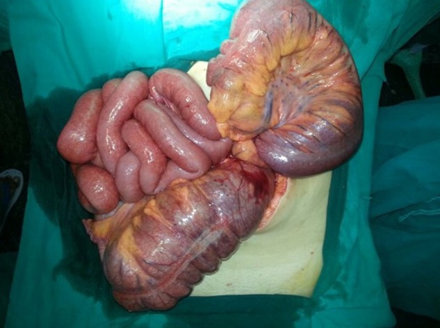
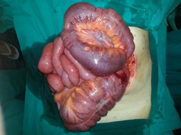
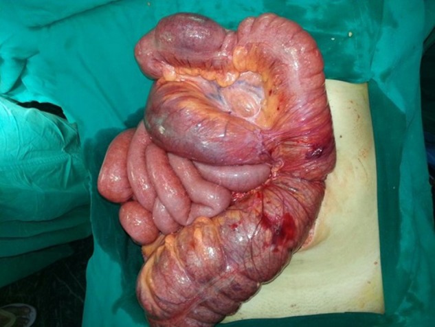
At surgery a 360° twist (anticlock wise) in a massively dilated transverse colon was observed (figures 1 and 2). The hepatic flexure appeared free and the transverse colon was extremely long and had a long mesocolon which was thickened at the base. The distal transverse colon appeared bound down to this band. De-rotation of the colon with adhesiolysis was performed (figure 3). The entire length of the bowel was traced and found viable. There were no Ladd's bands across the duodenum. The large bowel was repositioned into the left side and small bowel on right side of the abdomen with caecum and ascending colon anchored to lateral abdominal wall with interrupted sutures.
Figure 1.

Intraoperative image showing large bowel on left side of abdomen with non-fixation of caecum and ascending colon with transverse colon volvulus (anticlockwise). Small bowel is on right side.
Figure 2.

Intraoperative image showing the same findings.
Figure 3.

Viable large and small bowel after de-rotation of volvulus.
Outcome and follow-up
Postoperative period was uneventful, she was symptom free and her pregnancy continued uneventfully.
Discussion
An Intestinal obstruction (IO) in pregnancy is rare. Major causes include adhesions, ventral hernias, cancer of colon, Meckel's diverticulum, volvulus and intussusception.3 7 8 Although uncommon, IO in pregnancy carries a significant maternal mortality (6%) and fetal (26%) mortality.9 Often this is due to delay in diagnosis and treatment, because the symptoms mimic pregnancy associated problems and radiological investigations (X-ray and C CT scans) are generally contraindicated or used with extreme caution in pregnancy.
Intestinal volvulus is responsible for 25% of acute bowel obstructions in pregnant women and 3–5% in non-pregnant women. Malrotation occurs as a result of an arrest of rotation of the intestine during fetal development. The malrotation of the bowel makes it susceptible for volvulus as they are redundant and have defect in fixation as happened in our case where there was non-fixation of caecum and ascending colon making them susceptible to volvulus.10
Most patients are symptomatic early in life, with 75–80% presenting with a midgut volvulus during the first month of life. Less frequently, patients may present with symptoms later in life.11 Older patients usually present with vague, chronic symptoms rather than the acute symptoms seen in infancy and childhood .The most common symptom is vomiting, intermittent crampy abdominal pain from torsion or bands or volvulus.
Complications from intestinal malrotation including volvulus, ischaemia and infarction of the bowel can be life threatening or cause marked morbidity. In a study of volvulus occurring during pregnancy,12 2 patients presented during the first trimester, 9 during the second trimester and 25 in the third trimester. The diagnosis of malrotation is not often considered in pregnant patients as symptoms are often non-specific and avoidance of radiological studies during pregnancy may also delay the diagnosis. The diagnostic modalities include X-ray, sonography and MRI.
In our patient, the diagnosis was delayed because initial clinical picture and ultrasound were suggestive of ureteric colic, but subsequently she developed acute IO and was taken up for exploratory laparotomy. There was always a reluctance in using radiological studies due to her early pregnancy.
Treatment is often surgical, if there is concomitant volvulus, it should be relieved and viability of the bowel should be assessed. If there is non-fixation of the caecum and ascending colon, the caecum should be stabilised.
Management is similar to non-pregnant women. Clinical suspicion is vital and joint management between surgeons and obstetricians is crucial. The basis of treatment is timely surgery and minimising delays in decision-making. In our patient, as conservative measures failed, she was planned for an emergency exploratory laparotomy where the actual cause was diagnosed, and appropriate surgical procedure could be accomplished.
Learning points.
Malrotation of the gut leading to volvulus of pregnancy complicating pregnancy is an uncommon and potentially devastating development and should be recognised as a surgical emergency.
Diagnosis requires a high index of suspicion in a patient who presents with problem of abdominal pain and evidence of bowel obstruction. Since, the radiological investigations are very limited, in early pregnancy, the diagnosis becomes even more difficult.
Delay in diagnosis results in strangulation of bowel with perforation, peritonitis and sepsis and increased fetal and maternal morbidity and mortality.
Prompt intervention is necessary to minimise these complications and achieve a definitive cure.
An additional learning point from this case would be that in an obstetric patient with an unremarkable medical history, presenting with abdominal pain, one should also consider surgical causes other than merely obstetric or gynaecological causes of pain.
Footnotes
Competing interests: None.
Patient consent: Obtained.
Provenance and peer review: Not commissioned, externally peer reviewed.
References
- 1.Ventura-Braswell AM, Satin AJ, Higby K. Delayed diagnosis of bowel infarction secondary to maternal midgut volvulus at term. Obstet Gynecol 1998;2013:801–10 [DOI] [PubMed] [Google Scholar]
- 2.Iwamoto I, Miwa K, Fujino T, et al. Perforated colon volvulus coiling around the uterus in a pregnant woman with a history of severe constipation. J Obstet Gynaecol Res 2007;2013:731–3 [DOI] [PubMed] [Google Scholar]
- 3.Kolusari A, Kurdoglu M, Adali E, et al. Sigmoid volvulus in pregnancy and puerperium: a case series. Cases J 2009;2013:article 9275 [DOI] [PMC free article] [PubMed] [Google Scholar]
- 4.Allen JC. Sigmoid volvulus in pregnancy. J R Army Med Corps 1990;2013:55–6 [DOI] [PubMed] [Google Scholar]
- 5.Fraser JL, Eckert LA. Volvulus complicating pregnancy. CMAJ 1983;2013:1045–8 [PMC free article] [PubMed] [Google Scholar]
- 6.Coughlan BM, O'Herlihy C. Acute intestinal obstruction during pregnancy. J R Coll Surg 1978;2013:175–7. [PubMed] [Google Scholar]
- 7.Perdue PW, Johnson HW, Stafford PW. Intestinal obstruction complicating pregnancy. Am J Surg 1992;2013:384–8 [DOI] [PubMed] [Google Scholar]
- 8.Damore LJ, II, Damore TH, Longo WE, et al. Congenital intestinal malrotation causing gestational intestinal obstruction. A case report. J Repord Med 1997;2013:805–8 [PubMed] [Google Scholar]
- 9.Perdue PW, Johnson HW, Jr, Stafford PW. Intestinal obstruction complicating pregnancy. Am J Surg 1992;2013:384–8 [DOI] [PubMed] [Google Scholar]
- 10.Lopez Carral JM, Esen UI, Chandrashekar MV, et al. Volvulus of the right colon at pregnancy. Int J Clin Pract 1998;2013:270–1 [PubMed] [Google Scholar]
- 11.Gilbert HW, Armstrong CP, Thompson MH. The presentation of malrotation of the intestine in adults. Ann R Coll Surg Engl 1990;2013:239–42 [PMC free article] [PubMed] [Google Scholar]
- 12.Malkasian GD, Jr, Welch JS, Hallenbeck GA. Volvulus associated with pregnancy. Am J Obstet Gynecol 1959;2013:112–23 [DOI] [PubMed] [Google Scholar]


