Abstract
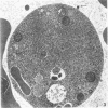
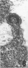
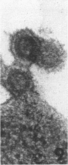
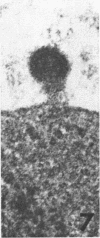
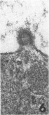
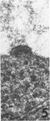
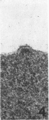
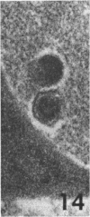
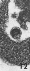
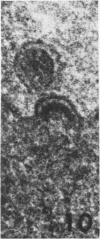
After 30 and 78 hr, Friend murine leukemia virus (FLV) particles were detected by electron microscopy in the mid-gut lumen of the mosquitoes Aedes aegypti (Linnaeus) and Anopheles stephensi Liston which had fed on leukemia BALB/c mice infected with FLV. Various developmental stages of the virions were observed within and on the surface of ingested blood cells, particularly young erythroblasts, as well as free in the lumen after budding. These preliminary findings indicate that FLV continues to multiply in the mid-gut of these species for at least 3 days despite the action of digestive enzymes. Detailed studies are in progress to determine the fate of FLV in these mosquito species.
Full text
PDF


Images in this article
Selected References
These references are in PubMed. This may not be the complete list of references from this article.
- BERTRAM D. S., BIRD R. G. Studies on mosquito-borne viruses in their vectors. I. The normal fine structure of the midgut epithelium of the adult female Aedes aegypti (L.) and the functional significance of its modification following a blood meal. Trans R Soc Trop Med Hyg. 1961 Sep;55:404–423. doi: 10.1016/0035-9203(61)90085-2. [DOI] [PubMed] [Google Scholar]
- CHIRIGOS M. A., LUBER E., MARCH R., PETTIGREW H. ANTIVIRAL CHEMOTHERAPEUTIC ASSAY WITH FRIEND LEUKEMIA VIRUS IN MICE. Cancer Chemother Rep. 1965 Apr;45:29–33. [PubMed] [Google Scholar]


