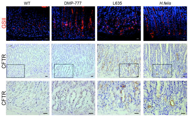
Figure 4. Expression of CFTR in normal murine gastric mucosa and SPEM models.

Frozen sections of C57BL/6 mouse fundic mucosa were immunostained with either GSII lectin (a mucous neck cell and SPEM marker) or an antibody against CFTR to investigate CFTR protein expression in SPEM. Top Panel: GSII (green) labels mucous neck cells in WT stomach mucosa and SPEM at the bases of glands in each of the SPEM models. Middle and Bottom Panels: Frozen sections immunohistochemical staining for CFTR showed no CFTR expression was detected in either WT mucosa or non-inflammatory SPEM (DMP-777-induced SPEM). The inset shows magnification of the bases of glands. CFTR expression was detected on the apical membranes of SPEM cells accompanied by inflammation (3 day L635 administration and 12 month H. felis infection). Scale bars = 20 μm.
