Abstract
White-nose syndrome (WNS) has claimed the lives of millions of hibernating insectivorous bats in North America. Its etiologic agent, the psychrophilic fungus Geomyces destructans, causes skin lesions that are the hallmark of the disease. The fungal infection is characterized by a white powdery growth on muzzle, ears and wing membranes. While WNS may threaten some species of North American bats with regional extinction, infection in hibernating bats in Europe seems not to be associated with significant mortality. We performed histopathological investigations on biopsy samples of 11 hibernating European bats, originating from 4 different countries, colonized by G. destructans. One additional bat was euthanized to allow thorough examination of multiple strips of its wing membranes. Molecular analyses of touch imprints, swabs and skin samples confirmed that fungal structures were G. destructans. Additionally, archived field notes on hibernacula monitoring data in the Harz Mountains, Germany, over an 11-year period (2000–2011) revealed multiple capture-recapture events of 8 banded bats repeatedly displaying characteristic fungal colonization. Skin lesions of G. destructans-affected hibernating European bats are intriguingly similar to the epidermal lesions described in North American bats. Nevertheless, deep invasion of fungal hyphae into the dermal connective tissue with resulting ulceration like in North American bats was not observed in the biopsy samples of European bats; all lesions found were restricted to the layers of the epidermis and its adnexae. Two bats had mild epidermal cupping erosions as described for North American bats. The possible mechanisms for any difference in outcomes of G. destructans infection in European and North American bats still need to be elucidated.
Introduction
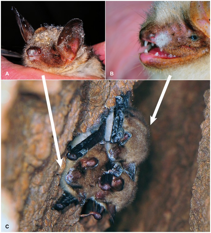
Since 2006 North America has experienced mass mortalities of estimated more than 6.7 million hibernating bats [1] caused by the cold-loving keratinophilic fungus, Geomyces destructans [2], [3]. Epidermal fungal colonization of affected bats is characterized by distinct white fungal patches on snout, ears and wing membranes [4]–[6]; the facial distribution of the fungus lead to the name of “white-nose syndrome” (WNS). Signs of WNS in affected hibernacula include aberrant hibernation behavior (day flight) and increased mortality rates, whereas to confirm the diagnosis of WNS the identification of typical histopathologic lesions and genetic identification of the fungus is required [1], [6], [7]. Since its first detection in New York state the fungal infection has spread through hibernacula in most neighboring states along the east coast. It has also moved further west, to Tennessee and Oklahoma, as well as crossed the Canadian border into Ontario, Quebec and Nova Scotia [8]. Seven bat species are currently known to be affected by WNS, but it is feared that more species will become involved since the habitat range of additional hibernating bat species is located in western areas. As a result of WNS, many North American hibernacula that previously contained thousands to hundreds of thousands of bats have been decimated [9]. Population viability analyses predict that Myotis lucifugus (the little brown bat), formerly one of the most abundant bat species in many regions of North America, could become extinct in 16–20 years [9]. The impact of WNS on biodiversity and on agricultural economy remains unknown, but will most likely be of major significance [10].
Fungal infectious agents can either serve as a primary pathogen or they may invade secondary to predisposing factors like co-infections by other pathogens. Bats dying of WNS had no consistent significant pathologic changes in their internal organs neither bacteriological nor virological analyses revealed consistent evidence for any known pathogen [4], yet experimental infections with G. destructans resulted in the same lesions that occur in natural infections [2], [3]. Many of the dead bats are found in an emaciated state which is thought to be due to the noted increased frequency of arousal cycles in affected bats, resulting in premature consumption of fat reserves [3], [11]. A recent study of naturally infected M. lucifigus supports this hypothesis, as WNS affected bats showed decreased torpor bout duration [7]. Furthermore, wing membrane lesions associated with WNS are hypothesized to cause an imbalance of homeostasis of body fluids ultimately leading to the death of the affected animals [11].
Dermatophyte infections are restricted to superficial skin structures, i.e. corneal stratum or hair cuticle. In contrast, infections by G. destructans in North American bats range from cup-like intraepidermal colonies with erosions to severe ulceration of the affected skin and deep invasion by fungal hyphae into the underlying dermal connective tissue. However, even extensive G. destructans invasion lacks noticeable cellular inflammatory response in its early stages [6]. As hibernation has been shown to reduce body functions, including the immune system to a minimal level in other hibernators [12], it is assumed that the lack of inflammation against invading fungal hyphae reflects this temporary physiologic unresponsiveness in bats.
In Europe, a wide distribution of G. destructans was found in 13 European countries involving 7 different species of hibernating bats [13]. Despite a long standing tradition of annual hibernacula census in a number of European countries, no fungus-associated mass mortalities have been recorded. Furthermore, bats which were notably colonized with the fungus left their hibernacula uneventfully in spring alongside their unaffected colony members [13]. Growth requirements, morphologic characteristics and the 100% sequence similarity within the ITS and SSU rRNA gene segments indicate that European G. destructans isolates are closely related to the US type strain [13]–[16].
In contrast to the management of bat populations in North America, all bat species in Europe are protected under the European Union’s 1992 Conservation of Wild Flora and Fauna Directive (http://ec.europa.eu/environment/nature/legislation/habitatsdirective/index_en.htm) (92/43/EEC) and the Agreement on the Conservation of Populations of European Bats (www.eurobats.org) both of which prohibit invasive sampling of bats. Owing to these legislations, until recently it was not possible in most European countries to investigate fungal colonization of hibernating bats beyond touch imprints of fungal structures from the surface of the skin. With the emergence of WNS and the pressing need to shed light on the question of whether hibernating bats from Europe also had inflammatory reactions to G. destructans, governmental authorities granted limited permission for invasive sampling such as wing punch biopsies. Here, we describe histopathologic investigations of wing punch biopsies from bat skin colonized by G. destructans as well as long term field observations on capture-recapture events in hibernating bats with fungal growth.
Materials and Methods
Permits
For this study, exemption permits for the collection of punch biopsies of wing membranes from hibernating bats were given to designated bat activists registered for annual hibernacula monitoring, by the responsible regional governmental authorities (General Directorate for Agriculture, Natural Resources and Environment, Namur, Belgium (F. Forget), Préfecture du Cher, France (L. Arthur), Hanover region nature conservation agency, Hanover, Germany (K. Passior), Saxony-Anhalt Ministry for Agriculture and Environment, Magdeburg, Germany (B. Ohlendorf), Hungarian National Inspectorate for Environment, Nature and Water (T. Görföl)). Further permission under the Flora and Fauna Act was given to P. Lina of the Netherlands Centre for Biodiversity ‘Naturalis’ by the Netherlands Ministry of Economic Affairs, Agriculture and Innovation (FF/75/2003/169b, valid 5th February 2007 through 12th April 2010) to euthanize a single bat with visible fungal growth.
Field Sampling
The aim of the study was to investigate the pathogenic effect of G. destructans colonizing the skin of hibernating European bats. Bat populations in most hibernacula in Europe are significantly smaller than those in North America. The average number of individual bats in the hibernacula in this study ranged from 20 to 200. So if G. destructans colonization were to be found in hibernacula, it is usually limited to one or a few affected animals. Of these bats only the most visibly severe cases of fungal growth were chosen for sampling in this study. Sampling dates were set towards the end of the hibernation period when the first bats of the respective colonies had already left the hibernacula. In this way sampled animals, unavoidably aroused during the handling process, would be minimally affected by the procedure. Body condition was estimated by the palpable thickness of subcutaneous brown fat tissue dorsal between the shoulders. Prior to biopsy sampling, adhesive tape touch imprints were taken for the German and Hungarian bats for microscopic fungal spore identification while swab samples were taken from the bat from France. A total of 10 live hibernating M. myotis with gross evidence of extensive fungal colonization on snout, ears and wing membranes were sampled by wing punch biopsies (see Table 1) including one bat from Belgium (BE 1), one bat from France (FR 1), 7 bats from Germany (GE 1–7) and one bat from Hungary (HU 1). Punch biopsies of 3 mm in diameter were taken at the border region of visible fungal growth and adjacent tissue and were stored in 70% ethanol. The Belgium sample (BE 1) was submitted to the Institut de Pathogénétique, Gosselies, Belgium. The French, German and Hungarian biopsies (FR 1, GE 1–7, HU 1) were sent to the Leibniz Institute for Zoo and Wildlife Research, Berlin, Germany. Wing punch biopsies are a widely accepted standard method [17] and routinely used in bat biology research for retrieving DNA samples for molecular analyses [18]. Resulting perforations of a similar size to those in our study have been shown to heal within a 1 to 4 weeks [19].
Table 1. Details on bat samples regarding bat species, origin, collection date, sampled tissue, histology result and accession numbers for ITS gene sequences.
| Bat species | Country | Location | Collection date | Sample | Sample ID | Histology result | GenBank Acc. No. |
| M. myotis | Belgium | Wallonia | 22/07/2011 | W | BE 1 | Gd restricted to str. corneum | n.a. |
| M. myotis | France | Cher | 15/02/2012 | W+uro | FR 1 | Gd restricted to str. corneum | n.a.* |
| M. myotis | Germany | Lower Saxony | 21/03/2011 | W | GE 1 | Gd in str. corneum+single cupping erosion filled withGd hyphae | JQ342818 |
| M. myotis | Germany | Lower Saxony | 21/03/2011 | W | GE 2 | Gd restricted to str. corneum | JQ342819 |
| M. myotis | Germany | Saxony-Anhalt | 29/03/2011 | W | GE 3 | Gd restricted to str. corneum | JQ342820 |
| M. myotis | Germany | Saxony- Anhalt | 21/03/2011 | W | GE 4 | Gd restricted to str. corneum | JQ342821 |
| M. myotis | Germany | Saxony- Anhalt | 22/03/2011 | W | GE 5 | Gd in str. corneum+hair shaft encased by Gd hyphae | JQ342822 |
| M. myotis | Germany | Saxony- Anhalt | 21/03/2011 | W | GE 6 | Gd restricted to str. corneum | JQ342823 |
| M. myotis | Germany | Saxony- Anhalt | 22/03/2011 | W | GE 7 | Gd restricted to str. corneum+aerial hyphae | JQ342824 |
| M. myotis | Hungary | Kislőd | 24/03/2010 | W+uro | HU 1 | Gd in str. corneum+intraepidermal microabscesseswith Gd hyphae | JF502405 |
| M. daubentonii | The Netherlands | Gelderland | 09/03/2010 | Euthanized bat | NL 1 | Wing: Very few cupping erosions+intraepidermalpustules with Gd hyphae; snout: some hair folliclesfilled with Gd hyphae+Gd hyphae in mucosalepithelium of nasal orifice | JF502411 |
Samples refer to biopsy punches from wing or uropatagial membranes. One bat from the Netherlands was euthanized and multiple strips of wing membrane were investigated. If touch imprints or tissue samples, corresponding to the histological examined sites, were taken for molecular analysis, GenBank accession numbers (Acc. No.) are included; W = wing; Urop = uropatagium; Gd = Geomyces destructans; str. = stratum; n.a. = not applicable; * = sequence obtained was 100% identical to all other ITS sequences of this study.
Further, a single M. daubentonii from the Netherlands was euthanized for this study, which had multifocal fungal colonization on its wing membranes, its ears and around its snout. Euthanasia was performed by cervical dislocation in accordance to the guidelines for euthanasia of small mammals for technically skilled personnel [18], [20]. The carcass of the euthanized M. daubentonii (NL 1) was immediately frozen after death at –20°C for transport to allow subsequent molecular biology methods as well as histopathology.
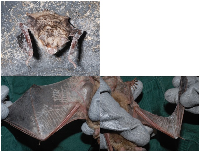
On February 15, 2012, the French M. myotis was detected in its hibernacula in poor body condition. As the left wing membrane was extensively torn, it can be assumed its pre-hibernation hunting success must have been markedly impaired, resulting in an insufficient fat storage for hibernation. Of all bats sampled this animal had the most visible evidence of fungal growth (Fig. 1). It was taken into a rehabilitation center to be sustained throughout the winter. Immediately upon arriving at the center a swab sample and a wing punch biopsy (Fig. 1A) were taken from one of the most affected areas.
Figure 1. Emaciated Myotis myotis from a hibernaculum in France covered by Geomyces destructans.
1A: Old laceration of the left wing identified on the day of collection. 1B: Bat with improved body condition after some weeks of rehabilitation. 1C+D: Wing membranes after cleaning with notable depigmentation.
Molecular-biology Investigations
All adhesive tape or swab samples (no sample from the Belgian bat (BE 1) was collected for genetic analysis), as well as tissue samples of wing membrane, muzzle and ear of the Dutch M. daubentonii were investigated by PCR amplification of the fungal rRNA gene internal transcribed spacer (ITS) region DNA (ITS1, 5.8S, and ITS2). Total nucleic acids were extracted from all samples using PrepMan Ultra reagent (Applied Biosystems, Darmstadt, Germany) following the manufacturer’s instructions. Ribosomal RNA gene ITS region DNA was PCR amplified using primers ITS4 and ITS5 [21] and GoTaq DNA polymerase (Promega, Madison, Wisconsin). Cycling parameters were an initial 2 min denaturation at 98°C followed by 30 cycles of denaturation at 98°C for 10 s, annealing at 50°C for 30 s, and extension at 72°C for 1 min, with a final extension at 72°C for 7 min. All PCR products were further processed for direct sequencing and retrieved sequences were blasted against sequences of G. destructans in GenBank. Samples with 100% sequence similarity to the respective gene segments of the G. destructans type isolate NWHC 20631–21 (GenBank accession no. EU884921) were considered positive for G. destructans and their sequences were submitted to GenBank.
Histopathology Investigations
The skin of the entire muzzle of the euthanized Dutch M. daubentonii (NL 1) was removed and dissected in cross sectional planes vertical to the longitudinal axis of the skull. Numerous long strips of the wing membranes, particularly between the fourth and fifth fingers, were cut and fixed in formalin for histology investigations.
Punch biopsies of the 10 live bats were examined with a dissection microscope to identify the site of fungal growth. Upon detection of fungal colonization, biopsies were cut diagonally to ensure that the maximum length of an affected wing membrane biopsy was sectioned for histological examination. Biopsy halves as well as the skin samples of muzzle, ear and wing membranes of the Dutch M. daubentonii were subsequently processed for histology investigations, embedded in liquid paraffin and serially sectioned at 3 µm. All sample material was completely used for these sequential sections and slides were stained alternating with hematoxylin-eosin and periodic acid Schiff stain.
Banding and Recapture
Hibernacula located in the southeastern Harz Mountains, Germany, are annually monitored and census data, i.e. number of bats present, bat species, gender and banding numbers as well as unusual observations are recorded. Unmarked bats receive a new banding ring. All capture-recapture data were archived in field books and also submitted to the Dresden Bat Banding Center, Germany, to be included in a general banding database. Field books from 2000 onwards were searched for bat capture-recapture entries in connection with noted fungus colonization on live bats. Additionally, during the annual hibernacula census in Lengefeld, Saxony, Germany, 2 banded M. myotis (A45309, male; A45392, female) were noticed with fungal growth on the 5th February 2011. Adhesive tape samples and photographs were taken. Three weeks later, on the 26th February 2011, the same animals were re-observed and new photographs taken (E.& R. Francke).
Results
All sampled bats, except FR 1, had the appearance of a healthy animal with round body shape, fluffy fur coat and moderate amounts of palpable subcutaneous brown fat tissue between their shoulders and were therefore considered to be of good to moderate body condition. Only FR 1 was of poor appearance with a very thin body outline and no palpable brown fat tissue and it was therefore classified emaciated (Fig. 1).
Molecular Investigations and Dissection Microscopy
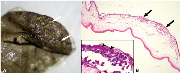
Samples from a total of 11 bats from 4 countries were investigated (see Table 1). All samples, except the sample from Belgium (which was not analyzed genetically), had an ITS sequence 100% identical to Gd type isolate (GenBank accession no. EU884921). The hibernating M. myotis from Hungary was heavily colonized by fungus on its snout, ears, forearms, wing membranes, and uropatagium. The latter was biopsied at the site of superficial fine strands of white fungal growth. Upon arrival to the lab, the ethanol fixed uropatagium sample (HU 1) still had the fungal strands attached, but unfortunately sloughed off when the biopsy was removed from the vial a second time for photography. The fungal strands were associated with minute, well demarcated, circular, light grey lesions visible by dissection microscopy (Fig. 2A). In contrast to the Hungarian biopsy the wing punches from the 7 M. myotis from Germany (GE 1–7) did not reveal distinct lesions by low power dissection microscopy examination. Similarly, the dissection microscopy examination of the M. daubentonii carcass from the Netherlands did not reveal any evidence of fungal growth. The Belgian wing punch biopsy (BE 1) was not examined by dissection microscopy.
Figure 2. Biopsy of uropatagial membrane of Myotis myotis with acute inflammatory response against Geomyces destructans.
2A (sample HU 1): Dissection microscope image of punch biopsy with distinct circular lesions (arrows), where G. destructans had sloughed off during preparation. 2B: Histological cross section of 2A with multiple well-demarcated intraepidermal microabscesses (arrows). Hematoxylin-eosin staining. Inset: Hyphae of G. destructans invading the microabscesses (arrow heads). PAS staining.
Histopathologic Examination
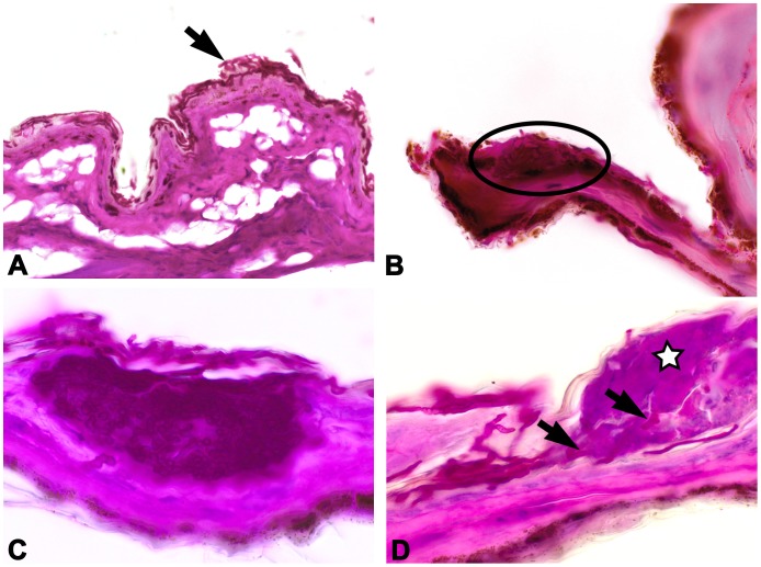
All wing punch biopsies from the Belgian (BE 1) and 7 German bats (GE 1–7) contained fungal hyphae superficially located in the stratum corneum of the epidermis (Fig. 3A). In one biopsy (GE 1), a small focus of densely interwoven fungal hyphae, similar to the described intradermal cup-shaped colonies in North American bats [6], was located within the epidermal layers, but without dermal invasion (Fig. 3B). Another biopsy (GE 6) contained a hair follicle with intraluminal colonization by fungal hyphae growing in perpendicular orientation to the hair shaft and extending into the neighboring sebaceous gland. None of these fungal hyphae were associated with cellular inflammatory response. In contrast, the Hungarian biopsy of the uropatagium (HU 1) contained multiple intraepidermal microabscesses corresponding to the light grey circular discolorations visible via dissection microscopy. Microabscesses consisted of a mixed inflammatory cell population dominated by neutrophils (Fig. 2B). The covering epidermis was intact but colonized by fungal hyphae, which reached further into the underlying abscessations (Fig. 2B, inset). The adjacent dermis was moderately edematous with mild infiltration by neutrophils and plasma cells.
Figure 3. Geomyces destructans colonization of superficial epidermis occasionally associated with epidermal pustules.
3A: A representative wing biopsy punch (GE 7). 3B (wing punch GE 1): Small epidermal cluster of G. destructans hyphae (encircled). 3C (sample NL 1): Wing membrane of euthanized bat with cupping erosion and densely packed hyphae of G. destructans restricted to the epidermis. 3D (sample NL 1): Fungal hyphae (arrows) invasion into intraepidermal pustules (star). PAS staining.
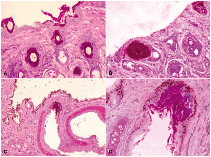
Examination of the euthanized Dutch M. daubentonii (NL 1) revealed numerous hair follicles of the skin of the snout densely filled with fungal hyphae. Occasionally the entire follicular epidermal isthmus was replaced by hyphae, but there was no inflammatory reaction (Fig. 4A+B). Similar to the wing biopsy punches described above, there was multifocal superficial colonization of fungal hyphae in the stratum corneum. Hyphae also extended above the haired skin bearing asymmetrical curved conidia characteristic for G. destructans [5] and corresponding with the macroscopically visible fungal growth. One of the nares had intraluminal fungal colonization of its dorsal cutaneous epithelium similar to the cupping erosions of the wing membranes. Here, fungal hyphae reached the bordering basement membrane and invoked a localized suppurative inflammation limited to the epithelial layer. There was no evidence for cellular response in the underlying connective tissue (Fig. 4C+D). Investigation of the euthanized M. daubentonii wing membranes showed abundant superficial colonization with fungal hyphae limited to the stratum corneum, but extending above the surface to create a dense network of aerial mycelium. There was one focal cluster of cup-shaped intradermal fungal hyphae in the wing membrane of this bat (Fig. 3C). Occasionally, remnants of asymmetrical curved conidia were detected amongst these fungal hyphae. To a lesser extent, multiple intracorneal and intraepidermal pustules were noted, which were associated with invading fungal hyphae (Fig. 3D). Interestingly, despite the marked neutrophilic accumulation within the microabscesses no further inflammatory cells were found in the adjacent dermis, even if abscesses and hyphae bulged into the deeper tissue. All internal organs of this bat were unremarkable and without evidence of disease.
Figure 4. Skin of the muzzle of a Geomyces destructans infected Myotis daubentonii (sample NL 1).
4A: Multiple hair follicles with intraluminal colonization of G. destructans hyphae. 4B: Wall of a hair follicle replaced by marked growth of G. destructans hyphae. 4C+D: Cutaneous epithelium of one nare severely colonized by G. destructans with mild localized suppurative inflammation limited to the epithelial layer. PAS staining.
Similar findings to the other biopsies were encountered in the uropatagial biopsy of the French bat (FR 1), which had marked superficial fungal growths with focally numerous hyphae intermingled with each other. Additionally, formation of superficial suppurative pustules was detected within the outer layer of the epidermal corneal stratum containing neutrophilic granulocytes and being covered by fungal hyphae. There was neither indication of deep tissue invasion by the fungus nor ulceration. The macroscopic images of the French bat (Fig. 1) show the extent of fungal colonization and depigmentation of the wing. The delineated crisscross pattern of the elastic wing membranes could suggest that these edges, as the most superficially located areas when the wings are folded up, are sites most easily covered by fungal spores.
Recapture Data
Archived bat banding and census data books from 2000 to 2011 contained notes on 8 M. myotis (4 males, 4 females) banded and recaptured in subsequent years and displaying fungal growth, particularly around their muzzle, at different times of detection (Table 2). The longest capture-recapture entry of a single individual covers 11 years. Four bats were found twice in subsequent years with fungal growth. A further 4 animals were found once with fungus; one of these was recaptured 3 winters later without fungus, another one was found in a maternity colony 3 months later. During this period of 11 years, the bats remained philopatric to their chosen hibernaculum, except one individual.
Table 2. Retrospective data on capture/recapture of banded Myotis myotis with visible fungal colonization.
| Banding-ID | Sex | Capture/recapture date | Hibernaculum (location, gallery) | Fungus |
| A 17581 | f | 21.02.2000 | Rübeland, “Trinkwasserstollen” | no |
| 08.03.2005 | same place | yes | ||
| 31.08.2008 | same place | no | ||
| A 17586 | f | 21.02.2000 | Rübeland, “Trinkwasserstollen” | no |
| 08.03.2005 | same place | yes | ||
| A 21750 | m | 24.02.2000 | Tresburg, “Luppbodestollen” | no |
| 06.02.2001 | same place | no | ||
| 31.03.2010 | same place | yes | ||
| 30.03.2011 | same place | yes | ||
| A 22024 | f | 17.02.2000 | Elbingerode, “Augustenstollen” | yes |
| 18.01.2001 | same place | no | ||
| 06.03.2002 | Elbingerode, “Pinge Charlotte” | no | ||
| 18.01.2006 | same place | yes | ||
| A 22209 | m | 09.03.2000 | Elbingerode, “Büchenbergstollen” | no |
| 12.03.2001 | same place | yes | ||
| 28.12.2001 | same place | no | ||
| 02.03.2002 | same place | yes | ||
| 30.12.2009 | same place | no | ||
| 22.03.2011 | same place | no | ||
| A 22213 | m | 09.03.2000 | Elbingerode, “Büchenbergstollen” | yes |
| 12.03 2001 | same place | yes | ||
| 28.12.2001 | same place | no | ||
| A 88088 | m | 15.01.2009 | Silberhütte, “Fürst-Viktor-Stollen” | no |
| 29.03.2011 | same place | yes | ||
| A 93149 | f | 29.03.2011 | Treseburg, “Falkenklippenstollen” | yes |
| 12.06.2011 | Bleicherode (maternity colony) | no |
Bats were detected during hibernacula monitoring from 2000–2011 in galleries of the southern Harz Mountains, Germany.
In 2011, 2 banded M. myotis with very small patches of fungal growth on nose and ear rims were observed in a hibernaculum in Saxony on the 5th February (Fig. 5A+B). Three weeks later the same animals were found again with their wing membranes, ears and muzzle markedly covered with numerous coalescent fungal patches (Fig. 5C).
Figure 5. Development of Geomyces destructans colonization over 3 weeks’ time on naturally infected bats.
5A+B: Two banded Myotis myotis with mild facial fungal patches on 5th February 2011. 5C: Recapture of these bats on 26th February 2011 with marked fungal colonization over most of the glabrous skin parts and the snout.
Discussion
One of the features of North American bats suffering and dying from WNS is the invasion by fungal hyphae of G. destructans reaching deeply into the subcutaneous tissue, causing ulcerative necrotic areas and the destruction of wing membranes [6]. The mildest microscopic changes seen in the wing membranes are cup-like epidermal erosions filled with fungal hyphae. Ulceration occurs commonly, i.e. the epidermal basal membrane is destroyed by fungal invasion spanning the full thickness of the wing membrane. When the muzzle is involved, fungal hyphae fill hair follicles, invad sebaceous and apocrine glands, and extend into the regional connective tissue obscuring epithelial boundaries of the adnexa. While typically these hibernating bats have an absence of inflammatory reaction in their skin even with extensive fungal invasion, in some animals edema and neutrophilic granulocytes as well as occasional intradermal abscesses can be observed in the regional connective tissue [6]. The severe injuries are thought to provoke increased arousal frequencies in the bats with significantly shortened torpor bouts [3]. Presumably, the ulcerated epidermal surface of the wing membranes loses its barrier function to maintain homeostasis of body fluids, thus accelerated fluid evaporation gives rise to severe dehydration and likely causes the bats to arouse [11]. This increase in arousal frequency and its associated high energy cost prematurely reduces the fat storage of the bats, inducing emaciation before the end of the hibernation period. Dehydration and emaciation then accounts for the death of the affected hibernating bats.
In contrast to North America, European hibernating bats infected with G. destructans are found in many countries but to date no mortalities are known to be associated with the fungus [8]. Similarly, changes in hibernation behavior such as frequent day flights in cold winter have not been observed [13]–[16]. Recently Pikula et al. [22] reported on histological investigations of 2 M. myotis found dead in a hibernaculum in the Czech Republic. The examination of these animals showed fungal colonization consistent with G. destructans. While most fungal hyphae on these 2 animals were found in the superficial skin, some hyphae were also invading the connective tissue of the dermis. Unfortunately, as the bats were found some time after their death and their organs were rendered by autolysis it was not possible to conclude the cause of death.
In the present study, wing punch biopsies of 11 hibernating live bats with macroscopic visible fungal growth on their wing membranes were collected and immediately fixed for microscopic examination. Additional skin samples were taken from one euthanized bat and were also examined by microscopy. Histologically, in this animal fungal hyphae were clearly visible in the epidermis of muzzle and wing membranes, but they were mostly restricted to the very superficial layer, i.e. stratum corneum, or the lumen of hair follicles often encasing the hair shafts. Using the histopathologic scoring system implemented by Reeder et al. [7] this bat would receive severity score 1. However, 2 other animals we investigated revealed multifocal localized intraepidermal neutrophilic infiltration associated with intralesional fungal hyphae. In their investigations on bats recovered from WNS, Meteyer et al. [6], [23] describe the presence of similar inflammatory reactions in the regional connective tissue of the skin of North American bats. In European bats such inflammatory reactions seem limited to the epidermis and do not extend beyond the epidermal basement membrane. Similarly, in the examined biopsies fungal hyphae invasion appeared restricted to the epidermis and adnexae without deep invasion into the underlying connective tissue, in contrast to the invasion of dermal connective tissue as is well documented in North American bats. Histological images of a euthanized bat from the Czech Republic with visible fungal colonization show epidermal changes similar to our findings [22]. Interestingly, even a severely compromised animal like the French bat (FR 1) described above, seemingly a prime candidate for an overwhelming fungal invasion, evidenced no ulceration of the wing membranes by connective tissue invasion of the fungus. However, although we tried to sample one of the most affected areas the limited size of the biopsy prevents conclusive decisions about the entire wing surface.
One of the morphologic structures for assessing skin diseases is the basal membrane, a dense laminar formation of collagen which separates the cell layers of the outer epidermis and its adnexae from the underlying connective tissue of the dermis. The basal membrane is regarded as one of the most important defensive barrier against injuries. Once this structure is breached, e.g. by an invading infectious agent, severe damage can result as the physiologic function of the skin, like body fluid homeostasis, is at risk. Many studies have shown that hibernating bats in North America can suffer from extensive necrosis of their wing membranes [2], [3], [6] and resulting scarring can be observed in animals surviving the infection [23], [24]. However, it was also shown that bats were able to recover and heal these lesions [23], [25]. Although the number of analyzed European samples has been very limited compared to North America, extensive skin damages with destruction of the basal membrane has not been observed in these European samples (this study, [22]), supporting the hypothesis that infection by G. destructans does not have the same serious consequences for bats in Europe compared to North America.
The reasons for the differences between North American and European bats are not clear, but evidence supports the hypothesis that bats in Europe might have co-evolved with the fungus while bats in North America have only been recently exposed to it [3], [8], [13], [14]. Parallel experimental infections conducted on North American M. lucifigus with G. destructans isolates from North America and Europe resulted in similar lesions, strengthening the hypothesis that G. destructans was introduced from Europe into North America [3]. Histological examination of the wing membrane biopsies of European bats with G. destructans showed that fungal hyphae remain mainly in the superficial corneal stratum. In cases where the hyphae invaded deeper layers of the epidermis, they were embraced by pustular neutrophilic infiltration. One biopsy (HU 1) seemed to represent a very early stage of such neutrophilic epidermal infiltration as well-demarcated microabscesses occurred within the epidermal layers. It could be speculated that over time, European bats might have developed an unspecific epidermal immune response, which restricts G. destructans colonization to the epidermis.
Regardless of these findings, multiple epidermal cup-like clusters of fungal hyphae, identical to cupping erosions described in North American bats [6], could particularly be found in the skin of the snout, where multiple hair follicles were largely expanded by densely interwoven hyphae. Such follicular hideouts could serve as a preservative source for repeated fungal growth over multiple hibernation seasons. However, Meteyer et al. [23] investigated G. destructans infected bats 70 days after recovery and found no evidence of remaining fungal structures on wing membranes or snout. Experiments under laboratory conditions showed that G. destructans samples stored on dry cotton swabs for 8 months at 24°C were not able to germinate [26], suggesting that G. destructans might not be able to survive on bats that are active (with temperatures >24°C) through the summer season (6–8 month).
Nothing has yet been reported about the growth rate of G. destructans on hibernating European bats under natural conditions. But the observation that within 3 weeks the wings and snouts of 2 banded bats from Saxony were covered with white fungus hints towards a fast growth rate under ideal conditions. Regardless of whether characteristic facial fungal patches are newly acquired or re-emerging in each hibernation season, recaptures of banded M. myotis with repeated detection of characteristic facial fungal patches provide evidence that European bats can survive repeated G. destructans infections for several years. Despite the variations in the observation dates, archival field data of annual hibernacula monitoring from southern Harz Mountains, Germany, are in accordance with other publications showing that in most years the fungus was not visible to the naked eye before mid February [13]–[15].
Differing lengths of winter and hibernation periods between North America and Europe are often discussed as a possible reason for the differing impact of G. destructans on the respective bat populations. In North America, WNS associated aberrant hibernation behavior as well as WNS lesions are already detectable after January [27] and recent experimental G. destructans infections in M. lucifigus confirm that WNS with deep invading fungal hyphae can be reproduced after 3 months of hibernation [3]. In our study, all bats investigated hibernated for at least 90–120 days without developing deep fungal tissue invasion. The microscopic features of superficial G. destructans infection in the skin of European bats could point towards a less severe fungal growth as compared to North American bats.
While it has been experimentally shown that bat to bat contact results in the transmission of G. destructans between the animals [2], it is unknown how common this transmission pathway is under natural conditions. In spring, immediately after their arousal, bats start to rigorously groom themselves and remove all visible fungi from their body, while some bats achieve this already in between torpor bouts during hibernation [13]. In contrast to the temperature of euthermic bats, most cavernous hibernacula have a rather constant low temperature and remaining infectious spores of G. destructans only need contact to a suitable substrate to start to grow. In autumn, during the swarming season when large numbers of bats aggregate to mate, they begin to enter the hibernacula and it seems likely that conidia can be swept up from cave walls by their bodies and subsequently exchanged between animals during mating [13]. The detection of G. destructans in caves has been shown for North America and Europe likewise [13], [28]. An alternative route of infection might take place during hibernation, when aerial hyphae on wing membranes and snouts produce innumerable conidia. Many bat species, like M. myotis, will occasionally move between torpor bouts and choose a different hanging place, often in close proximity to conspecifics, easily transmitting fungal conidia.
Unless European bats have evolved specific behaviors to limit Gd transmission between individuals, transmission pathways are likely to be the same in North American and European bats; however, the impact of G. destructans infections on bats seems to differ drastically between the continents.
Conclusions
Numerous hibernating bats from North America succumb to infections with G. destructans. In contrast, in European bats even severely covered by fungal mycelia, fungal hyphae seem to be restricted to the epidermis with occasional formation of neutrophilic pustules. Invasion of the dermis and ulceration could not be found despite including several heavily infected animals. However, with the limited size of wing punch biopsies, areas of deeper invasion might have been missed. The recapture of banded bats over several years with repeated detection of fungal growth characteristic for G. destructans further supports the hypothesis that G. destructans has a minor impact on hibernating bats in Europe.
Acknowledgments
The authors would like to thank to Imbre Dombi, Elke Francke, Reimund Francke, Markus Fritze, Peter Lina, Julia Prüger, Chris Rupsch, Wigbert Schorcht, Christine Teumer for their tireless help in obtaining samples for this project, as well as Nadine Jahn, Claudia Kohl, Doris Krumnow and Dagmar Viertel for their excellent technical assistance. We are grateful to Laurent Arthur and the Muséum d’Histoire naturelle de la ville de Bourges, France, Peter Lina and the Netherlands Centre for Biodiversity, The Netherlands, and Thierry Kervyn and the Service Public de Wallonie, Direction Générale Agriculture, Ressources Naturelles et Environnement, Belgium, for their support in gaining sampling permissions. We wish to thank the anonymous reviewers for their helpful and thoughtful comments, and we are thankful to Nancy Allison for copyediting the manuscript.
Funding Statement
The authors have no support or funding to report.
References
- 1.U.S. Fish and Wildlife Service (2012) Information on White-nose in bats. http://www.fws.gov/whitenosesyndrome/about.html.
- 2. Lorch JM, Meteyer CU, Behr MJ, Boyles JG, Cryan PM, et al. (2011) Experimental infection of bats with Geomyces destructans causes white-nose syndrome. Nature 480: 376–378. [DOI] [PubMed] [Google Scholar]
- 3. Warnecke L, Turner JM, Bollinger TK, Lorch JM, Misra V, et al. (2012) Inoculation of bats with European Geomyces destructans supports the novel pathogen hypothesis for the origin of white-nose syndrome. Proc Natl Acad Sci U S A 109: 6999–7003. [DOI] [PMC free article] [PubMed] [Google Scholar]
- 4. Blehert DS, Hicks AC, Behr M, Meteyer CU, Berlowski-Zier BM, et al. (2009) Bat white-nose syndrome: an emerging fungal pathogen? Science 323: 227. [DOI] [PubMed] [Google Scholar]
- 5. Gargas A, Trest MT, Christiensen M, Volk TJ, Blehert DS (2009) Geomyces destructans sp. nov. associated with bat white-nose syndrome. Mycotaxon 108: 147–154. [Google Scholar]
- 6. Meteyer CU, Buckles EL, Blehert DS, Hicks AC, Green DE, et al. (2009) Histopathologic criteria to confirm white-nose syndrome in bats. J Vet Diagn Invest 21: 411–414. [DOI] [PubMed] [Google Scholar]
- 7. Reeder DM, Frank CL, Turner GG, Meteyer CU, Kurta A, et al. (2012) Frequent arousal from hibernation linked to severity of infection and mortality in bats with white-nose syndrome. PLoS ONE 7: e38920. [DOI] [PMC free article] [PubMed] [Google Scholar]
- 8. Puechmaille SJ, Frick W, Kunz TH, Racey PA, Voigt CC, et al. (2011) White-Nose Syndrome: is this emerging disease a threat to European bats? Trends Ecol Evol 26: 570–576. [DOI] [PubMed] [Google Scholar]
- 9. Frick WF, Pollock JF, Hicks AC, Langwig KE, Reynolds DS, et al. (2010) An emerging disease causes regional population collapse of a common North American bat species. Science 329: 679–682. [DOI] [PubMed] [Google Scholar]
- 10. Boyles JG, Cryan PM, McCracken GF, Kunz TH (2011) Economic importance of bats in agriculture. Science 332: 41–42. [DOI] [PubMed] [Google Scholar]
- 11. Cryan PM, Meteyer CU, Boyles JG, Blehert DS (2010) Wing pathology of white-nose syndrome in bats suggests life-threatening disruption of physiology. BMC Biol 8: 135. [DOI] [PMC free article] [PubMed] [Google Scholar]
- 12. Bouma HR, Carey HV, Kroese FGM (2010) Hibernation: the immune system at rest? J Leukocyte Biol 88: 1–6. [DOI] [PubMed] [Google Scholar]
- 13. Puechmaille SJ, Wibbelt G, Korn V, Fuller H, Forget F, et al. (2011) Pan-European distribution of white-nose syndrome fungus (Geomyces destructans) not associated with mass mortality. PLoS ONE 6: e19167. [DOI] [PMC free article] [PubMed] [Google Scholar]
- 14. Wibbelt G, Kurth A, Hellmann D, Weishaar M, Barlow A, et al. (2010) White-nose syndrome fungus (Geomyces destructans) in bats, Europe. Emerg Infect Dis 16: 1237–1242. [DOI] [PMC free article] [PubMed] [Google Scholar]
- 15. Martínková N, Bačkor P, Bartonička T, Blažková P, Červený J, et al. (2010) Increasing incidence of Geomyces destructans fungus in bats from the Czech Republic and Slovakia. PLoS ONE 5: e13853. [DOI] [PMC free article] [PubMed] [Google Scholar]
- 16. Puechmaille SJ, Verdeyroux P, Fuller H, Ar Gouilh M, Bekaert M, et al. (2010) White-nose syndrome fungus (Geomyces destructans) in bat, France. Emerg Infect Dis 16: 290–293. [DOI] [PMC free article] [PubMed] [Google Scholar]
- 17. Gannon WL, Sikes RS (2007) Guidelines of the American Society of Mammalogists for the Use of Wild Mammals in Research. J Mammal 88: 809–823. [DOI] [PMC free article] [PubMed] [Google Scholar]
- 18.Simmons NB, Voss RS (2009) Collection, preparation and fixation of bat specimens and tissues. In: Kunz TH, Parsons S, eds. Ecological and Behavioural Methods for the Study of Bats, Second Edition. Johns Hopkins University Press. 863–867.
- 19. Faure PA, Re DE, Clare BL (2009) Wound healing in the flight membranes of big brown bats. J Mammal 90: 1148–1156. [Google Scholar]
- 20.AVMA Guidelines on Euthanasia (2007). Available: http://www.avma.org/issues/animal_welfare/euthanasia.pdf.
- 21.White TJ, Bruns T, Lee S, Taylor JW (1990) Amplification and direct sequencing of fungal ribosomal RNA genes for phylogenetics. In: Innis MA, Gelfand DH, Sninsky JJ, White TJ, eds. PCR protocols: a guide to methods and applications. Academic Press, Inc., New York. 315–322.
- 22. Pikula J, Bandouchova H, Novotný L, Meteyer CU, Zukal J, et al. (2012) Histopathology confirms white-nose syndrome in bats in Europe. J Wildl Dis 48: 207–211. [DOI] [PubMed] [Google Scholar]
- 23. Meteyer CU, Valent M, Kashmer J, Buckles EL, Lorch JM, et al. (2011) Recovery of little brown bats (Myotis lucifugus) from natural infection with Geomyces destructans, white-nose syndrome. J Wildl Dis 47: 618–626. [DOI] [PubMed] [Google Scholar]
- 24. Reichard JD, Kunz TH (2009) White-nose syndrome inflicts lasting injuries to the wings of little brown myotis (Myotis lucifugus). Acta Chiropt 11: 457–464. [Google Scholar]
- 25. Fuller NW, Reichard JD, Nabhan ML, Fellows SR, Pepin LC, et al. (2011) Free-ranging little brown Myotis (Myotis lucifugus) heal from wing damage associated with white-nose syndrome. Ecohealth 8: 154–162. [DOI] [PubMed] [Google Scholar]
- 26. Puechmaille SJ, Fuller H, Teeling EC (2011) Effect of sample preservation methods on the viability of Geomyces destructans, the fungus associated with white-nose syndrome in bats. Acta Chiropt 13: 217–221. [Google Scholar]
- 27. Foley J, Clifford D, Castle K, Cryan PM, Ostfeld RS (2011) Investigating and managing the rapid emergence of white-nose syndrome, a novel, fatal, infectious disease of hibernating bats. Conserv Biol 25: 223–231. [DOI] [PubMed] [Google Scholar]
- 28. Lindner DL, Gargas A, Lorch JM, Banik MT, Glaser J, et al. (2010) DNA-based detection of the fungal pathogen Geomyces destructans in soils from bat hibernacula. Mycologia 103: 241–245. [DOI] [PubMed] [Google Scholar]


