

Abstract
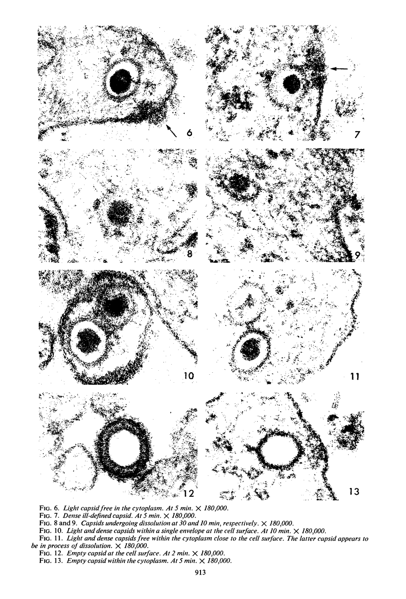
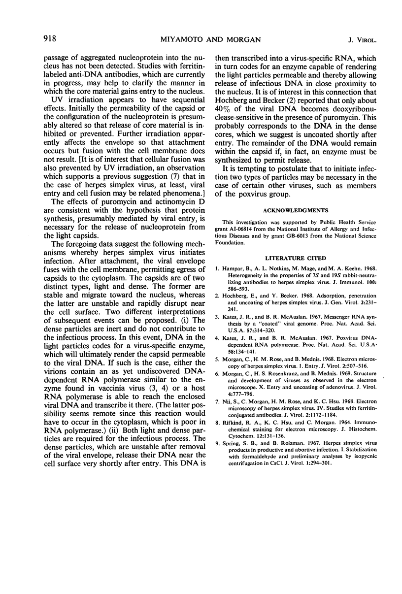
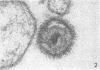
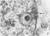
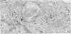
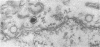
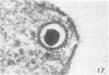
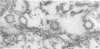
Two morphologically distinct types of capsids are described. The dense capsid appeared to be disrupted near the cellular membrane with release of core material. The light capsid was more stable and was frequently encountered close to the nucleus, where empty capsids were also found. Pretreatment of cells before infection with either puromycin or actinomycin D markedly decreased the percentage of empty capsids. It is suggested that the two types of capsids play different roles in the process of initiating infection. One (the dense capsid) releases deoxyribonucleic acid (DNA) shortly after entry. This DNA is transcribed into a virus-specific ribonucleic acid, which codes for an enzyme capable of altering the permeability of the second type of capsid (the light capsid). In proximity to the nucleus, the infectious DNA then escapes without gross disruption of the capsid.
Full text
PDF


Images in this article


Selected References
These references are in PubMed. This may not be the complete list of references from this article.
- Hampar B., Notkins A. L., Mage M., Keehn M. A. Heterogeneity in the properties of 7 S and 19S rabbit-neutralizing antibodies to herpes simplex virus. J Immunol. 1968 Mar;100(3):586–593. [PubMed] [Google Scholar]
- Hochberg E., Becker Y. Adsorption, penetration and uncoating of herpes simplex virus. J Gen Virol. 1968 Mar;2(2):231–241. doi: 10.1099/0022-1317-2-2-231. [DOI] [PubMed] [Google Scholar]
- Kates J. R., McAuslan B. R. Messenger RNA synthesis by a "coated" viral genome. Proc Natl Acad Sci U S A. 1967 Feb;57(2):314–320. doi: 10.1073/pnas.57.2.314. [DOI] [PMC free article] [PubMed] [Google Scholar]
- Kates J. R., McAuslan B. R. Poxvirus DNA-dependent RNA polymerase. Proc Natl Acad Sci U S A. 1967 Jul;58(1):134–141. doi: 10.1073/pnas.58.1.134. [DOI] [PMC free article] [PubMed] [Google Scholar]
- Morgan C., Rose H. M., Mednis B. Electron microscopy of herpes simplex virus. I. Entry. J Virol. 1968 May;2(5):507–516. doi: 10.1128/jvi.2.5.507-516.1968. [DOI] [PMC free article] [PubMed] [Google Scholar]
- Morgan C., Rosenkranz H. S., Mednis B. Structure and development of viruses as observed in the electron microscope. V. Entry and uncoating of adenovirus. J Virol. 1969 Nov;4(5):777–796. doi: 10.1128/jvi.4.5.777-796.1969. [DOI] [PMC free article] [PubMed] [Google Scholar]
- Nii S., Morgan C., Rose H. M., Hsu K. C. Electron microscopy of herpes simplex virus. IV. Studies with ferritin-conjugated antibodies. J Virol. 1968 Oct;2(10):1172–1184. doi: 10.1128/jvi.2.10.1172-1184.1968. [DOI] [PMC free article] [PubMed] [Google Scholar]
- RIFKIND R. A., HSU K. C., MORGAN C. IMMUNOCHEMICAL STAINING FOR ELECTRON MICROSCOPY. J Histochem Cytochem. 1964 Feb;12:131–136. doi: 10.1177/12.2.131. [DOI] [PubMed] [Google Scholar]
- Spring S. B., Roizman B. Herpes simplex virus products in productive and abortive infection. I. Stabilization with formaldehyde and preliminary analyses by isopycnic centrifugation in CsCl. J Virol. 1967 Apr;1(2):294–301. doi: 10.1128/jvi.1.2.294-301.1967. [DOI] [PMC free article] [PubMed] [Google Scholar]