Abstract
A total of 520 overwintered sclerotia were collected from surface of soil under mulberry trees in six locations in Korea during February in 2006 and 2007. The collected sclerotia were tested for their germination in vitro and identified based on their morphological characteristics. Out of all sclerotia tested, 52.3% of the sclerotia germinated and produced two types of apothecia. The two types of fungi occurred from the sclerotia at the ratio of 49.8 vs. 50.2. The fungal type with cup-shaped apothecia was identified as Ciboria shiraiana and another type of fungus with club-shaped apothecia as Scleromitrula shiraiana. Taxonomy and distribution of the two sclerotial fungi were described and discussed.
Keywords: Ciboria shiraiana, Fruit, Mulberry tree, Scleromitrula shiraiana
Mulberry trees have been grown worldwide as a crop to rear silkworm for a long time. Recently, several parts of the tree are being used as uncooked or processed foods for health care. Especially, mulberry fruits were proved to include several substances with medical action. Due to increase in demand for the fruits, growing area of the tree has been remarkably increasing. However, mummified white fruits of the tree severely occurred in the fields and much damaged production of normal fruits. The disease in other countries has been known as popcorn disease, swollen fruit disease or shrunken fruit disease (Kishi, 1998; Kohn and Nagasawa, 1984; Whetzel and Wolf, 1945). Ciboria carunculoides (Siegler and Jenkins) Whetzel, Ciboria shiraiana (Henn.) Whetzel and Scleromitrula shiraiana (Henn.) Imai have been reported separately as causal agents of the fruit diseases (Kishi, 1998; Kohn and Nagasawa, 1984; Whetzel and Wolf, 1945). In Korea, only Sclerotinia shiraiana Hennings was recorded as a causal agent of the disease (Cho and Shin, 2004), but there is no description of the fungus in detail. The present study was conducted to identify the causal agents of sclerotial disease on mulberry fruits and to investigate their distribution in Korea.
Materials and Methods
Overwintered sclerotia were collected from surface of soil under mulberry trees in six locations in Korea during February in 2006 and 2007. The collected sclerotia were washed with distilled sterile water and dried at room temperature. The sclerotia were placed in 250 ml-flasks with sterile wet sand and incubated at 15℃ for 10~30 days in alternating cycles of 12-hr fluorescent light and 12-hr darkness to induce apothecial formation. Germination rates of the sclerotia tested and apothecial types from the germinated sclerotia were examined during the incubation. Apothecia were identified based on their morphological and cultural features. The distribution of apothecial types for each location was also compared. To culture ascospores released from apothecia, they were suspended with distilled sterile water, streaked on water agar (WA) and then incubated at 25℃ for 18-hr. The germinating spores were isolated and transferred to WA and then incubated for 4~7 days. The fungi grown from the single spores were cultured on potato dextrose agar (PDA).
Results and Discussion
Description of Species
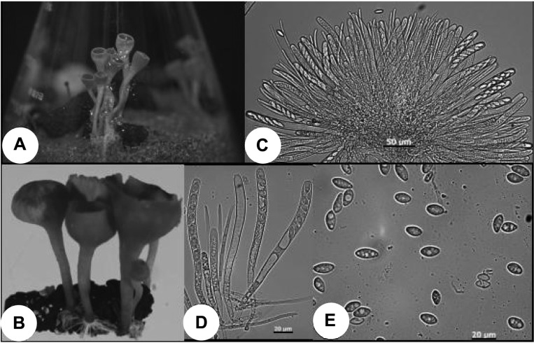
Ciboria shiraiana (Henn.) Whetzel, Mycologia 37: 489. 1945. (Fig. 1).
Fig. 1.
Morphological features of Ciboria shiraiana. A and B, cup-shaped apotheica induced from overwintered sclerotia in vitro; C~E; asci, ascospores and paraphyses.
Sclerotia are black, hard and variable in size but usually large like mummified fruits. Apothecia are induced one to several from a sclerotium. The disks are snuff brown, cup-shaped and measured 8~23 mm in diameter and 0.3~1.6 mm in thickness. Stipes are pale brown, cylindrical, straight or flexuous and measured 8~36 mm in length and 0.5~2.5 mm in width. Asci are cylindrical or clavate and measured 200~155 × 12.5~7.5 µm. Ascospores are uniseriate, ovoid or ellipsoid, hyaline, one-celled and measured 16.0~11.3 × 7.5~6.0 µm. Ascospores germinate on WA and PDA but do not grow after producing short germ tubes.
The causal agent of swollen fruit disease of mulberry tree in Asia including China and Japan was recorded as C. shiraiana (Ling, 1948; Kishi, 1998), and that of popcorn disease of mulberry tree in southeastern United States was known as C. carunculoides (Siegler and Jenkins) Whetzel. Whetzel and Wolf (1945) reported that the two species were strikingly similar in external characteristics of apothecia but distinct in shape and size of asci and ascospores. In this study, we tried to culture C. shiraiana several times using several kinds of media but the fungus did not grow on artificial media such as PDA and malt extract agar. Whetzel and Wolf (1945) reported that C. carunculoides was easily cultured on PDA except that the fungus did not grow rapidly. Sclerotinia shiraiana was first described by Hennings (1900) but transferred to C. shiraiana by Whetzel (Whetzel and Wolf, 1945).
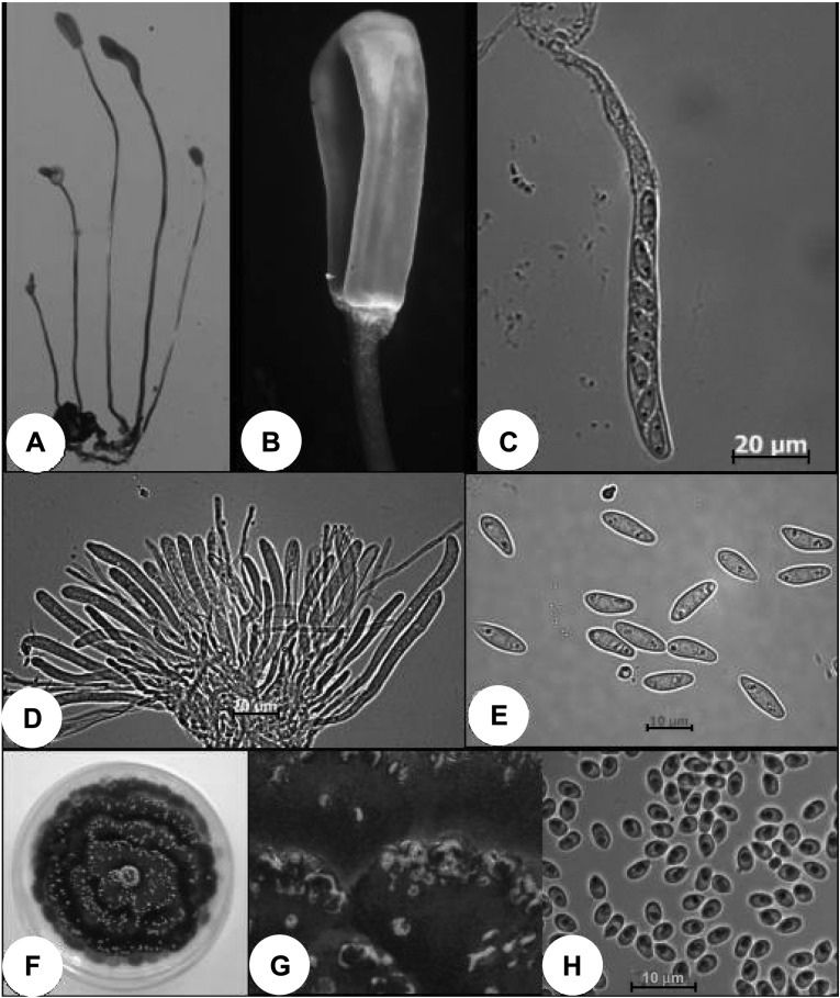
Scleromitrula shiraiana (Henn.) Imai, J. Fac. Hokkaido Univ. 45: 177. 1941. (Fig. 2).
Fig. 2.
Morphological and cultural features of Scleromitrula shiraiana. A and B, club-shaped apothecia induced from overwintered sclerotia; C~E, asci, ascospores and paraphyses; F, colony on PDA; G, pustules containing milk colored microconidial mass on stromata; H, microconidia.
Sclerotia are black, hard and variable but usually small like drupelets in size. Apothecia are produced one to several from a sclerotium. The heads are ochraceous to umber, ovoid to ellipsoid-fusiform with surface marked by 3~6 longtudinal ridges and measured (2~) 4.2~17.3 mm in length and 2.1~4.1(~8.7) mm in width. Stipes are brown, straight, slender, cylindrical and 27~108 mm long and 0.4~0.8 mm wide. Asci are cylindrical or clavate and measured 80.0~73.0 × 7.5~6.3 µm. Ascospores are uniseriate, hyaline, one-celled, ovoid-reniform and measured 12.5~9.0 × 5.3~3.0 µm. Paraphyses are filiform, slightly apically inflated, 3~4 septate. Ascospores are germinated well on water agar. Colony on PDA is olive grey on surface and dark olive brown on reverse and aerial mycelium is very rare. As the culture is older, pustules containing microconidial mass are produced in concentric zone on PDA. Microconidial mass released from them are milk colored, water soluble and mucilaginous. Microconidia are ellipsoid to obovoid, one celled and measured 4.8~3.2 × 2.7~2.1 µm (av. 3.7 × 2.4 µm).
S. shiraiana was first identified from sclerotia of mulberry fruits in Korea. In China and Japan, the species has been known to cause shrunken fruit disease of mulberry tree (Kishi, 1998; Ling, 1948).
Distribution of C. shiraiana and S. shiraiana in Korea
A total of 520 overwintered sclerotia were collected from six locations. Out of them, 52% of sclerotia in average were germinated in vitro and two types of apothecia were produced in the ratio of about five to five from the sclerotia germinated (Table 1). The fungus with cup-shaped apothecia was identified as C. shiraiana, and that with club-shaped apothecia as S. shiraiana. The two species were distributed in all locations investigated. Occurrence frequency of C. shiraiana in Icheon, Wonju and Jeonju was higher than that of S. shiraiana, but lower in Suwon, Yangpyung and Jangsung. The occurrence frequency of C. shiraiana was higher than that of S. shiraiana in locations where large sclerotia were mainly collected, while lower in locations where small sclerotia were mainly collected.
Table 1.
Formation of apothecia from overwintered sclerotia collected from six locations in Korea

In Japan, swollen fruit disease caused by C. shiraiana was known as the most widespread fruit diseases of mulberry tree, while shrunken fruit disease caused by S. shiraiana as less frequently occurred disease in the same localities (Kohn and Nagasawa, 1984).
References
- 1.Cho WD, Shin HD. List of plant diseases in Korea. Fourth edition. Suwon, Korea: Korean Society of Plant Pathology; 2004. p. 779. [Google Scholar]
- 2.Imai S. Geoglossaceae Japoniae. J Fac Hokkaido Univ. 1941;45:1–264. [Google Scholar]
- 3.Kishi K. Plant disease in Japan. Tokyo, Japan: Zenkaku Noson Kyoiku Kyokai Co., Ltd.; 1998. p. 943. [Google Scholar]
- 4.Kohn LM, Nagasawa E. The genus Scleromitrula (Sclerotiniaceae), Episclerotium gen. nov. (Leotiaceae) and allied stipitate-capitate species with reduced ectal excipula. Trans Mycol Soc Japan. 1984;25:127–148. [Google Scholar]
- 5.Ling L. Host index of parasitic fungi of Szechwan, China. Pl Dis Rep Suppl. 1948;173:23–38. [Google Scholar]
- 6.Whetzel HH, Wolf FA. The cup fungus, Ciboria carunculoides, pathogenic on mulberry fruits. Mycologia. 1945;37:476–491. [Google Scholar]


