Abstract
Context:
The neurobiological effect of yoga on the cortical structures in the elderly is as yet unknown.
Materials and Methods:
Seven healthy elderly subjects received yoga intervention as an add-on life-style practice. Magnetic resonance imaging scans were obtained before and 6 months later. Voxel-based-morphometric analyses compared the brains before and after the yoga.
Results:
Yoga group was found to have increases in hippocampal, but not in occipital gray matter.
Conclusion:
Yoga has potential to reduce neuro-senescence. Small sample size and absence of the control group prevent generalization of the findings limiting its translational value.
Keywords: Elderly, hippocampus, magnetic resonance imaging, yoga
INTRODUCTION
Hippocampus is the vulnerable structure for loss of grey matter with aging.[1] Volume reductions in the hippocampus are the earliest indicators of Alzheimer's dementia (AD).[2] Exercise has neuroprotective effects in patients with dementia.[3] Exercise increases regional blood flow and helps enhance neuronal connectivity to hippocampus.[4] Contextually, it is interesting to note that aerobic exercise has been shown to increase hippocampal grey matter volume.[5] Among the life-style practices as alternative to exercise, yoga is practiced in the east and in many western populations. Yoga comprises of various domains of practices such as physical postures, regulated breathing, meditation and several other related techniques. One study found that gray matter volumes were higher in those who were practicing hatha yoga and meditation for a long time as compared with controls not practicing yoga.[6] There is some evidence that meditation alone too provides neuroprotective effects by the way of increasing cortical thickness and that older people have advantage in getting this benefit of meditation.[7,8] We found elevations in serum brain-derived neurotrophic factor (BDNF), a neuroprotective chemical, after 3-month Yogāsana and Prāṇāyāma therapy in adults with depression.[9] BDNF is highly expressed in the hippocampus.[10] Hence, it is possible that yoga practice might result in changes in the hippocampus. In this study, the effect of yoga practice on hippocampus volume is examined in healthy elderly subjects.
MATERIALS AND METHODS
Subjects and yoga procedure
The sample for this study comes from a larger randomized controlled trial comparing cognitive and other effects of yoga and wait-listing in the elderly. Seven consenting healthy elderly (age range 69-81 years; 4 males) living in old age homes who participated in this magnetic resonance imaging (MRI) study before and after 6 months of yoga formed the sample. The National Institute of Mental Health and Neuroscience Ethics Committee approved this study. All subjects were clinically screened for AD, depression and cerebrovascular disease using appropriate instruments and a detailed clinical examination. They were also screened for any contraindications for MRI or yoga. None used alcohol or any other drugs of abuse that could potentially affect brain function or structure. They were not on any psychotropic medication.
The yoga module was prepared and validated[11] specifically to provide benefits against age-related cognitive loss. The practices included Yogāsanas (e.g., Bhujaṅgāsana,, Paścimottānāsana etc.), Prāṇāyāma (e.g., Naḍiśuddhi, Bhastrikā etc.) and OM chanting [Reference 11 published in this issue has more details of the yoga practices used in this module]. These were taught 5 days a week for 3 months by a researcher formally trained in yoga. After the 3 months of training, the subjects received a manual describing these practices. The subjects were to continue the same with its help on a daily basis. They received “booster” training sessions at monthly intervals and on any other day if the participants requested. Each day's session would last about an hour.
MRI acquisition
MRI was performed with 3.0 Tesla Scanner. T1 weighted three-dimensional magnetization prepared rapid acquisition gradient echo sequence was performed (time repetition=8.1 ms, time echo=3.7 ms, nutation angle=8°, field of view=256 mm, slice thickness=1 mm without interslice gap, NEX=1, matrix=256 × 256). MRI was obtained before and after the 6-month yoga intervention.
Image processing and analysis
Voxel based morphometry analysis was performed using Statistical Parametric Mapping (SPM8) (http://www.fil.ion.ucl.ac.uk/spm) and MATLAB 7.8 (Math-Works, Natick, MA, USA). MRI images were initially segmented into gray matter (GM), white matter and cerebrospinal fluid using the standard unified segmentation model in SPM8.[7] GM population templates were then generated from the entire image dataset using the Diffeomorphic Anatomical Registration Using Exponentiated Lie Algebra (DARTEL) technique.[12] Then, after an initial affine registration of the GM DARTEL templates to the tissue probability maps in Montreal Neurological Institute (MNI) space (http://www.mni.mcgill.ca/), non-linear warping of GM images was performed to the DARTEL GM template in MNI space. Next, images were modulated to ensure that relative volumes of GM were preserved following the spatial normalization procedure. Lastly, images were smoothed with an 8 mm full width at half maximum Gaussian kernel. After spatial pre-processing, the smoothed, modulated and normalized GM datasets were used for statistical analysis. The masks for the region of interests (ROIs) (hippocampus as the apriori ROI and superior occipital gyrus as the control brain region) were constructed using Wake Forest University Pick Atlas (version 3.0.4).[13,14] Paired sample t-test within the SPM interface was performed to assess the impact of yoga practice on hippocampus. Superior occipital gyrus was chosen as the control brain region. Given the focused hypothesis on specific apriori regions, significance was set at uncorrected P<0.05.
RESULTS
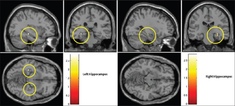
All participants completed the 6-month yoga intervention. They spontaneously reported no adverse event. Effect on hippocampus volume paired sample t-test of GM images with apriori ROI of hippocampus revealed a significant increase in bilateral hippocampus volume (posterior region) following 6 months of yoga in elderly subjects (N=7) (left hippocampus: X = −33, Y=−30, Z=−11; T=2.9; uncorrected P=0.01; right hippocampus: X =32, Y=−22, Z=−18; T=2.3; uncorrected P=0.03) [Figure 1]. No change in volume was observed in the control brain region (superior occipital gyrus).
Figure 1.

Voxel based morphometric analysis of the effects of yoga practice in healthy elderly (demonstrates significant increase in bilateral hippocampus gray matter volume after 6 months of yoga practice [indicated by the yellow circle])
DISCUSSION
In this study, 6 months of yoga practice was associated with the increase in volume of bilateral hippocampus; however, this finding has to be considered preliminary and has to be interpreted with caution in the context of following critical limitations: (1) Small sample size (N=7); (2) absence of a control group scanned twice at an interval of 6 months; (3) we did not perform multiple comparison correction; (4) lack of concurrent assessment of certain related neurobiological parameters like BDNF.
It is known that in healthy aging too, the elderly obtain a 1-2% loss in hippocampus volume annually.[1] In contrast, after 6 months of yoga intervention, the hippocampus gained volume in this study. Such change did not occur in occipital cortex. Hippocampus and occipital cortices were chosen as the target and control regions due to their differential susceptibility to neuro-senescence; with the occipital cortex being the least affected in the age-related brain atrophy.[15] This neuroplastic effect in the hippocampus is in keeping with similar reports in the literature; aerobic exercise[5] and meditations[7] too produced increases in the volume of the hippocampus. Demonstrating this differential effect of an intervention using a control group as done in an earlier study[16] would have been more convincing. A behavioral/functional correlate to the observed neuroplastic effect too was desirable. In this small sample of seven subjects, such correlation was not attempted. Similarly, correlation with other neuroplasticity measures could strengthen the finding.
In summary, 6 months of practice of yoga obtained greater hippocampal, but not occipital cortex volumes in non-demented elderly. Yoga has the potential to be a barrier to age-related neuro-senescence. The findings merit confirmation in the larger sample, as well as with a control intervention and/or aerobic exercise. Given the cultural acceptance of yoga, this should be exploited as a public health intervention to fight neuro-senescence and diseases including AD.
ACKNOWLEDGMENTS
We thank Mr. Sushrutha and Mr. Bhagath of Swami Vivekananda Yoga Anusandhana Samsthana for their help with transliteration.
Footnotes
Source of Support: The research was done under the Advanced Centre for Yoga - Mental Health and Neurosciences, a collaborative centre of NIMHANS and the Morarji Desai Institute of Yoga, New Delhi
Conflict of Interest: None declared.
REFERENCES
- 1.Raz N, Lindenberger U, Rodrigue KM, Kennedy KM, Head D, Williamson A, et al. Regional brain changes in aging healthy adults: General trends, individual differences and modifiers. Cereb Cortex. 2005;15:1676–89. doi: 10.1093/cercor/bhi044. [DOI] [PubMed] [Google Scholar]
- 2.Jack CR, Jr, Wiste HJ, Vemuri P, Weigand SD, Senjem ML, Zeng G, et al. Brain beta-amyloid measures and magnetic resonance imaging atrophy both predict time-to-progression from mild cognitive impairment to Alzheimer's disease. Brain. 2010;133:3336–48. doi: 10.1093/brain/awq277. [DOI] [PMC free article] [PubMed] [Google Scholar]
- 3.Ahlskog JE, Geda YE, Graff-Radford NR, Petersen RC. Physical exercise as a preventive or disease-modifying treatment of dementia and brain aging. Mayo Clin Proc. 2011;86:876–84. doi: 10.4065/mcp.2011.0252. [DOI] [PMC free article] [PubMed] [Google Scholar]
- 4.Burdette JH, Laurienti PJ, Espeland MA, Morgan A, Telesford Q, Vechlekar CD, et al. Using network science to evaluate exercise-associated brain changes in older adults. Front Aging Neurosci. 2010;2:23. doi: 10.3389/fnagi.2010.00023. [DOI] [PMC free article] [PubMed] [Google Scholar]
- 5.Erickson KI, Prakash RS, Voss MW, Chaddock L, Hu L, Morris KS, et al. Aerobic fitness is associated with hippocampal volume in elderly humans. Hippocampus. 2009;19:1030–9. doi: 10.1002/hipo.20547. [DOI] [PMC free article] [PubMed] [Google Scholar]
- 6.Froeliger B, Garland EL, McClernon FJ. Yoga meditation practitioners exhibit greater gray matter volume and fewer reported cognitive failures: Results of a preliminary voxel-based morphometric analysis. Evid Based Complement Alternat Med 2012. 2012 doi: 10.1155/2012/821307. 821307. [DOI] [PMC free article] [PubMed] [Google Scholar]
- 7.Hölzel BK, Carmody J, Vangel M, Congleton C, Yerramsetti SM, Gard T, et al. Mindfulness practice leads to increases in regional brain gray matter density. Psychiatry Res. 2011;191:36–43. doi: 10.1016/j.pscychresns.2010.08.006. [DOI] [PMC free article] [PubMed] [Google Scholar]
- 8.Lazar SW, Kerr CE, Wasserman RH, Gray JR, Greve DN, Treadway MT, et al. Meditation experience is associated with increased cortical thickness. Neuroreport. 2005;16:1893–7. doi: 10.1097/01.wnr.0000186598.66243.19. [DOI] [PMC free article] [PubMed] [Google Scholar]
- 9.Naveen GH, Thirthalli J, Rao MG, Varambally S, Christopher R, Gangadhar BN. Positive therapeutic and neurotropic effects of yoga in depression: A comparative study. Indian J Psychiatry. 2013;55:S400–4. doi: 10.4103/0019-5545.116313. [DOI] [PMC free article] [PubMed] [Google Scholar]
- 10.Pezawas L, Verchinski BA, Mattay VS, Callicott JH, Kolachana BS, Straub RE, et al. The brain-derived neurotrophic factor val66met polymorphism and variation in human cortical morphology. J Neurosci. 2004;24:10099–102. doi: 10.1523/JNEUROSCI.2680-04.2004. [DOI] [PMC free article] [PubMed] [Google Scholar]
- 11.Hariprasad VR, Varambally S, Sivakumar PT, Thirthalli J, Basavaraddi IV, Gangadhar BN. Designing, validation and feasibility of a yoga-based intervention for elderly. Indian J Psychiatry. 2013;55:S344–9. doi: 10.4103/0019-5545.116302. [DOI] [PMC free article] [PubMed] [Google Scholar]
- 12.Ashburner J, Friston KJ. Unified segmentation. Neuroimage. 2005;26:839–51. doi: 10.1016/j.neuroimage.2005.02.018. [DOI] [PubMed] [Google Scholar]
- 13.Ashburner J. A fast diffeomorphic image registration algorithm. Neuroimage. 2007;38:95–113. doi: 10.1016/j.neuroimage.2007.07.007. [DOI] [PubMed] [Google Scholar]
- 14.Maldjian JA, Laurienti PJ, Kraft RA, Burdette JH. An automated method for neuroanatomic and cytoarchitectonic atlas-based interrogation of fMRI data sets. Neuroimage. 2003;19:1233–9. doi: 10.1016/s1053-8119(03)00169-1. [DOI] [PubMed] [Google Scholar]
- 15.Peters R. Ageing and the brain. Postgrad Med J. 2006;82:84–8. doi: 10.1136/pgmj.2005.036665. [DOI] [PMC free article] [PubMed] [Google Scholar]
- 16.Erickson KI, Voss MW, Prakash RS, Basak C, Szabo A, Chaddock L, et al. Exercise training increases size of hippocampus and improves memory. Proc Natl Acad Sci U S A. 2011;108:3017–22. doi: 10.1073/pnas.1015950108. [DOI] [PMC free article] [PubMed] [Google Scholar]


