Abstract
Endo-β-1, 4-xylanases is thought to be of great significance for several industries namely paper, pharmaceuticals, food, feed etc. in addition to better utilization of lignocellulosic biomass. The present investigation was aimed to develop an easy, simple and efficient assay technique for endo-β-1, 4-xylanases secreted by the aerobic fungi. Under the proposed protocol, 9 g/L xylan containing agar was prepared in 100 mM phosphate buffer at different pH (4.5, 5.5 and 6.5). The sterilized xylan agar was dispensed in 90 mm petri dishes. 100 µl of culture supernatant of 12 fungal isolates was added to the wells and left overnight at 31±10C. The petri dishes were observed for zone of clearance by naked eye and diameter was measured. Congo red solution (1 g/L) was applied over the petri dishes as per the established protocol and thereafter plates were flooded with 1M Sodium chloride solution for the appearance of zone of clearance. The diameter for zone of clearance by the proposed method and the established protocol was almost identical and ranged from 21 to 42 mm at different pH depending upon the activity of endo-β-1, 4-xylanases. Change of pH towards alkaline side enabled similar or marginal decrease of diameter for the zone of clearance in most of the fungal isolates. The specific activities of these fungal isolates varied from 1.85 to 11.47 IU/mg protein. The present investigation revealed that the proposed simple diffusion technique gave similar results as compared to the established Congo red assay for endo-β-1, 4-xylanases. Moreover, the present technique avoided the cumbersome steps of staining by Congo red and de-staining by sodium chloride.
Keywords: Endo-β-1, 4-xylanase, Diffusion technique, Congo red assay
INTRODUCTION
The agricultural crop residues and byproducts are renewable, inexpensive and abundantly available raw materials for the sustainable production of clean and affordable bio-fuels, bio-power and high value biological products like nutraceuticals. Hemicellulose is the second most abundant biomolecule found to be present in the lignocellulsoic biomass and β-1,4-xylan is the major hemicellulose component (5). It represents approximately 25 to 35% of dry biomass of woody tissues of dicot and lignified tissues of monocot and its proportion may be as high as 50% in some tissues of cereal grains (11). Because of its availability in enormous quantity from diverse agricultural crop residues and agro-industrial wastes, disposal of this biomass is a huge problem (4, 15). Hydrolysis of xylan is an important step towards the proper utilization of lignocellulosic material in nature as well as for the production of certain emerging functional foods like xylooligosaccharides (13, 16, 17). Xylanases (EC 3.2.1.8; endo-β-1, 4-xylanase) are primary hydrolytic enzymes that can catalyze the breakdown of xylan having β-1,4-xylosidic linkages (9, 13, 14, 21).
Recently the endo-β-1, 4-xylanases have attracted considerable attention because of their biotechnological potential (23) in various industrial processes such as bleaching of pulp and paper industry, bioconversion of biomass wastes to fermentable sugars and clarification of fruit juices and wines (2, 6, 20), animal feed (18), emerging nutraceuticals i.e. xylooligosaccharides (1, 7). A number of methods are being followed amongst researchers in food and pharmaceutical industries to assay endo-β-1,4-xylanases activity. These assays differ not only in assay conditions (temperature, incubation time, substrate etc) but also in principle of quantification of the enzyme activity i.e. detection of reducing sugars from substrate, detection of dye released from covalently dyed xylan, measurement of viscosity or monitoring the turbidity. Presently the qualitative method of Congo red assay (3, 17) is popular among the researchers to confirm the endo-β-1,4-xylanases activity based on diffusion principle. Congo red binds strongly to xylan containing β-1,4-xylosidic linkages and provides the basis for the highly sensitive and qualitative test for the said enzyme. However, the method is cumbersome and time consuming since it uses large quantities of chemicals like Congo red and sodium chloride for staining and de-staining, respectively. On the other hand for precisely quantifying the enzyme activities of xylanase, reducing sugars liberated from xylan are measured through DNS method (10) or Nelson-Somogyi method (12, 19). To this end, we have developed a simple and easy qualitative technique to screen the aerobic fungi for endo-β-1, 4-xylanases by diffusion technique. Comparative studies with the established Congo red assay and colorimetric techniques were adopted for further substantiation of the findings.
MATERIALS AND METHODS
Twelve aerobic fungi (procured from ITCC, Indian Agricultural Research Institute, New Delhi) were grown in media containing oat spelt xylan (10.0 g/L), yeast extract (5.0 g/L), NaNO3 (1.0 g/L), KH2PO4 (1.0 g/L), peptone (1.0 g/L) and MgSO4 7H2O (0.3 g/L). The pH of the media was adjusted to 5.5±0.05. A 50 ml aliquot of above media was dispensed in 125 ml conical flask and autoclaved. The fungal isolates were inoculated in the media and incubated at 31±10C in a shaking incubator for 5 days. After the stipulated period of incubation, the extracellular enzymes were harvested by filtering through an ordinary filter paper and further clarified by centrifuging at 10,000 rpm for 20 minutes at 40C. The harvested enzyme (100 µl) was subjected to proposed simple and easy diffusion method for endo-β-1, 4-xylanases.
According to the new protocol, 9.0 g/L oat spelt xylan (Sigma, USA) was dissolved in 100 mM phosphate buffer having pH 4.5, 5.5 and 6.5. Agar was added at 18.0 g/L in the above xylan solutions and sterilized by autoclaving at 15 psi for 20 minutes. After cooling (500C), approximately 25 to 30 ml solution was poured into sterilized petri dishes (90 mm). The petri dishes were incubated overnight at 31±10C in an incubator to check any contamination. Wells were prepared with the help of sterilized cork borer with 10 mm diameter. Using a micropipette, 100 µl of harvested enzyme was added to each well of the plates. Each plate had a control well filled with 100 µl sterile distilled water. The plates were incubated overnight at 31±10C in an upright position. The diameter for zone of clearance was measured in mm and results were recorded. To compare the zone of clearance with the established Congo red assay, the same plates were flooded with Congo red solution (1.0 g/L) and kept at room temperature for 30 minutes. Subsequently, the plates were de-stained with 1 M sodium chloride solution and zone of clearance (diameter) was measured again.
The Nelson-Somogyi method (12, 19) was followed to measure the fungal endo-β-1, 4-xylanases activity. The assay mixture comprised of 975 µl of 10 g/L oat spelt xylan (Sigma, USA) dissolved in 100 mM of sodium phosphate buffer (pH 6.5) and 25 µl of appropriately diluted enzyme. Incubation was carried out at 500C for 15 minutes in a shaking water bath. Xylose (Sigma, USA) was used for preparation of standard curve. One unit of activity was defined as the amount of enzyme required to liberate 1 μmol of reducing sugars (xylose) per minute per milliliter. Protein was quantified according to the standard method (8). Specific activity of xylanase was expressed as units of activity per mg of protein (IU/mg protein).
RESULTS AND DISCUSSION
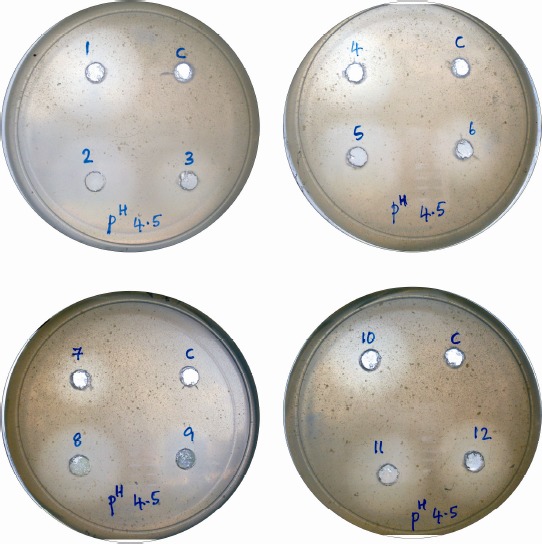
A novel enzyme diffusion technique was applied to qualitatively assess the activity of endo-β-1,4-xylanase within a short period of time without involving the use of a spectrophotometer or colorimeter. It is an easy and reproducible protocol for rapid screening of endo-β-1,4-xylanase secreted by a wide group of microbes. Following overnight incubation, petri dishes were observed for the appearance of zone of clearance without any stains. It revealed that except for control, there was appearance of transparent zone (clearly visible with naked eye) against opaque zone of intact xylan agar media (Plate 1). The diameters of zone of clearance as measured are presented in Table 1. The same petri dishes were subjected to the endo-β-1,4-xylanases activity through established Congo red protocol for appearance of yellow zone of clearance against red background of undigested xylan. The diameter for zone of clearance by proposed simple method and Congo red assay (Table 1) was almost identical. From the results it appeared that the proposed new simple diffusion technique was as efficient as already reported and tested Congo red assay (3) for qualitatively detection of the endo-β-1,4-xylanases activity of fungi. Moreover, the present technique did not involve the steps of staining and de-staining by any chemicals. Further, the qualitative assay of Nelson – Somogyi also confirmed the presence of the endo-β-1,4-xylanases in the fungal isolates (Table 1). The activity ranged from 3.02 to 14.89 IU. The simple diffusion technique can be easily considered for rapid screening of endo-β-1,4-xylanases producing microorganisms which bypass the staining and de-staining of Congo red assay along with time involved in the same.
Plate 1.

Zone of clearance produced by fungal endo-β-1, 4-xylanase in simple diffusion assay: 1 Aspergillus Japonicus 4371, 2 Aspergillus oryzae 4010, 3 Penicillium citrinum 4009, Penicillium purpurogenum 4248, 5 Penicillium purpurogenum 5252, 6 Aspergillus oryzae 2398, 7 Aspergillus oryzae 2624, 8 Aspergillus oryzae 4712, 9 Penicillium purpurogenum 2029, 10 Penicillium purpurogenum 2433, 11 Aspergillus oryzae 4714, 12 Aspergillus oryzae 4964.
Table 1.
Endo-β-1, 4-xylanase activity measured by modified diffusion technique and congo red assay vis a vis specific activity produced by aerobic fungi
| Sl No | Fungal spp. with ITCC no. | Diameter of zone of clearance (mm) at different pH | Endoxylanase activity (IU/ml) | Specific activity (IU/mg protein) | |||||
|---|---|---|---|---|---|---|---|---|---|
| Simple diffusion | Congo red assay | ||||||||
| 4.5 | 5.5 | 6.5 | 4.5 | 5.5 | 6.5 | ||||
| 1 | Aspergillus japonicus 4371 | 42 | 40 | 30 | 42 | 40 | 30 | 7.17 | 6.76 |
| 2 | Aspergillus oryzae 4010 | 36 | 36 | 36 | 36 | 36 | 36 | 11.67 | 7.48 |
| 3 | Penicillium citrinum 4009 | 25 | 25 | 25 | 25 | 25 | 25 | 3.02 | 1.85 |
| 4 | Penicillium purpurogenum 4248 | 32 | 36 | 35 | 32 | 36 | 36 | 6.76 | 5.04 |
| 5 | Penicillium purpurogenum 5252 | 35 | 36 | 32 | 35 | 36 | 32 | 7.23 | 4.88 |
| 6 | Aspergillus oryzae 2398 | 30 | 30 | 27 | 30 | 30 | 27 | 14.89 | 9.13 |
| 7 | Aspergillus oryzae 2624 | 25 | 25 | 25 | 25 | 25 | 25 | 9.96 | 6.00 |
| 8 | Aspergillus oryzae 4712 | 35 | 29 | 29 | 35 | 29 | 29 | 6.89 | 6.43 |
| 9 | Penicillium purpurogenum 2029 | 29 | 21 | 21 | 29 | 21 | 21 | 4.10 | 2.99 |
| 10 | Penicillium purpurogenum 2433 | 33 | 33 | 30 | 33 | 33 | 30 | 5.07 | 3.59 |
| 11 | Aspergillus oryzae 4714 | 35 | 35 | 33 | 35 | 35 | 33 | 6.05 | 5.04 |
| 12 | Aspergillus oryzae 4964 | 35 | 35 | 35 | 35 | 35 | 35 | 11.36 | 11.47 |
Earlier, both Congo red and colorimetric methods were considered to assay endo-β-1,4-xylanases activity of anaerobic fungal isolates of rumen (17). The present investigation confirmed that the simple diffusion technique could be adopted to assess the presence of endo-β-1,4-xylanases activity in the culture supernatant of aerobic fungi; but did not assess its quantity. Moreover, increase in pH of xylan agar resulted identical or decreased zone of clearance except Penicillium purpurogenum 4248. This reflected that the ideal pH for endo-β-1,4-xylanases of fungi used in the present investigation was around 4.5. However, the diameter of zone of clearance exhibited a non-linear relationship with the enzyme activity. This could be attributed to the multiple occurrence of β-1,4-endoxylanase in microorganisms and also its several isomeric forms (14, 17, 22). Moreover, the rate of diffusion is inversely proportional to the molecular weight. The endo-β-1,4-xylanases produced by different species of fungi used in our present study may differ considerably in their molecular weights. As well diffusion technique is only qualitative; one should follow the colorimetric assay to get quantitative values as well. Hence, the present investigation concluded that the simple diffusion technique could be followed for rapid screening of fungi for their ability to secrete endo-β-1,4-xylanase.
ACKNOWLEDGEMENT
The authors are highly grateful to Department of Biotechnology, Government of India for financial support of the project (BT/PR10518/AAQ/01/361/2008). The authors are indebted to Dr. KT Sampath, Director for constant support and encouragement.
REFERENCES
- 1.Akpinar O., Erdogan K., Bosanci S. Enzymatic production of xylooligosaccharide from selected agricultural wastes. Food Bioprod. Process. 2009;87:145–151. [Google Scholar]
- 2.Bajpai P., Bhardwaj P.K., Bajpai P.K., Jauhari M.B. The impact of xylanases on bleaching of eucalyptus kraft pulp. J. Biotechnol. 38:1–6. [Google Scholar]
- 3.Beguin P. Detection of cellulase activity in polyacrylamide gels using Congo red stained agar replicas. Anal. Chem. 1983;131:333–336. doi: 10.1016/0003-2697(83)90178-1. [DOI] [PubMed] [Google Scholar]
- 4.Biely P. Microbial xylanolytic systems. Trends Biotechnol. 1985;3:286–290. [Google Scholar]
- 5.Dobrev G., Zhekova B., Delcheva G., Koleva L., Tziporkov N., Pishtiyski I. Purification and characterization of endoxylanase Xln-1 from Aspergillus niger B03. World J. Microbiol. Biotechnol. 2009;25:2095–2102. [Google Scholar]
- 6.Kumar R., Singh S., Singh O.M.V. Bioconversion of lignocellulosic biomass: Biochemical and molecular perspectives. J. Ind. Microbiol. Biotechnol. 2008;35:377–391. doi: 10.1007/s10295-008-0327-8. [DOI] [PubMed] [Google Scholar]
- 7.Liu M.Q., Liu. G.F. Expression of recombinant Bacillus licheniformis xylanase A in Pichia pastoris and xylooligosaccharides released from xylans by it. Protein Expression and Purif. 2008;57:101–107. doi: 10.1016/j.pep.2007.10.020. [DOI] [PubMed] [Google Scholar]
- 8.Lowry O.H., Rosebrough N.J., Farr A.L., Randall R.J. Protein measurement with the Folin-Phenol reagents. J. Biol. Chem. 1951;193:265–275. [PubMed] [Google Scholar]
- 9.Maheshwari R., Bharadwaj G., Bhat M. Thermophilic fungi: Their physiology and enzymes. Microbiol Mol. Rev. 2000;64:461–488. doi: 10.1128/mmbr.64.3.461-488.2000. [DOI] [PMC free article] [PubMed] [Google Scholar]
- 10.Miller G.L. Use of dinitrosalicylic acid reagent for determination of reducing sugar. Anal. Chem. 1959;31:426–428. [Google Scholar]
- 11.Moura A., Patricia G., Herminia D., Parajo J.C. Advances in the manufacture, purification and applications of xylo-oligosaccharides as food additives and nutraceuticals. Process Biochem. 2006;41:1913–1923. [Google Scholar]
- 12.Nelson N. A photometric adaptation of the Somogyi method for the determination of glucose. J. Biol. Chem. 1944;153:375–380. [Google Scholar]
- 13.Pham P.L., Taillandier P., Delmas M., Strehaiano P. Optimization of a culture medium for Xylanase production by Bacillus spp. using statistical experimental designs. World J. Microbiol. Biotechnol. 1998;14:185–190. [Google Scholar]
- 14.Poorna C. A., Prema P. Production of cellulase-free endoxylanase from novel alkalophilic thermotolerent Bacillus pumilus by solid-state fermentation. Bioresource Technol. 2007;98:485–490. doi: 10.1016/j.biortech.2006.02.033. [DOI] [PubMed] [Google Scholar]
- 15.Prade R.A. Xylanases, from biology to biotechnology. Biotechnol. Gen. Eng. Rev. 1995;13:101–131. doi: 10.1080/02648725.1996.10647925. [DOI] [PubMed] [Google Scholar]
- 16.Samanta A.K., Kolte A.P., Chandrasekhariah M., Thulasi A., Sampath K.T., Prasad C.S. Prebiotics: The rumen modulator for enhancing the productivity of dairy animals. Indian Dairyman. 2007;59:58–61. [Google Scholar]
- 17.Samanta A.K., Walli T.K., Rajput Y.S., Batish V.K., Grover S., Mohanty A.K. Fractionation and partial purification of endoglucanase and xylanase from Piromyces sp. isolated from rumen of riverine buffalo. Microbiol. Aliments Nutr. 1999;17:81–91. [Google Scholar]
- 18.Samanta A.K., Senani S., Kolte A. P., Sridhar Manpal, Rao S.B.N. Importance of exogenous fibrolytic enzymes in ruminants diet. Indian J Dairy Biosci. 2008;19(1&2):52–54. [Google Scholar]
- 19.Somogyi M. Notes on sugar determination. J. Biol. Chem. 1952;195:19–23. [PubMed] [Google Scholar]
- 20.Sonia K.G., Chadha B.S., Saini H.S. Sorghum straw for xylanase hyper-production by Thermomyces lanuginosus (D2W3) under solid state fermentation. Bioresource Technol. 2005;96:1561–1569. doi: 10.1016/j.biortech.2004.12.037. [DOI] [PubMed] [Google Scholar]
- 21.Sunna A., Antranikian G. Xylanolytic enzymes from fungi and bacteria. Crit. Rev. Biotechnol. 1997;17:39–67. doi: 10.3109/07388559709146606. [DOI] [PubMed] [Google Scholar]
- 22.Wang K.K.Y., Tan L.U., Saddlar J.N. Multiplicity of beta-1,4 xylanase in microorganisms: functions and applications. Microbiol. Rev. 1988;52:305–317. doi: 10.1128/mr.52.3.305-317.1988. [DOI] [PMC free article] [PubMed] [Google Scholar]
- 23.Yi X., Shi Y., Xu H., Li W., Xiel J., Yu R., Zhu J., Caol Y., Qiao D. Hyperexpression of two Aspergillus niger xylanase genes in Escherichia coli and characterization of the gene products. Braz. J. Microbiol. 2010;41:778–786. doi: 10.1590/S1517-83822010000300030. [DOI] [PMC free article] [PubMed] [Google Scholar]


