Abstract
Background: Fluorosis is a condition resulting from excessive ingestion of fluoride during early childhood leading to the formation of defective enamel. The increased fluoride content is thought to result in a metabolic alteration of ameloblasts, which results in defective matrix, and improper calcification of teeth.
Materials and Methods: A total of 6244 patients between the ages of 6yrs to 60yrs, who presented to our outpatient clinic from October 2009 to December 2010 were included in the study. The study subjects were grouped according to their age into the following groups- 6-14 yrs, 15-25 yrs, 26-40 yrs, and 40-60yrs. Only permanent dentition was taken into consideration in this study.
Results: The overall prevalence of fluorosis in this study was 63.34% (3955 of 6244 patients). Men had a slightly higher prevalence of 64.27% compared to 62.28% among women.
Conclusion: Prevention of fluorosis would require efforts at raising awareness among the people about the harmful effects of their dietary choices on their teeth. They also need to be educated about adequate and proper oral hygiene, such as brushing their teeth at least two times daily.
How to cite this article: Sunil T K L, Shetty S, Annapoorna B M, Pujari S C, Reddy P S, Nandlal B. A Pioneering Study of Dental Fluorosis in the Libyan Population. J Int Oral Health 2013; 5(3):67-72.
Key Words: : Fluorosis, Pitting, Hypomineralization.
Introduction
In its moderate and severe form, fluorosis results in a significant increase in enamel porosity. This causes the enamel to take up stain after eruption into the oral cavity, leading to brown and black discolouration of the teeth. Teeth with moderate to severe fluorosis are prone to attrition and wear resulting in pitting, chipping and decay.1
As evidenced by epidemiological findings, the distribution of dental fluorosis is highly variable among individuals exposed to a drinking water source containing fluoride ions at a concentration greater than 0.1 mg/L during the period of tooth development. This variation depends on the waterborne fluoride level, climatic factors, availability of other dietary sources of fluoride, and individual variation in fluoride metabolism (Whitford, 1989). It has also been observed that incisor fluorosis is more apparent in the presence of a short or open upper lip that results in drying of exposed enamel. 2,3
The present study was carried out in the city of Zawia in Libya to record the prevalence of dental fluorosis. To date there are no reported studies to the best of our knowledge.
Materials and Method:
Our study was performed as a survey of male and female patients presenting to the outpatient department of the faculty of dentistry at the Al-Jabal-Gharby-University, Zawia, Libya.
A total of 6244 patients between the ages of 6yrs to 60yrs, who presented to our outpatient clinic from October 2009 to December 2010 were included in the study. The study subjects were grouped accor-ding to their age into the following groups- 6-14 yrs, 15-25 yrs, 26-40 yrs, and 40-60yrs. Only perm-anent dentition was taken into consideration in this study. Pedodontic patients were considered as having permanent teeth. A single dentist carried
Fig. 3: Tooth Surface Index of Fluorosis (TSIF) Type 3.

out the examination to standardize all the readings.
Each tooth was carefully examined under natural light after it was thoroughly dried.
Tooth Surface Index of Fluorosis (TSIF) was used to determine the prevalence of dental fluorosis 2. TSIF identifies seven types of fluorosis as described below:
Enamel shows no evidence of fluorosis.
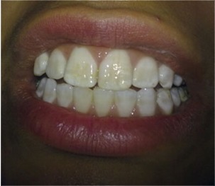
Enamel shows definite evidence of fluorosis, namely areas with parchment-white colour that total less than 1/3 of the visible enamel surface. This category includes fluorosis confined only to the incisal edges of anterior teeth or cusp tips of posterior teeth.
Parchment-white fluorosis totals at least 1/3 but less than 2/3 of the visible surface.
Parchment-white fluorosis totals at least 2/3 of the visible surface.
Enamel shows staining in conjunction with any of the preceding levels of fluorosis. Staining is defined as an area of definite discoloration that may range from light to very dark brown.
Discrete pitting of the enamel exists, unaccompanied by evidence of staining of intact enamel. A pit is defined as a definite physical defect in the enamel surface with a rough floor that is surrounded by a wall of intact enamel. The pitted area is usually stained or differs in colour from the surrounding enamel.
Both discrete pitting and staining of the intact enamel exists.
Confluent pitting of the enamel surface exists. Large areas of the enamel may be missing and the anatomy of the tooth may be altered. Dark brown stain is usually present.
Results:
The overall prevalence of fluorosis in this study was 63.34% (3955 of 6244 patients). Men had a slightly higher prevalence of 64.27%(Table 1) compared to 62.28% (Table 2) among women. The 6-14 age group had the least prevalence, while patients in the 41-60 year age group had the highest rate of fluorosis. (Tables 1 & 2)
| Table 1: Chart for Male Patients Examined | |||||||||||||||||
| Age ears) | No. of Patients Examined | No. of Patients with Fluorosis | % | Class I | % | Class II | % | Class III | % | Class IV | % | Class V | % | Class VI | % | Class VII | % |
| 6-14 yrs | 296 | 139 | 46.99 | 28 | 20.14 | 31 | 22.30 | 24 | 17.26 | 23 | 16.55 | 16 | 11.50 | 6 | 4.31 | 11 | 7.91 |
| 15-25 yrs | 1702 | 1055 | 61.9 | 58 | 5.50 | 123 | 11.66 | 158 | 14.98 | 418 | 39.62 | 97 | 9.19 | 48 | 4.56 | 153 | 14.50 |
| 26-40 yrs | 763 | 511 | 66.97 | 32 | 6.26 | 63 | 12.32 | 81 | 15.85 | 183 | 35.81 | 52 | 10.18 | 36 | .05 | 64 | 12.52 |
| 41-60y | 174 | 123 | 70.69 | - | - | 15 | 12.20 | 18 | 14.63 | 18 | 26.83 | 17 | 13.82 | 12 | 28 | 28 | 22.76 |
| Table 2: Chart for Female Patients Examined | |||||||||||||||||
| Age ears) | No. of Patients Examined | No. of Patients with Fluorosis | % | Class I | % | Class II | % | Class III | % | Class IV | % | Class V | % | Class VI | % | Class VII | % |
| 6-14 yrs | 296 | 139 | 46.99 | 28 | 20.14 | 31 | 22.30 | 24 | 17.26 | 23 | 16.55 | 16 | 11.51 | 6 | 4.31 | 11 | 7.91 |
| 15-25 yrs | 1702 | 1055 | 61.9 | 58 | 5.50 | 123 | 11.66 | 158 | 14.98 | 418 | 39.62 | 97 | 9.19 | 48 | 4.56 | 153 | 14.50 |
| 26-40 yrs | 763 | 511 | 66.97 | 32 | 6.26 | 63 | 12.32 | 81 | 15.85 | 183 | 35.81 | 52 | 10.18 | 36 | .05 | 64 | 12.52 |
| 41-60y | 174 | 123 | 70.69 | - | - | 15 | 12.20 | 18 | 14.63 | 33 | 26.83 | 17 | 13.82 | 12 | 9.76 | 28 | 22.76 |
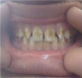
Majority of the patients who presented to our clinic were in the 15-24 year age group (38.91%). Among both men and women, class IV (corresponding to TSIF score 4), was the most common form of fluorosis.(Fig 4)
Fig. 4: Tooth Surface Index of Fluorosis (TSIF) Type 4.

Interestingly, none of the female patients in the 41-60 year age group had Type I fluorosis (Fig 1)
Fig. 1: Tooth Surface Index of Fluorosis (TSIF) Type 1.

(TSIF score 0, which corresponds with no enamel showing no evidence of fluorosis). This meant that every female patient in the 41-60 year age group had some definitive evidence of fluorosis.
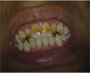
Type VI was more prevalent among males in the age group between 26 to 40 (fig 6)
Fig 6: Tooth Surface Index of Fluorosis (TSIF) Type 6.

Type VII was more prevalent among males in the age group between 41 to 60 (fig 7)
Fig 7: Tooth Surface Index of Fluorosis (TSIF) Type 7.

Type VI & VII was more prevalent among females in the age group between 40 to 60 (fig 6 & 7)
Discussion:
The increased prevalence of fluorosis in Libya can be attributed to the predominant consumption of
ground water, which has been shown to contain higher fluoride content than surface water sources such as river or lake water4. The amount of fluo-ride content in groundwater was found to be 5 ppm by researchers in Al-Fateh University of Tripoli.
Type IV fluorosis was the most common type in general (Fig 4). But among children and adolescents (the 6-14 years age group), Type I and II were more common(Fig 1 & Fig 2). But as the population aged, more advanced classes of fluorosis (IV and V) were seen (Fig 4 & Fig 5). This is likely due to continued exposure to fluoride via water, food and beverages with high fluoride content 5-7. The food intake of Libyan population mainly contains of a non-vegetarian diet, mainly consisting of beef, goat, chicken and seafood. The vegetables that are consumed are potato, brinjal, greens, radish, carrot, groundnuts, cucumber, beets, onions, etc. The fruits that are usually cultivated are oranges, peaches, lemons, strawberries, etc. The usual vegetables consumed include potatoes, onions, carrots and spinach, usually on a daily basis8. Since potatoes, onions and carrots grow below the ground, they are exposed to a higher concentration of fluoride9,10. Another staple of Libyan diet is their bread known as kubbs, which is usually harder and abrasive in nature than the normal bread. There is also a high intake of sweets, chocolates and cheese. There is an enormous intake of carbonated drinks such as Pepsi and Coke on a daily basis. Type VI & VII fluorosis can be attributed to the constant intake of abrasive food along with acidic beverages (Fig 6 & 7).
Fig. 2: Tooth Surface Index of Fluorosis (TSIF) Type 2.

FIG 5: Tooth Surface Index of Fluorosis (TSIF) Type 5.

The low level of education makes it is difficult to explain to them the importance of brushing the teeth & oral hygiene maintenance. The Libyan population in the city brush their teeth only once in the morning11. They usually prefer a toothbrush that is hard and abrasive in nature and use non-fluorinated toothpaste. In rural areas people have a habit of brushing with Meswak stick, which is very abrasive in nature9.
On dental examination, the population usually has high deposits of plaque and calculus with gingival recession. The degree of porosity (hypominera-lization) of teeth in older age group results in a diminished physical strength of the enamel, and parts of the superficial enamel may break away12. Such loss of outermost enamel is particularly frequent along incisal edges and cusp tips. In the latter case, this will also involve the occlusal surfaces, which become rapidly worn out. There is loss of tooth morphology in older patients because of the loss of normal enamel maturation and due to the high occlusal forces acting on it7. The loss of enamel can also be attributed to the type of food consumed, as it is very abrasive in nature. The poor oral hygiene also contributes to enamel damage. There is also enormous amount of caries reported due to the high intake of sticky foods, cheese, and carbonated drinks daily and lack of proper oral hygiene.
Conclusion:
There is a relatively high prevalence of fluorosis in this part of the country. Fluorosis not only harms the teeth, but also dents an individuals self confidence by affecting their looks and smile. Dietary choices, and lack of adequate oral hygiene are at the root of this problem. Prevention of fluorosis would require efforts at raising awareness among the people about he harmful effects of their dietary choices on their teeth. They also need to be educated about adequate and proper oral hygiene, such as brushing their teeth at least two times daily. Public health agencies should make efforts to decrease the fluoride content in drinking water sources. People who are already affected by fluorosis can be treated with a range of available techniques depending on degree of fluorosis. Treatments include cosmetic interventions or providing crowns, laminates or venners.
Footnotes
Source of Support: Nil
Conflict of Interest: None Declared
Contributor Information
Sunil Tejaswi K L, Department of Conservative Dentistry and Endodontics, JSS Dental College and Hospital, Mysore, Karnataka, India.
Suneeth Shetty, Department of Conservative Dentistry and Endodontics, JSS Dental College and Hospital, Mysore, Karnataka, India.
Annapoorna B M, Department of Conservative Dentistry and Endodontics, JSS Dental College and Hospital, Mysore, Karnataka, India.
Sudarshan C Pujari, Department of Conservative Dentistry and Endodontics, JSS Dental College and Hospital, Mysore, Karnataka, India.
Sarveshwar Reddy P, Department of Conservative Dentistry and Endodontics, JSS Dental College and Hospital, Mysore, Karnataka, India.
B Nandlal, JSS Dental College and Hospital, Mysore, Karnataka, India..
References:
- 1.Koleoso DC Umesi. Dental fluorosis and other enamel disorders in 12 year-old Nigerian children Journal of community medicine and primary health care. 2004;16(1):25–28. [Google Scholar]
- 2.Horowitz, S Herschel. Dental Fluorosis. Encyclopedia of Public Health. 2002 [Google Scholar]
- 3.Khalid A. The presence of dental fluorosis in the permanent dentition in Doha. East Mediterr Health J. 2004;10(3):425–428. [PubMed] [Google Scholar]
- 4.H Horowitz, W Driscoll, R Meyers, S Heifitz, Kingman New method for assessing the prevalence of dental fluorosis-the tooth surface index of fluorosis. J Am Dent Assoc. 1984;109(1):37–41. doi: 10.14219/jada.archive.1984.0268. [DOI] [PubMed] [Google Scholar]
- 5.Heilman Jr, MC Kiritsy, SM Lew, JS Wefel. Fluoride concentrations of infant foods. J Am Dent Assoc. 1997;128(7):857–863. doi: 10.14219/jada.archive.1997.0335. [DOI] [PubMed] [Google Scholar]
- 6.Heilman Jr, MC Kiritsy, SM Lew, JS Wefel. Assessing fluoride levels of carbonated soft drinks. J Am Dent Assoc. 1999;130(11):1593–1599. doi: 10.14219/jada.archive.1999.0098. [DOI] [PubMed] [Google Scholar]
- 7.O Fejerskov, F Manji, V Baelum. The nature and mechanisms of dental fluorosis in man. J Dent Res. 1990;69 Spec No:692–700. doi: 10.1177/00220345900690S135. [DOI] [PubMed] [Google Scholar]
- 8.Alarcón-Herrera M Teresa, Martín-Domínguez Ignacio R, Trejo-Vázquez Rodolfo, Chihuahua b Sandra Rodriguez-Dozalc. Well water fluroide,dental fluorosis, and bone fracture in the Guadiana valley of mexico. Fluoride Vol. 2001;34(2):139–149. [Google Scholar]
- 9.Baskaradoss, Jagan, Clement, Roger, Narayanan, Aswath Prevalence of dental fluorosis and associated risk factors in 11-15 year old school children of Kanyakumari District, Tamilnadu, India: A cross sectional survey.(Original Research)(Clinical report) Indian Journal of Dental Research. 2008;19(4):297–303. doi: 10.4103/0970-9290.44531. [DOI] [PubMed] [Google Scholar]
- 10.Gonini Cde A, MC Morita. Dental fluorosis in children attending basic health units. J. Appl. Oral Sci. 2004;12(3):189–194. doi: 10.1590/s1678-77572004000300005. [DOI] [PubMed] [Google Scholar]
- 11.SM Levy, B Brofitt, TA Marshall. Associations Between Fluorosis of Permanent Incisors and Fluoride Intake From Infant Formula, Other Dietary Sources and Dentifrice During Early Childhood. J Am Dent Assoc. 2010;141(10):1190–1201. doi: 10.14219/jada.archive.2010.0046. [DOI] [PMC free article] [PubMed] [Google Scholar]
- 12.A Watts, M Addy. Tooth discolouration and staining: A review of the literature. Br Dent J. 2001;190(6):309–316. doi: 10.1038/sj.bdj.4800959. [DOI] [PubMed] [Google Scholar]


