Laparoscopic evaluation can be useful in a setting of clinical and radiographic diagnostic uncertainty.
Keywords: Gallbladder torsion, Gallbladder ischemia
Abstract
Introduction:
Torsion of the gallbladder in the pediatric population is rare. A nonspecific clinical presentation is characteristic, which frequently contributes to diagnostic uncertainty.
Case Description:
We report a case of gallbladder torsion in a 6-y-old male with abdominal distention and lethargy accompanied by a low-grade leukocytosis. An abdominal computed tomography scan suggested the finding of acute appendicitis suspicious for perforation.
Discussion:
This case highlights the utility of laparoscopic evaluation in the setting of clinical and radiographic diagnostic uncertainty.
INTRODUCTION
Torsion of the gallbladder (GB) in the pediatric population is rare. A nonspecific clinical presentation is characteristic, which frequently contributes to diagnostic uncertainty. We report a case of GB torsion identified in a 6-y-old male with abdominal distention and lethargy accompanied by low-grade leukocytosis. A preoperative abdominal computed tomography (CT) scan suggested the finding of appendicitis suspicious for perforation. This case highlights the utility of laparoscopic evaluation in the setting of clinical and radiographic diagnostic uncertainty.
CASE REPORT
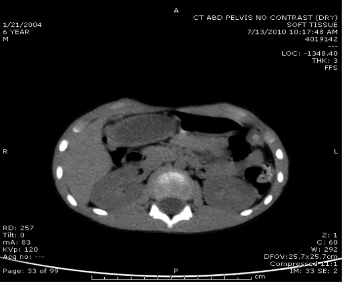
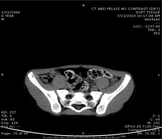
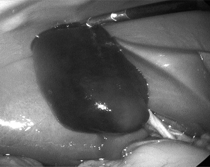
A 6-y-old previously healthy male presented to the emergency department with 1 d of worsening nausea, vomiting, and abdominal pain. The pain was crampy in character and extended across the lower abdomen. Physical examination findings included lethargy and abdominal distention without peritonitis. Laboratory evaluation was remarkable for a leukocytosis of 16,000 with a left shift. An abdominal CT scan (Figures 1 and 2) was remarkable for a thickened, horseshoe-shaped appendix with fluid in the pelvis suggestive of acute appendicitis with possible perforation. Preoperative discussion focused on laparoscopic or, possibly, open appendectomy with the possible need for a pelvic drain, if a pelvic abscess was identified. Two lower midline 5-mm trocars were placed as well as a 10-mm periumbilical trocar. Operative findings were remarkable for a small amount of turbid fluid adjacent to a healthy-appearing appendix. A markedly distended bladder was decompressed with an 8-F catheter to better evaluate the pelvis, which was otherwise unremarkable aside from the previously identified turbid fluid. The remainder of the abdomen was surveyed, at which time an ischemic GB was identified (Figure 3). The GB was completely peritonealized and appeared to have rotated on a pedicle comprised by the cystic duct and artery. Laparoscopic cholecystectomy and appendectomy were performed, and the patient's recovery was unremarkable. The GB pathology report noted the entire GB to have a dark red/purple appearance with a thickened wall. Dissection revealed thick, green bile and blood clot measuring up to 4 × 3cm filling the entire GB lumen with evidence of infarction. The appendix was healthy.
Figure 1.

Abdominal CT without contrast. The GB was interpreted as healthy on CT scan.
Figure 2.

Abdominal/pelvic CT. Appendicitis is suspected. It has likely ruptured, because the caliber is relatively small (5 mm), and there is a fairly large amount of fluid; some of the small fluid contains dots of free peritoneal air in the dependent-most portion of the pelvic free fluid.
Figure 3.

Intraoperative view of ischemic peritonealized GB demonstrating counterclockwise rotation on a GB pedicle comprised of the cystic duct and artery.
DISCUSSION
GB torsion is rare, with less than 400 reported cases in the literature and few treated laparoscopically.1,2,3 The youngest reported patient was 2 y old, with the peak incidence in the pediatric population occurring between the ages of 6 and 13 y with a 4:1 male-to-female preponderance.4 Specific clinical signs or symptoms are typically absent, and laboratory data may reveal nonspecific inflammatory changes. The challenge therefore is to correctly identify this unusual cause of abdominal pain that is commonly misdiagnosed. Levard et al.5 describe 2 of 9 patients in whom the diagnosis was missed and who subsequently died. Even with early diagnosis and surgical intervention, the mortality rate is estimated to be 5%.5
The differential diagnoses that should be considered for right upper quadrant abdominal pain include acute cholecystitis, peptic ulcer, intussusception, intestinal volvulus, and high retrocecal appendicitis as well as GB torsion.6 Although there have been reports in the literature that suggest conventional imaging modalities, such as ultrasound (US) and CT, are useful in the preoperative diagnosis of GB torsion, most cases are diagnosed intraoperatively.7 These reports suggest that radiographic features of GB torsion on abdominal CT scan include a fluid collection in the GB fossa, an unusual GB location with marked dilation of the GB, a well-enhanced cystic duct located on the right side of the GB, and inflammatory changes, such as edema with GB wall thickening.6,8 In this case, even in retrospect, the GB was distended, but not necessarily pathologic appearing on abdominal CT scan, highlighting the difficulty in diagnostic specificity. A preoperative US was not obtained because findings such as GB wall thickening or even an emphysematous gallbladder wall were not identified by abdominal CT scan. The pathologic evaluation identified full-thickness wall ischemia with impending necrosis with no evidence for perforation.
Multiple hypotheses have been proposed as the mechanism of GB torsion. From an anatomic perspective, torsion of the GB in children may be related to perturbation of the embryological migration of the GB, which leaves the organ abnormally mobile on a stalk consisting only of the cystic duct and artery.6 Two anatomic variants have been described: (1) a torsion-prone mesentery and (2) a mesentery supporting only the cystic duct allowing a completely peritonealized GB to hang free.9 In conjunction with an anomalous anatomic configuration, an inciting may even occur to initiate torsion of the cystic duct and artery pedicle.10 It has been suggested that events, such as violent peristaltic movements of neighboring organs, sudden body movements, or abdominal trauma may play a role in precipitating GB torsion. Carter et al.4 described 2 types of torsion: (1) incomplete torsion (rotation < 180 degrees) with gradual onset and (2) complete torsion (rotation > 180 degrees), with acute onset. Further, cholelithiasis is an infrequent cause of GB torsion, because one large study of 245 patients found gallstones in 24.4%.11 Torsion of the gallbladder leads to occlusive obstruction of biliary drainage and blood flow with subsequent ischemia and gangrenous changes.
CONCLUSION
This case report highlights the importance of laparoscopic evaluation of nonspecific abdominal pain when there is clinical and radiographic diagnostic uncertainty. Although the diagnosis of GB torsion with ischemia is rare, a delayed or missed diagnosis will contribute to increased morbidity and mortality.
Contributor Information
Trevor C. Farnsworth, Department of Physician Assistant Studies, SUNY Upstate Medical University, Syracuse, NY, USA..
Carl A. Weiss, III, General Surgery, Auburn Community Hospital, Auburn, NY, USA..
References:
- 1. Amarillo HA, Pirchi ED, Mihura ME. Complete gallbladder and cystic pedicle torsion. Laparoscopic diagnosis and treatment. Surg Endosc. 2003;17:832–833 [DOI] [PubMed] [Google Scholar]
- 2. Matsuda A, Sasajima K, Miyamoto M, et al. Laparoscopic treatment for torsion of the gallbladder in a 7-year-old female. JSLS. 2009;13:441–444 [PMC free article] [PubMed] [Google Scholar]
- 3. Chilton CP, Mann CV. Torsion of the gallbladder in a nine-year-old boy. J R Soc Med. 1980;73:141–143 [PMC free article] [PubMed] [Google Scholar]
- 4. Carter R, Thompson RJ, Jr., Brennan LP, Hinshaw DB. Volvulus of the gallbladder. Surg Gynecol Obstet. 1963;116:105–108 [PubMed] [Google Scholar]
- 5. Levard G, Weil D, Barret D, Barbier J. Torsion of the gallbladder in children. J Pediatr Surg. 1994;29:569–570 [DOI] [PubMed] [Google Scholar]
- 6. Kitagawa H, Nakada K, Enami T, et al. Two cases of torsion of the gallbladder diagnosed preoperatively. J Pediatr Surg. 1997;32:1567–1569 [DOI] [PubMed] [Google Scholar]
- 7. Kimura T, Yonekura T, Yamauchi K, Kosumi T, Sasaki T, Kamiyama M. Laparoscopic treatment of gallbladder volvulus: a pediatric case report and literature review. J Laparoendosc Adv Surg Techn A. 2008;18:330–334 [DOI] [PubMed] [Google Scholar]
- 8. Merine D, Meziane M, Fishman EK. CT diagnosis of gallbladder torsion. J Comput Assist Tomogr. 1987;11:712–713 [DOI] [PubMed] [Google Scholar]
- 9. Mouawad NJ, Crofts B, Streu R, Desrochers R, Kimball BC. Acute gallbladder torsion—a continued pre-operative diagnostic dilemma. World J Emerg Surg. 2001;6:13. [DOI] [PMC free article] [PubMed] [Google Scholar]
- 10. Shaikh AA, Charles A, Domingo S, Schaub G. Gallbladder volvulus: report of two original cases and review of the literature. Am Surg. 2005;71:87–89 [PubMed] [Google Scholar]
- 11. Nakao A, Matsuda T, Funabiki S, et al. Gallbladder torsion: case report and review of 245 cases reported in the japanese literature. J Hepatobiliary Pancreat Surg. 1999;6:418–421 [DOI] [PubMed] [Google Scholar]


