



Abstract
The Enterobacter cloacae species includes an extremely diverse group of bacteria that are associated with plants, soil and humans. Publication of the complete genome sequence of the plant growth-promoting endophytic E. cloacae subsp. cloacae ENHKU01 provided an opportunity to perform the first comparative genome analysis between strains of this dynamic species. Examination of the pan-genome of E. cloacae showed that the conserved core genome retains the general physiological and survival genes of the species, while genomic factors in plasmids and variable regions determine the virulence of the human pathogenic E. cloacae strain; additionally, the diversity of fimbriae contributes to variation in colonization and host determination of different E. cloacae strains. Comparative genome analysis further illustrated that E. cloacae strains possess multiple mechanisms for antagonistic action against other microorganisms, which involve the production of siderophores and various antimicrobial compounds, such as bacteriocins, chitinases and antibiotic resistance proteins. The presence of Type VI secretion systems is expected to provide further fitness advantages for E. cloacae in microbial competition, thus allowing it to survive in different environments. Competition assays were performed to support our observations in genomic analysis, where E. cloacae subsp. cloacae ENHKU01 demonstrated antagonistic activities against a wide range of plant pathogenic fungal and bacterial species.
Introduction
Enterobacter cloacae is a gram-negative Proteobacterium belonging to the Enterobacteriaceae family. Within this family, Enterobacter is most closely related to, and is grouped in a sub-clade with, Klebsiella . The two groups of bacteria diverged from a sub-clade consisting of Escherichia , Citrobacter and Salmonella [1].
The E. cloacae species comprises an extremely diverse group of bacteria that has been found in diverse environments, ranging from plants to soil to humans. Plant pathogenic strains of E. cloacae have been reported to cause Enterobacter bulb decay in onion plants and bacterial wilt in mulberry [2,3]; endophytic E. cloacae strains have been shown to colonize and benefit plant growth in various crops, such as soybean, cucumber, corn, rice and ginger [4,5]. Previous biological studies of several plant-origin isolates have shown that E. cloacae has antagonistic effects against the oomycete pathogen Pythium ultimum [6], the fungal pathogens Fusarium moniliforme and Fusarium oxysporum [5,7] and the bacterial pathogen Ralstonia solanacearum [8]. Additionally, several strains of E. cloacae are considered to be plant growth-promoting rhizobacteria (PGPR). The genomic features that underlie the antagonistic characteristics have been demonstrated in previous genome analyses of model PGPR species, such as the gram-negative Pseudomonas and the gram-positive Bacillus [9,10,11], but these features have not been studied in Enterobacter spp.
E. cloacae is best known as a human opportunistic pathogen that is commonly found in hospitals and causes a wide range of infections, such as lower respiratory tract infections, urinary tract infections and meningitis [12]. Outbreaks usually occur in Intensive Care Units, primarily affecting patients in vulnerable age groups and patients who are hospitalized for a prolonged period. E. cloacae is clinically significant, particularly because its strains usually carry multiple antibiotic resistance genes [13,14]. The complete genome sequence was published for E. cloacae subsp. cloacae ATCC13047, a classic strain isolated in the last century from human brain fluids, but no detailed analysis or supporting data were reported [15].
Compared with other genera in the family of Enterobacteriaceae such as Escherichia , Salmonella and Yesinia, which are well-known for their association with pathogenicity in humans and animals, there is scarcity of genomic data for Enterobacter . Eight complete Enterobacter genomes have been reported to NCBI (http://www.ncbi.nlm.nih.gov/genome/1219): E. aerogenes KCTC2190, E. cloacae subsp. cloacae ATCC13047, E. cloacae subsp. dissolvens SDM, E. cloacae subsp. cloacae NCTC9394, E . lignolyticus SCF1 (named as E. cloacae SCF1 in NCBI database), E. asburiae LF7a, E. cloacae EcWSU1 and Enterobacter sp. 638. These genome sequencing projects mostly emphasized the potential for the use of Enterobacter in industrial applications, such as lignin degradation [1,16] and 2,3-butanediol production [17,18,19]. A detailed genome study illustrated the synergistic interactions between the poplar tree host and the growth promoting endophyte Enterobacter sp. 638 [18]. Four of the available Enterobacter genomes belong to E. cloacae, which include human opportunistic pathogens E. cloacae subsp. cloacae NCTC9394 and E. cloacae subsp. cloacae ATCC13047 [15], a plant pathogen E. cloacae EcWSU1 [20] and a 2,3-butanediol producing E. cloacae subsp. dissolvens SDM [19].
Using the complete genome sequence of the plant growth-promoting endophytic E. cloacae subsp. cloacae ENHKU01 and the available complete genome sequence data from different strains of E. cloacae [15,19,20,21], we have performed a comparative genome analysis between E. cloacae strains that originated from diverse environments. In this study, we identified crucial conserved genomic factors that support the antagonistic functions of E. cloacae. Genomic factors differentiating the various strains of E. cloacae were also investigated. Also, this study is the first comprehensive comparative genome analysis for E. cloacae genomes.
Materials and Methods
Discovery of E. cloacae subsp. cloacae ENHKU01
E. cloacae subsp. cloacae ENHKU01 was isolated from a pepper plant infected by R. solanacearum in Hong Kong in May 2010. Three single colony subcultures were performed to obtain a pure isolate on TTC (2,3,5-triphenyl- tetrazolium chloride) medium w/v 1.5% agar plates at 28°C. An inoculation and re-isolation experiment using 4-week old tomato and pepper seedlings was carried out to confirm its endophytic characteristic in plants. Unlike R. solanacearum, the bacterial isolate did not cause wilting symptoms in these plants following inoculation. Inoculation on potato tubers and onion bulbs also indicated that the isolate did not cause disease in these plants. Sequencing and BLAST analysis of partial 16S rRNA and housekeeping genes (fusA, gyrB, hsp60, rpoB) [22] was conducted for classification, and the bacterial strain was shown to belong to E. cloacae and was most closely related to E. cloacae subsp. cloacae ATCC13047.
Genome sequencing and comparative analysis
Whole genome sequencing and annotation of E. cloacae subsp. cloacae ENHKU01 was performed as described in Liu et al., 2012 [21]. In brief, de novo shotgun sequencing and paired-end sequencing strategies were applied to produce the whole genome sequence of ENHKU01 using the 454 GS Junior platform (454 Life Sciences, Branford, CT, USA). Newbler Assembler software (454 Life Sciences, Branford, CT, USA) was used to construct a draft genome with one scaffold containing 36 contigs [23]. To complete the whole-genome sequence of E. cloacae subsp. cloacae ENHKU01, sequence gaps were filled by PCR, primer walking and Sanger sequencing.
Gene annotation and analysis was performed using NCBI Prokaryotic Genomes Automatic Annotation Pipeline (PGAAP) [24]. Annotation was performed using BLASTP, and the protein sequences of predicted genes were searched against all proteins from complete microbial genomes and aligned with the best BLAST-hit [25]. Genome sequence data have been deposited to NCBI, GenBank accession number is CP003737.1, and it is available for download at NCBI: ftp://ftp.ncbi.nlm.nih.gov/genomes/Bacteria/Enterobacter_cloacae_ENHKU01_uid172463/.
For comparative genome analysis, complete genome sequence data of Enterobacter species: E. aerogenes KCTC2190 (GenBank accession CP002824), E. cloacae subsp. cloacae ATCC13047 (GenBank accession CP001918, CP001919 and CP001920), E. cloacae subsp. dissolvens SDM (GenBank accession CP003678), E . lignolyticus SCF1 (named as E. cloacae SCF1 in NCBI database) (GenBank accession CP002272), E. asburiae LF7a (CP003026, CP003027 and CP003028), E. cloacae EcWSU1 (GenBank accession CP002886 and CP002887) and Enterobacter sp. 638 (GenBank accession CP000653 and CP000654) were obtained from NCBI (ftp://ftp.ncbi.nlm.nih.gov/genomes/Bacteria/). E. cloacae subsp. cloacae NCTC9394 sequence data were not available for download; thus, it was not included in this study. Scaffolds of the Enterobacter species involved in this study were also uploaded to the Rapid Annotations using Subsystems Technology (RAST) server for SEED-based automated annotation, whole-genome sequence-based comparative analysis and Kyoto Encyclopedia of Genes and Genomes (KEGG) metabolic pathway for comparative analysis [26]. Efficient Database framework for comparative Genome Analyses using BLAST score Ratios (EDGAR) was used for core genome, pan genome and singleton analysis, and Venn diagram construction using E. cloacae subsp. cloacae ENHKU01 as a reference genome [27]. Further comparative analysis was performed for specific regions and genes-of-interest by BLASTN, BLASTX and BLASTP.
Phylogenomic analysis of Enterobacter spp
An in-house pipeline has been developed for the phylogenomic analysis of Enterobacter spp. Scaffolds and genome data of the chromosome of eight Enterobacter genomes and three Pantoea genomes: Pantoea sp. At-9b (NC_014837), P . vagans C9-1 (NC_014562) and P . ananatis LMG20103 (NC_013956), were downloaded from NCBI (ftp://ftp.ncbi.nlm.nih.gov/genomes/). Gene clustering was performed using OrthoMCL 2.0 under default parameters [28]. A minimum length coverage filter of 85% was applied to further confirm orthology and the families that passed were aligned by MUSCLE v3.8.31 [29]. The corresponding amino acid alignments of 1732 core genes were created and then concatenated to construct a “supergene” for the reconstruction of the phylogenomic tree of Enterobacter spp. by MrBayes v3.2 using the WAG model. Ten million generations were performed with four chains; burn-in was set to 10,000 generations [30].
Phylogenetic analysis. To analyze the phylogeny of specific genes-of-interest, partial- or full-length CDS of E. cloacae subsp. cloacae ENHKU01 was used as a bait to search for orthologs in the NCBI database by BLASTP/ BLASTX using an E-value cut-off of 1e-10. Protein sequences of the top 100 hits were obtained. MEGA5 was used to perform phylogenetic analysis, multiple alignments of protein sequences were built by CLUSTALW and a 100 bootstrap replicate Neighbor-joining tree was constructed for phylogenetic analysis [31].
Phylogenetic analysis of the Type VI Secretion System (T6SS) was performed using a similar strategy as described in previous studies with modifications [32]. T6SS component genes were searched in the Integrated Microbial Genome database version IMG 3.4 in Joint Genome Institute (http://img.jgi.doe.gov) with 1350 finished bacterial genomes. T6SS clusters containing core component genes COG0542 (clpV) with at least four of the following loci: COG3516 (ImpB), COG3517 (ImpC), COG3519 (VasA), COG3520 (VasB) and COG3522 (ImpJ/VasE) were included for further analysis. T6SS clusters with fewer than five core component genes were discarded. As a result, a total of 346 T6SS clusters were identified in 230 bacterial genomes, mostly belonging to the five subdivisions of Proteobacteria. Two T6SSs of ENHKU01 were manually identified and reconfirmed by BLAST and included in the analysis. MEGA5 was used to perform phylogenetic analysis for the ClpV orthologs in a total of 348 T6SS [31]. Multiple alignments of ClpV protein sequences were built by CLUSTALW, and a Neighbor-joining tree was constructed for evolutionary analysis.
Isolation of other bacterial, fungal isolates and antagonistic bioassays
Bioassays were designed to investigate the antagonistic activities of E. cloacae subsp. cloacae ENHKU01 against various plant bacterial and fungal pathogens. All biological samples involved in this study were collected within a 200 km zone in Hong Kong SAR and Guangdong, China. Plant pathogenic fungi Alternaria sp., Choanephorainfundibulifera , Colletotrichum capsici , Didymellabryoniae , Fusarium oxysporum, Sclerotinia sclerotiorum and Sclerotinia rolfsii were isolated from infected host plants and purified by subsequent subculture on potato dextrose agar (PDA) plates at 28°C; next, pure isolates were obtained and strain identification was confirmed by cloning and sequencing (Clover Seed Co., Ltd., Hong Kong). For fungal antagonistic assays, each challenging fungus was grown for 4 days at 28°C. A sample composed of 0.5 cm of agar with hyphae was cut and placed at 2 cm from the edge of a new potato dextrose agar (PDA) plate. An overnight culture of ENHKU01 was streaked across the middle of the plate [33]. Controls were set up for challenging fungus without E. cloacae subsp. cloacae ENHKU01. Fungal growth was monitored for two weeks. Three independent experiments were performed for each E. cloacae subsp. cloacae ENHKU01-fungal antagonistic assay.
The bacterial isolate, Ralstonia solanacearum, was isolated from the same pepper plant as the E. cloacae subsp. cloacae ENHKU01 isolate. Three single-colony subcultures were performed to obtain a pure isolate on TTC medium w/v 1.5% agar plates at 28°C. Identification of R. solanacearum was confirmed by cloning and sequencing of partial 16S rRNA and fulfilling Koch’s Postulates (Clover Seed Co., Ltd., Hong Kong) [34]. E. cloacae subsp. cloacae ENHKU01- R. solanacearum competition assays were performed in both planktonic and biofilm cultures. To prepare for the competition assay, bacterial cultures were grown overnight in TTC medium and adjusted to OD600=0.1(~1 x 10⁸ c.f.u/ml). For the planktonic culture, equal volumes (25 ml) of ENHKU01 and R. solanacearum were mixed in a 1:1 ratio in a 250 ml flask and incubated in a shaking incubator (250rpm at 28°C). One milliliter of culture was collected at 2, 4, 6 and 24 hours after incubation for quantification. For the biofilm culture, ENHKU01 and R. solanacearum were mixed in a 1:10 ratio. 20 µl of ENHKU01 culture was placed on a TTC w/v 1.5% agar plate and incubated at 28°C. Biofilm samples were collected for quantification using 1 ml pipette tips and re-suspended in water after 24 hours of incubation. Controls consisted of samples containing only ENHKU01 or R. solanacearum for both planktonic and biofilm competition assays. For quantification (including initial concentration of ENHKU01 and R. solanacearum), serial dilutions were prepared for each sample to a desirable concentration and plated on TTC w/v 1.5% agar. C.F.U.s were counted after incubation at 28°C for 48 hours. ENHKU01 and R. solanacearum can be distinguished by morphological differences in size, color and texture. Four replications were performed for each assay.
Results and Discussion
Phylogenomic analysis of Enterobacter
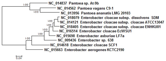
The classification of Enterobacter spp. using 16S rRNA and house-keeping genes has often been inconsistent [22]. It has also been a great challenge for researchers to distinguish the environmental and the pathogenic strains of E. cloacae in previous studies [22]. Phylogenomic analysis was performed using 1732 core genes of eight Enterobacter spp. and three Pantoea spp . The four functionally distinctive strains of E. cloacae (the plant growth promoting E. cloacae subsp. cloacae ENHKU01 (ENHKU01) ,the plant pathogen E. cloacae EcWSU1 (EcWSU1), the opportunistic human pathogen E. cloacae subsp. cloacae ATCC13047 (ATCC13047) and the 2,3-butanediol producing E. cloacae subsp. dissolvens SDM (SDM)) were clustered together, and this observation is consistent with previous studies of the phylogenetic relationships of E. cloacae [22](Figure 1).
Figure 1. Phylogenomic analysis of Enterobacter spp. Bayesian tree with posterior p-values of the genomes of Enterobacter spp. using 1732 core genes of eight Enterobacter and three Pantoea genomes.
General properties of the E. cloacae genomes
The general genome properties of ENHKU01, EcWSU1, ATCC13047 and SDM are shown in (Table 1). The genome of E. cloacae subsp. cloacae ENHKU01 is composed solely of a single 4.72-Mbp chromosome, and a total of 4338 protein coding regions (CDS) were predicted, with 87% being connected to Clusters of Orthologous Groups (COGs). The genome size, total number of genes and predicted CDS of ENHKU01 is slightly smaller but similar to that of EcWSU1 and SDM, while ATCC13047 appears to have a larger genome of ~5.31 Mb. The genome expansion of ATCC13047 is due to the addition of two plasmids and more than 20 genomic variable regions in its chromosome. EcWSU01 contains one plasmid, and SDM lacks plasmids. There is an absence of sequence similarity between the plasmids in different strains of E. cloacae. The genomic GC content of the strains ranges between 54.5-55.1%. MAUVE analysis showed an overall collinear relationship across E. cloacae strains, despite a large scale chromosomal reorganization that occurred in ATCC13047 that was conferred by a single recombination event and resulted in the inversion of a genomic region (Figure 2) [35].
Table 1. Summary of genome sequence projects of E. cloacae.
| Strain | E. cloacae subsp. cloacae ENHKU01 | E. cloacae subsp. cloacae ATCC13047 | E. cloacae EcWSU1 | E. cloacae subsp. dissolvens SDM |
|---|---|---|---|---|
| Size | 4.73 | 5.6 | 4.8 | 4.97 |
| No. of Chromosome | 1 | 1 | 1 | 1 |
| No. of Plasmid | 0 | 2 | 1 | 0 |
| GC content % | 55.1 | 54.6 | 54.5 | 55.1 |
| Total genes | 4445 | 5639 | 4740 | 4646 |
| Predicted CDS | 4338 | 5518 | 4619 | 4542 |
| No. of tRNAs | 82 | 24 | 83 | 53 |
| No. of rRNA operons | 8 | 8 | 8 | 3 |
| Host | Plant | Human | Plant | Soil |
| Important feature | Endophyte | Human opportunistic pathogen | Plant pathogen | 2,3-butanediol production |
Figure 2. Genomic alignment of Enterobacter cloacae.
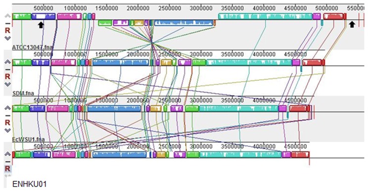
MAUVE [35] alignment of the genome sequences of E. cloacae subsp. cloacae ATCC13047, E. cloacae subsp. dissolvens SDM, E. cloacae EcWSU1 and E. cloacae subsp. cloacae ENHKU01. Same color boxes represent homologous regions of sequence, without rearrangement (locally collinear blocks or LCB), shared between E. cloacae genomes. Black arrows show the genomic position of the T4SS located in ATCC13047.
Major subsystems and metabolic pathways are conserved between E. cloacae strains; however, the number of genes is increased in ATCC13047 in several functional categories compared to other E. cloacae strains (Figure 3). E. cloacae strains, similar to other Enterobacter , are characterized by their ability to use a wide range of carbon sources through their diverse carbohydrate metabolic pathways and transport systems [18]. Over 640 of annotated genes, accounting for 13-15% of the E. cloacae genomes, had a designated role for carbohydrate utilization (Figure 3; Data S1), this number is comparable to that of the related genomes in the family of Enterobacteriacae [36,37,38].
Figure 3. Comparison of subsystem features between E. cloacae.
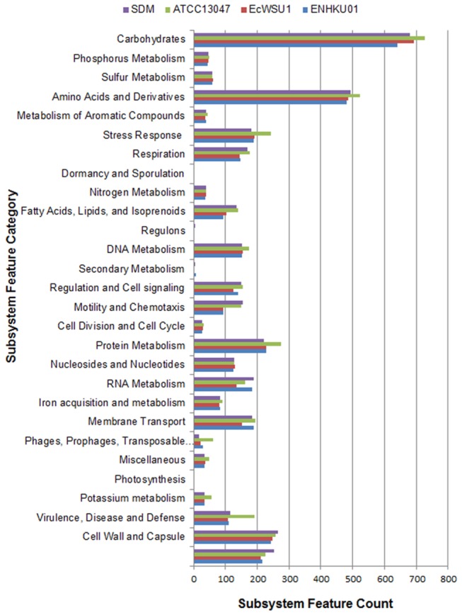
Genome sequences of ATCC13047, SDM, EcWSU1 and ENHKU01 were uploaded to the SEED Viewer server (http://rast.nmpdr.org/seedviewer.cgi) independently. Functional roles of RAST annotated genes were assigned and grouped in subsystem feature categories as shown in the figure [26], and colored bars indicate the number of genes assigned to each category. Details for subsystem and functional role assignment for the genes of each strain are listed in Data S1.
Pan-genome analysis was performed for eight Enterobacter species, and a further comparison was performed for E. cloacae strains (Table S1). As shown in the Venn diagram (Figure 4; Data S2), the four E. cloacae strains shared 3540 CDS in their core genome, corresponding to approximately 64%-82% of all CDS in these genomes. A relatively small portion of CDS were shared between two or three E. cloacae. Both ENHKU01 and SDM had approximately 6% of unique CDS that are absent in the other E. cloacae genomes evaluated, and the unique CDS percentage was 12% and 20% for EcWSU1 and ATCC13047, respectively. The majority of singletons were mostly associated with hypothetical proteins (Data S3), and this observation is consistent with singleton analysis of other species [39,40]. However, a larger number of functional singletons, most likely contributing to virulence, were identified in ATCC13047 (File 4).
Figure 4. Venn diagram of four E. cloacae strains.
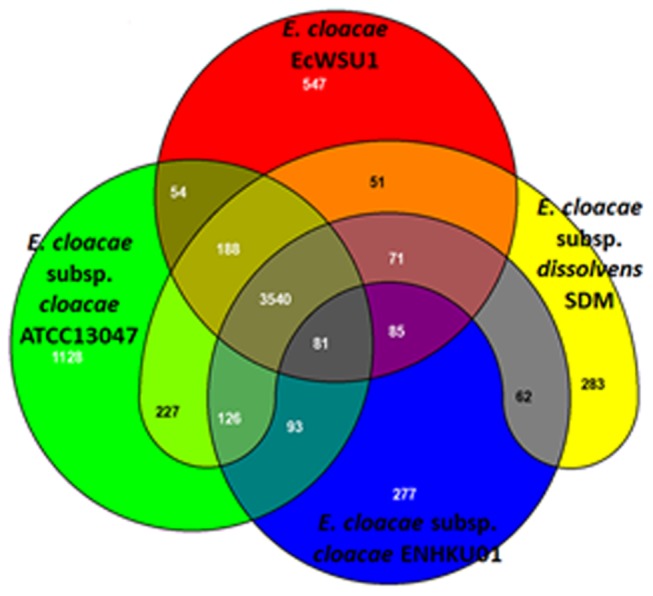
The Venn diagram shows the pan-genome of ATCC13047, SDM, EcWSU1 and ENHKU01 generated using EDGAR [27]. Overlapped regions represent common CDS shared between the E. cloacae genomes. The number outside the overlapped regions indicates the number of CDS in each genome without homologs in other sequenced E. cloacae genomes.
Virulence associated genes
The virulence genes of pathogenic bacteria are often associated with pathogenicity islands that encode a Type III secretion system (T3SS) or a Type IV secretion system (T4SS) acquired by horizontal gene transfer [41,42]. These genomic factors related to pathogenesis and virulence were identified in the plasmid and variable regions in the opportunistic human pathogen E. cloacae subsp. cloacae ATCC13047, and they are absent in the other E. cloacae strains. Two clusters of T4SS genes were found in the plasmid pECL_A and a 139-kb variable region of ATCC13047 in the chromosome, respectively (Figure 2; Table S1). T4SS is associated with pathogenesis in plants and mammalian bacterial pathogens and contributes to genome plasticity in bacteria [43,44]. DNA, effector proteins and virulence factors are delivered to the target host cells by the conjugative transfer machine, although several non-pathogenic strains also carry the membrane transfer system [43,45]. The plasmid pECL_A also contains multiple heavy metal resistance operons for copper, tellurium and mercury that are not conserved with other Enterobacter species but share notably high homology to Cronobacter sakazakii , Klebseilla pneumoniae and Escherichia coli (data not shown), which are all species commonly known as human pathogens or opportunistic pathogens. ATCC13047 is believed to acquire multiple heavy metal resistance genes horizontally from other human microflora associated bacteria, thereby contributing to the adaptation and fitness of the bacteria in heavy metal-rich environments, such as sewage [15].
The pathogenesis of many plant pathogens often involves T3SS and its associated effector proteins [46]. Although E. cloacae EcWSU1 was reported to be a plant pathogen that causes Enterobacter bulb decay in the onion, no T3SS or T4SS was observed in either the plasmid or the chromosome. It remains largely unclear how EcWSU1 became a plant pathogen.
Diversity of fimbriae contributes to variation in colonization and host determination of different E. cloacae strains
Adhesion to and colonization of the host is one of the key factors for host determination for symbiotic and endophytic bacteria and determining the success of pathogenesis for pathogenic bacteria [47,48]. Fimbriae, also known as pili, are widely distributed among the Proteobacteria and are known to be critical in adhesion and the specific binding to tissues of preferred hosts [49]. Located in the outer membrane, fimbriae subunits are usually assembled into filament structures using the chaperone/usher pathway [47]. We compared the fimbriae in different strains of E. cloacae and observed significant diversity. Nine to thirteen fimbrial protein-encoding loci were identified in each strain, but only four of them were conserved across all four E. cloacae strains (Table 2; Table S1). ATCC13047 and EcWSU1 each had two unique fimbrial clusters, and four of the ENHKU01 fimbrial clusters are absent in other E. cloacae (Table 2; File 2). The presence of multiple fimbriae genes indicates that E. cloacae are likely able to colonize a rather wide range of hosts or environments. Variation in fimbriae between different strains of E. cloacae is expected to alter the choice of hosts or environmental niches that E. cloacae strain can colonize and thus to contribute to the diversity of the species.
Table 2. Comparison of fimbriae in E. cloacae.
|
E. cloacae strain |
|||||
|---|---|---|---|---|---|
| Pan genome position | Representing Locus | ECENHK | EcWSU1 | ATCC13047 | SDM |
| 28-31 | ECENHK_00140-00155 | x | |||
| 625-629 | ECENHK_03165-03185 | x | x | ||
| 662-667 | ECENHK_03350-03375 | x | x | ||
| 809-815 | ECENHK_04095-04125 | x | x | x | |
| 827-830 | ECENHK_04185-04200 | x | |||
| 1096-1104 | ECENHK_05530-05570 | x | x | x | x |
| 1377-1381 | ECENHK_06955-06975 | x | x | x | x |
| 1691-1696 | ECENHK_08555-08580 | x | |||
| 1842-1846 | ECENHK_09330-09350 | x | x | x | x |
| 2718-2721 | ECENHK_13830-13845 | x | x | x | x |
| 3633-3636 | ECENHK_18495-18510 | x | |||
| 3728-3731 | ECENHK_18970-18985 | x | x | x | |
| 3900-3903 | ECENHK_19830-19845 | x | x | x | |
| 4417-4420 | ECL_04371-4368 | x | |||
| 4512-4516 | ECL_01105-01109 | x | |||
| 4918-4922 | ECL_03396-03400 | x | x | ||
| 5476-5479 | EcWSU1_00874-00877 | x | |||
| 5612-5616 | EcWSU1_02030-02034 | x | |||
| 6068-6072 | ECL_00070-00074 | x | x | ||
| 6079-6083 | ECL_00089-00093 | x | x | ||
| 6118-6122 | ECL_00370-00374 | x | x | ||
| Total number of fimbriae | 13 | 9 | 13 | 10 | |
One of the fimbrial loci in ATCC13047 and SDM, encoding the Colonization Factor Antigen I (CFA/I) fimbrial proteins (ECL_00070-00074), has drawn our attention. The locus is located in a cluster of genes encoding the virulence related resistance-nodulation-division (RND) efflux system within a variable region that is absent in the plant-associated ENHKU01 and EcWSU1strains [50]. CFA/I fimbriae, as a member of alpha-fimbriae belonging to Class 5 fimbriae, have been shown to play a critical role in colonizing the epithelia of the human intestine in the enterotoxigenic strains of Escherichia coli (ETEC) [51]. BLAST and alignment analyses further demonstrated that the fimbria are highly associated with human-related Enterobacter species [52] (Figure S1), but they are rarely found in plant-associated strains. The Enterobacter CFA/I fimbriae is most closely related to Escherichia coli (Figure S1), which illustrates that the CFA/I fimbrial gene cluster is a viable candidate for facilitating Enterobacter adhesion and colonization in humans. Such a specific correlation was not observed in the other fimbriae found in E. cloacae.
Antagonistic potential of E. cloacae: Microbial competition for resources
Microbes compete with each other for limited resources within their communities [53,54]. Certain microbes, primarily demonstrated for Pseudomonas spp., compete with other microbes for acquiring ferric ion (iron), an essential growth element from soil. These microbes produce higher affinity siderophores and indirectly suppress the growth of competing fungi, which produce lower affinity fungal siderophores, and non-siderophore producing bacteria [55,56]. Resource competition mechanisms thus lead to antagonistic effects in the rhizosphere. A conserved pathway for enterobacin synthesis was found across Enterobacter spp. In addition, E. cloacae strains possess an extra siderophore assembly kit for aerobactin siderophore biosynthesis, which is absent in certain other Enterobacter spp. (Table 3; Table S1).
Table 3. Key genes involved in potential antagonistic activities in Enterobacter .
|
E. cloacae strains |
Other Enterobacter
|
|||||||||
|---|---|---|---|---|---|---|---|---|---|---|
| Pan genome position | Description | Representing locus | ENHKU01 | EcWSU1 | ATCC13047 | SDM | LF7a | KCTC 2190 | SCF1 | 638 |
| Siderophores | ||||||||||
| 2810-2814 | Aerobactin siderophore biosynthesis | ECENHK_14300-14320 | + | + | + | + | - | - | - | - |
| 1196-1208 | Enterobactin synthesis | ECENHK_06040-06100 | + | + | + | + | + | + | + | + |
| Linoleic acid metabolic pathway | ||||||||||
| 4185 | Lysophospholipase L2 | ECENHK_21265 | + | + | + | + | + | + | + | + |
| 4189 | Phospholipase A | ECENHK_21285 | + | + | + | + | + | + | + | + |
| Type II secretion system | ||||||||||
| 1457-1468 | Type II secretion pathway (General secretion pathway) | ECENHK_07365-07425 | + | + | + | + | + | - | + | - |
| Chitinase | ||||||||||
| 1470 | Chitinase/ glycoside hydrolase family protein | ECENHK_07430 | + | + | + | + | + | - | + | - |
| 1472 | Chitinase | ECENHK_07440 | + | + | + | + | + | - | + | - |
| 1763 | Chitinase/ glycoside hydrolase family protein | ECENHK_08915 | + | + | + | + | + | + | - | - |
| Colicin V and Bacteriocin Production | ||||||||||
| 387-388 | Entericidin A/ B | ECENHK_01955-01960 | + | + | + | + | + | - | + | + |
| 570 | S-type Pyocin domain-containing protein | ECENHK_02880 | + | - | + | + | + | - | - | - |
| 681-684 | Tolerance to Colicin E2 | ECENHK_03445-03460 | + | + | + | + | + | + | + | + |
| 3073-3078 | Colicin V production protein & bacteriocin production cluster | ECENHK_15640-156470 | + | + | + | + | + | + | + | + |
Other resources subjected to microbial competition include fatty acids. Earlier functional studies demonstrated that E. cloacae competes for the seed exudate linoleic acid against P . ultimum , which suppresses sporangium germination of the oomycete pathogen and causes the damping-off of seedlings in many crop plants [57]. In this study, we have identified two candidate genes in the linoleic acid metabolic pathway that could play an important role in fatty acid competition. Phospholipase A1 Precursor (E.C. 3.1.1.4) and Lysophospholipase L2 (EC 3.1.1.5) are expected to hydrolyze lecithin (Table 3; File 2), which is an exudate lipid molecule composed of choline, linoleic acid, phosphorus and inositol, and initiate the fungal/ oomycete responses of seeds. Both genes are located adjunct to a multiple sugar transporter and are found across different Enterobacter genomes [57].
Chitinases
The ability of microbes to produce a wide range of antimicrobial compounds, including lytic agents, antibiotics, bacteriocins, protein exotoxins and other secondary metabolites, is critical to their success in antagonistic activities. In bacterial-fungal interactions, cell wall hydrolases, such as certain proteases and chitinases, are produced and secreted extracellularly by bacteria to target fungal cell walls [58,59,60]. Chitinases are lytic enzymes that break down glycosidic bonds in chitin, a major component of fungal cell walls, and have been demonstrated to effectively inhibit fungal growth. Three conserved putative chitinase genes have been identified across E. cloacae genomes. Two of these genes are located adjacent to a type II secretion system [61], while the third one is located elsewhere (Table 3; Table S1). The Type II secretion system and the associated chitinases are absent in Enterobacter sp. 638 and E . lignolyticus SCF1. Phylogenetic analysis has shown that ECENHK_07430 and the synthetic Enterobacter chitinases are associated with the well-characterized Chitinase A in Serratia marcescens [62,63] (Figure 5), which has been demonstrated to control several important plant pathogenic fungi including Botrytis spp., Rhizoctonia solani, Fusarium oxysporum f. sp. cyclaminis and Sclerotinia spp. In fact, a functional Chitinase A that effectively controls Rhizoctonia solani has been identified in E. agglomerans [64]. The other two chitinases have not been reported in any E. cloacae isolates. Orthologs of ECENHK_08915, can also be found in several related species of Klebsiella , Citrobacter and Serretia, but it is absent in other family members of Enterobacteriaceae. Interestingly, phylogenetic analysis further demonstrated the association between the Enterobacter ’s chitinase and Chitinase A1 of the gram-positive Bacillus spp. (Figure 5). It is notable that Bacillus spp. are well-known as plant growth-promoting rhizobacteria (PGPR), as they have antagonistic effects against other microorganisms and are used commercially as biological control agents for various fungal pathogens [65,66].
Figure 5. Phylogenetic analysis of chitinase genes.

Neighbor-joining tree with bootstrap values of chitinase genes associated with (A) ECENHK_07430 and (B) ECENHK_08915 constructed by twenty-five representing orthologs from each species using MEGA5. The blue box indicates the corresponding chitinase gene of ENHKU01. Chitinases that have been functionally characterized for their antifungal activities are highlighted in green boxes [87,88,89,90,91].
Production of other antimicrobial compounds
Several bacteria and fungi exhibit antagonistic effects in microbial competition through the production of antibiotics [67]. In microbial-microbial competition, other bacteria also develop detoxification mechanisms and/or antibiotic resistance to counteract anti-microbial compounds. Previous genome analysis demonstrated Klebsiella and Enterobacter possess a number of antibiotic resistance genes and multidrug efflux systems [37,68], these multiple antimicrobial mechanisms were also observed in E. cloacae (Table S2). One can expect, due to the wide range of antimicrobial resistance, multidrug resistance proteins and multidrug efflux systems benefit the survival of E. cloacae against both bacterial and fungal competitors in habitats with high microbial competition, and furthermore, this phenomenon explains how clinical strains of E. cloacae raise medical concerns [13,14].
The production of bacteriocins also plays an important role, particularly in bacteria-bacteria interactions [69]. Well-characterized mechanisms in gram-negative bacteria include the enteric bacteriocin, colicin [70] and the lipoprotein Entericidin [71]. Antimicrobial compounds identified in E. cloacae that are related to bacteriocin production involve the Colicin V and bacteriocin production cluster [69], tolerance to colicinE2, Entericidin A and B [71] and S-type Pyocin (Table 3; Table S1). Additionally, other bacteriocin production-related genes were found to be associated with the Type VI secretion system (T6SS) in E. cloacae.
Type VI secretion systems and their roles in microbial competition
The T6SS is widely distributed in gram-negative bacteria. A T6SS usually involves approximately 15 conserved genes in a cluster with genes encoding a functional secretory apparatus that penetrates cell membranes and translocates effector proteins into their eukaryotic hosts or recipient cells [72,73]. Earlier studies have related the T6SS to virulence in humans and animals [74]; however, recent studies have demonstrated the directed function of the T6SS toward microbial interaction and fitness in microflora [75,76,77,78]. It is common to find more than one cluster of T6SS genes within a genome [32,79,80]; both ATCC13047 and ENHKU01 contain two clusters of T6SS, while SDM and EcWSU1 have only one. Each T6SS gene cluster is composed of sets of conserved core component genes and variable regions distinguished by hypothetical proteins (Figure 6). A recent discovery allowed us to understand more about the roles of these hypothetical proteins in microbial competition as anti-bacterial effectors. Genetics and characterization of the bacteriolytic effectors Tse1, Tse2 and Tse3 in Psuedomonas aeruginosa revealed that T6SS effector proteins target the peptidoglycan of prokaryotic cells [76]. Within the variable region of E. cloacae T6SS, we found RhsB (a rhs-like genetic element involved in bacteriocin production) [81,82,83] and LysM (involved in bacterial cell wall degradation) [84,85] (Figure 6). It is believed that bacteria deliver different effector proteins to their specific target counterparts among the microflora using a conserved T6SS apparatus [76,83].
Figure 6. Genetic organization of T6SS in Enterobacter .

(A) and (B) show genetic organization of the two T6SS clusters commonly found in Enterobacter . ENHKU01 and ATCC13047 contain both T6SS, and EcWSU1, SDM and LF7a have one of the two, and other Enterobacter have none. The two clusters of T6SS in Enterobacter have different genetic organization but are aligned across the Enterobacter genomes at the corresponding loci. Each T6SS cluster is composed of conserved regions formed by conserved T6SS core component genes, which are indicated in solid blue/ green color boxes, and variable regions that are indicated by arrows. The variable regions contain a variable number of conserved genes (solid gray color) and unique genes (white color boxes). Most genes located in the variable regions are described as hypothetical proteins. Genes possibly involved in bacteriocin activity are shown in red. Details of the genetic organization of T6SSs in Enterobacter are listed in Data S4.
The sequences and genetic organization of core component genes in each T6SS cluster are aligned across the genomes of Enterobacter , but the T6SS clusters within a genome are not associated with one another (Figure 6; Data S4). There is also variation in the core component genes of each cluster (Figure 6). Such differentiation indicates that these T6SSs are not only evolutionarily distinct but also functionally and structurally different from each other [32]. Phylogenetic analysis of the representative T6SS component gene ortholog, ClpV, indicated that two evolutionary distinct T6SSs were introduced into E. cloacae independently before divergence, as ClpV orthologs of E. cloacae are distributed in two clades (Clade II and Clade III) (Figure 7A). In clade III, orthologs are clustered with those found in other plant and soil-associated bacteria of Enterobacteriaceae, such as the plant pathogens Erwinia amylovora and Pectobacteria wasabiae , and soil-borne bacteria, such as Pantoea vagans C9-1 and Erwinia pyrifoliae Ep1/96 (Figure 7B); also, there are many human and animal pathogenic/associated bacteria in this family, such as Escherichia coli, Klebsiella pneumoniae, Yersinia pestis are located in Clade II (Figure 7C). Our observation of the correlation between the phylogeny of T6SSs and the ecological niches of bacteria is in line with a previous T6SS phylogenetic analysis study [86].
Figure 7. Phylogenetic analysis of ClpV.

(A) The neighbor-joining tree of T6SSs using ClpV orthologs from 348 T6SS clusters in 231 species. The 231 species are grouped according to their class, which is indicated using color lines: α for the subdivision of Proteobacteria (Pink), β subdivision (Green), γ subdivision (Blue), δ/α subdivision (Black) and other bacteria unrelated to Proteobacteria (Gray). ClpV orthologs are distributed in five clades and named I-V. Naming of clades is according to Boyer et al [86]. Each clade is contributed by different bacterial families of Proteobacteria. Our result is consistent with a previous phylogenetic analysis of T6SS using 13 T6SS conserved component genes [86]. ClpV of E. cloacae are distributed in clades II and III of the phylogenetic tree and are clustered together with other strains and species in the family of Enterobacteriaceae possessing T6SS, thus forming sub-trees in each clade (indicated by gray circles). A simplified and enlarged version of the neighbor-joining tree with bootstrap values showing (B) the sub-tree of clade III formed by ClpV orthologs associated with ECENHK_13140 and (C) the sub-tree of clade II associated with ECENHK_15865. Color squares indicate the habitats of the corresponding bacterial species: plant/ soil-associated (light green), insect-associated (green) and Human/ animal associated (orange).
The presence of T6SS in E. cloacae strains is expected to allow the bacterial strain to enhance the survival of non-pathogenic strains in their habitats, thus competing with other microbes for the colonization of hosts [75]. E. cloacae strains possessing more than one distinct T6SS cluster, such as ATCC13047 and ENHKU01, are likely to have fitness advantages in a borader range of habitats and be able to survive in different environments.
Antagonistic activities of E. cloacae subsp. cloacae ENHKU01
Through genome analysis, the antagonistic potential of E. cloacae has been revealed in this study, and we further hypothesized that E. cloacae has advantages in microbial competition against other microbes within its environmental niche. To investigate the effect of antagonistic activities in E. cloacae strains further, we performed bacterial-fungal and bacterial-bacterial antagonistic assays using the plant growth-promoting endophytic ENHKU01 as a model. As expected, ENHKU01 showed significant antagonistic effects against a wide range of fungal species: Collectotricumcapsici . Sclerotinia sclerotiorum, Alternaria sp., Didymellabryoniae , and Fusarium oxysporum by suppressing their growth at different levels (Figure 8; Table S3). These fungal species are common plant pathogens, and C . capsici and S. sclerotiorum are direct competitors with ENHKU01, sharing the pepper plant as a host. Additionally, in our bacteria-bacteria competition assay, it was demonstrated that ENHKU01 effectively suppressed the growth of Ralstonia solanacearum, a devastating bacterial pathogen with a wide host range that is most well-known for causing bacterial wilt in Solanaceous crops, such as tomato, pepper and eggplant (Figure 9). Taken together, these bioassays provide further evidence, drawn from comparative genome analysis, demonstrating the antagonistic potential of E. cloacae.
Figure 8. Antagonistic activity of E. cloacae subsp. cloacae ENHKU01 against fungi.

(A) Visualization of fungal growth with and without ENHKU01: (from left to right, upper row) Alternaria sp., Colletotrichum capsici , Didymellabryoniae , (from left to right, lower row) Fusarium oxysporum and Sclerotinia sclerotiorum. Photos were taken 7 days after incubation; (B) Growth of Colletotrichum capsici (Col) and Sclerotiniascleotiorum (Scl) were closely monitored with and without ENHKU01. Challenging fungi were grown on PDA plates as described in Methods and Materials, the radius of growth of hyphae (in cm) was measured. Numbers show an average of 10 plates, and error bars represent the S.D. from the mean.
Figure 9. Antagonistic activity of E. cloacae subsp. cloacae ENHKU01 against R. solanacearum.
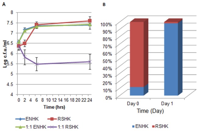
Figures show results of ENHKU01 – R. solanacearum competition assays: (A) the planktonic culture with the quantities of E. cloacae subsp. cloacae ENHKU01 (ENHK) and R. solanacearum (RSHK) in log C.F.U. per ml recorded at 0, 2, 4, 6 and 24 hours after incubation. Numbers show an average of 4 replications, and error bars represent the S.D. from the mean; (B) Biofilm culture, relative percentage of E. cloacae subsp. cloacae ENHKU01 (ENHK): R. solanacearum (RSHK) at day 0 and day 1 of incubation.
Conclusions
Comparative genome analysis reveals the antagonistic potential of E. cloacae. The multiple antagonistic mechanisms of E. cloacae are expected to contribute its success in competition against other microbes in various environmental niches, thus allowing different strains of E. cloacae to survive in diverse environments.
Supporting Information
The functional roles of RAST annotated genes were curated into subsystems by SEED Viewer, and the details are listed in this file. The numbers of genes grouped in each subsystem feature category for each E. cloacae strain were counted as shown in the summary page.
(XLSX)
List of singletons of ENHKU01, EcWSU1, ATCC13047 and SDM generated by comparing the genomes of the four of E. cloacae strains using E. cloacae subsp. cloacae ENHKU01 as the reference genome [27].
(XLSX)
List of genes associated with Type VI Secretion Systems in Enterobacter , corresponding to Figure 6A and Figure 6B.
(XLSX)
The comparative analysis of the CFA/I Fimbrial cluster.
(DOCX)
Pan-genome analysis of Enterobacter by EDGAR.In brief, E. cloacae subsp. cloacae ENHKU01 was selected as reference genome and its gene contents were taken as the base set for stepwise comparison of the seven Enterobacter genomes involved in this study. For each round of all-against-all comparison based on a bidirectional best BLAST hit, every gene in a Enterobacter genome that has no ortholog in the base set was added to the reference set and compared with the next genome. The process was repeated step by step to obtain the pan-genome of Enterobacter [27].
(XLSX)
List of genes involved in potential antibiotic resistance, multidrug resistance and multidrug efflux systems in Enterobacter .
(XLSX)
Table shows antagonistic activities of E. cloacae subsp. cloacae ENHKU01 against plant pathogenic fungal species.
(DOCX)
Funding Statement
This project was partially supported by the Strategic Research Theme of Infection and Immunology, Initiative of Clean Energy and Environment, The University of Hong Kong and a start-up fund for Bioinformatics Center, Nanjing Agricultural University. The funders had no role in study design, data collection and analysis, decision to publish, or preparation of the manuscript.
References
- 1. Deangelis KM, D’Haeseleer P, Chivian D, Fortney JL, Khudyakov J et al. (2011) Complete genome sequence of "Enterobacter lignolyticus" SCF1. Standards Genomics Sciences 5: 69-85. doi:10.4056/sigs.2104875. [DOI] [PMC free article] [PubMed] [Google Scholar]
- 2. Zhu B, Lou MM, Xie GL, Wang GF, Zhou Q et al. (2011) Enterobacter mori sp. nov., associated with bacterial wilt on Morus alba L. Int J Syst Evol Microbiol 61: 2769-2774. doi:10.1099/ijs.0.028613-0. [DOI] [PubMed] [Google Scholar]
- 3. Schroeder BK, Waters TD, du Toit LJ (2010) Evaluation of Onion Cultivars for Resistance to Enterobacter cloacae in Storage. Plant Dis 94: 236-243. doi:10.1094/PDIS-94-2-0236. [DOI] [PubMed] [Google Scholar]
- 4. Liu S, Hu X, Lohrke SM, Baker CJ, Buyer JS et al. (2007) Role of sdhA and pfkA and catabolism of reduced carbon during colonization of cucumber roots by Enterobacter cloacae . Microbiology 153: 3196-3209. doi:10.1099/mic.0.2006/005538-0. [DOI] [PubMed] [Google Scholar]
- 5. Hinton DM, Bacon CW (1995) Enterobacter cloacae is an endophytic symbiont of corn. Mycopathologia 129: 117-125. doi:10.1007/BF01103471. PubMed: 7659140. [DOI] [PubMed] [Google Scholar]
- 6. Nelson EB (1988) Biological Control of Pythium Seed Rot and Preemergence Damping-Off of Cotton with Enterobacter cloacae and Erwinia herbicola Applied as Seed Treatments. Plant Dis 72: 140-142. doi:10.1094/PD-72-0140. [Google Scholar]
- 7. Suárez-Estrella F, Vargas-García C, López MJ, Capel C, Morenoa J (2007) Antagonistic activity of bacteria and fungi from horticultural compost against Fusarium oxysporum f. sp. melonis . Crop Protect 26: 46-53. doi:10.1016/j.cropro.2006.04.003. [Google Scholar]
- 8. Xue QY, Chen Y, Li SM, Chen LF, Ding GC et al. (2009) Evaluation of the strains of Acinetobacter and Enterobacter as potential biocontrol agents against Ralstonia wilt of tomato. Biol Contr 48: 252-258. doi:10.1016/j.biocontrol.2008.11.004. [Google Scholar]
- 9. Chen XH, Koumoutsi A, Scholz R, Eisenreich A, Schneider K et al. (2007) Comparative analysis of the complete genome sequence of the plant growth-promoting bacterium Bacillus amyloliquefaciens FZB42. Nat Biotechnol 25: 1007-1014. doi:10.1038/nbt1325. PubMed: 17704766. [DOI] [PubMed] [Google Scholar]
- 10. Bloemberg GV, Lugtenberg BJJ (2001) Molecular basis of plant growth promotion and biocontrol by rhizobacteria. Curr Opin Plant Biol 4: 343-350. doi:10.1016/S1369-5266(00)00183-7. [DOI] [PubMed] [Google Scholar]
- 11. Paulsen IT, Press CM, Ravel J, Kobayashi DY, Myers GS et al. (2005) Complete genome sequence of the plant commensal Pseudomonas fluorescens Pf-5. Nat Biotechnol 23: 873-878. doi:10.1038/nbt1110. [DOI] [PMC free article] [PubMed] [Google Scholar]
- 12. Keller R, Pedroso MZ, Ritchmann R, Silva RM (1998) Occurrence of virulence-associated properties in Enterobacter cloacae . Infect Immun 66: 645-649. [DOI] [PMC free article] [PubMed] [Google Scholar]
- 13. Leverstein-van Hall MA, Blok HE, Paauw A, Fluit AC, Troelstra A et al. (2006) Extensive hospital-wide spread of a multidrug-resistant Enterobacter cloacae clone, with late detection due to a variable antibiogram and frequent patient transfer. J Clin Microbiol 44: 518-524. doi:10.1128/JCM.44.2.518-524.2006. [DOI] [PMC free article] [PubMed] [Google Scholar]
- 14. Paauw A, Verhoef J, Fluit AC, Blok HE, Hopmans TE et al. (2007) Failure to control an outbreak of qnrA1-positive multidrug-resistant Enterobacter cloacae infection despite adequate implementation of recommended infection control measures. J Clin Microbiol 45: 1420-1425. doi:10.1128/JCM.02242-06. [DOI] [PMC free article] [PubMed] [Google Scholar]
- 15. Ren Y, Zhou Z, Guo X, Li Y, Feng L et al. (2010) Complete genome sequence of Enterobacter cloacae subsp. cloacae type strain ATCC 13047. J Bacteriol 192: 2463-2464. doi:10.1128/JB.00067-10. [DOI] [PMC free article] [PubMed] [Google Scholar]
- 16. Manter DK, Hunter WJ, Vivanco JM (2011) Enterobacter soli sp. nov.: a lignin-degrading gamma-proteobacteria isolated from soil. Curr Microbiol 62: 1044-1049. doi:10.1007/s00284-010-9809-9. [DOI] [PubMed] [Google Scholar]
- 17. Shin SH, Kim S, Kim JY, Lee S, Um Y et al. (2012) Complete Genome Sequence of Enterobacter aerogenes KCTC 2190. J Bacteriol 194: 2373-2374. doi:10.1128/JB.00028-12. PubMed: 22493190. [DOI] [PMC free article] [PubMed] [Google Scholar]
- 18. Taghavi S, van der Lelie D, Hoffman A, Zhang YB, Walla MD et al. (2010) Genome sequence of the plant growth promoting endophytic bacterium Enterobacter sp.. PLOS Genet 638: 6: e1000943 [DOI] [PMC free article] [PubMed] [Google Scholar]
- 19. Xu Y, Wang A, Tao F, Su F, Tang H et al. (2012) Genome sequence of Enterobacter cloacae subsp. dissolvens SDM, an efficient biomass-utilizing producer of platform chemical 2,3-butanediol. J Bacteriol 194: 897-898. doi:10.1128/JB.06495-11. PubMed: 22275097. [DOI] [PMC free article] [PubMed] [Google Scholar]
- 20. Humann JL, Wildung M, Cheng C-H, Lee T, Steqart JE et al. (2011) Complete genome of the onion Enterobacter cloacae EcWSU1. Standards Genomics Sciences 5. [DOI] [PMC free article] [PubMed] [Google Scholar]
- 21. Liu WY, Chung KM, Wong CF, Jiang JW, Hui RK et al. (2012) Complete genome sequence of the endophytic Enterobacter cloacae subsp. cloacae strain ENHKU01. J Bacteriol 194: 5965. doi:10.1128/JB.01394-12. PubMed: 23045485. [DOI] [PMC free article] [PubMed] [Google Scholar]
- 22. Paauw A, Caspers MP, Schuren FH, Leverstein-van Hall MA, Delétoile A et al. (2008) Genomic diversity within the Enterobacter cloacae complex. PLOS ONE 3: e3018. doi:10.1371/journal.pone.0003018. PubMed: 18716657. [DOI] [PMC free article] [PubMed] [Google Scholar]
- 23. Li J, Jiang J, Leung FC (2012) 6-10x pyrosequencing is a practical approach for whole prokaryote genome studies. Gene 494: 57-64. doi:10.1016/j.gene.2011.11.051. PubMed: 22192914. [DOI] [PubMed] [Google Scholar]
- 24. Angiuoli SV, Gussman A, Klimke W, Cochrane G, Field D et al. (2008) Toward an online repository of Standard Operating Procedures (SOPs) for (meta)genomic annotation. Omics J Integr Biol 12: 137-141. doi:10.1089/omi.2008.0017. PubMed: 18416670. [DOI] [PMC free article] [PubMed] [Google Scholar]
- 25. McGinnis S, Madden TL (2004) BLAST: at the core of a powerful and diverse set of sequence analysis tools. Nucleic Acids Res 32: W20-W25. doi:10.1093/nar/gnh003. PubMed: 15215342. [DOI] [PMC free article] [PubMed] [Google Scholar]
- 26. Aziz RK, Bartels D, Best AA, DeJongh M, Disz T et al. (2008) The RAST Server: rapid annotations using subsystems technology. BMC Genomics 9: 75. doi:10.1186/1471-2164-9-75. PubMed: 18261238. [DOI] [PMC free article] [PubMed] [Google Scholar]
- 27. Blom J, Albaum SP, Doppmeier D, Pühler A, Vorhölter FJ et al. (2009) EDGAR: a software framework for the comparative analysis of prokaryotic genomes. BMC Bioinformatics 10: 154. doi:10.1186/1471-2105-10-154. PubMed: 19457249. [DOI] [PMC free article] [PubMed] [Google Scholar]
- 28. Chen F, Mackey AJ, Stoeckert CJ Jr., Roos DS (2006) OrthoMCL-DB: querying a comprehensive multi-species collection of ortholog groups. Nucleic Acids Res 34: D363-D368. doi:10.1093/nar/gkj123. PubMed: 16381887. [DOI] [PMC free article] [PubMed] [Google Scholar]
- 29. Edgar RC (2004) MUSCLE: multiple sequence alignment with high accuracy and high throughput. Nucleic Acids Res 32: 1792-1797. doi:10.1093/nar/gkh340. PubMed: 15034147. [DOI] [PMC free article] [PubMed] [Google Scholar]
- 30. Ronquist F, Huelsenbeck JP (2003) MrBayes 3: Bayesian phylogenetic inference under mixed models. Bioinformatics 19: 1572-1574. doi:10.1093/bioinformatics/btg180. PubMed: 12912839. [DOI] [PubMed] [Google Scholar]
- 31. Tamura K, Peterson D, Peterson N, Stecher G, Nei M et al. (2011) MEGA5: molecular evolutionary genetics analysis using maximum likelihood, evolutionary distance, and maximum parsimony methods. Mol Biol Evol 28: 2731-2739. doi:10.1093/molbev/msr121. PubMed: 21546353. [DOI] [PMC free article] [PubMed] [Google Scholar]
- 32. Schwarz S, West TE, Boyer F, Chiang WC, Carl MA et al. (2010) Burkholderia type VI secretion systems have distinct roles in eukaryotic and bacterial cell interactions. PLOS Pathog 6: e1001068 PubMed: 20865170. [DOI] [PMC free article] [PubMed] [Google Scholar]
- 33. Mela F, Fritsche K, de Boer W, van Veen JA, de Graaff LH et al. (2011) Dual transcriptional profiling of a bacterial/fungal confrontation: Collimonas fungivorans versus Aspergillus niger . ISME J 5: 1494-1504. doi:10.1038/ismej.2011.29. PubMed: 21614084. [DOI] [PMC free article] [PubMed] [Google Scholar]
- 34. Agrios GN (2005) Plant Pathology. In: Agrios GN. Elsevier Academic Press; pp. 26-27. [Google Scholar]
- 35. Darling AC, Mau B, Blattner FR, Perna NT (2004) Mauve: multiple alignment of conserved genomic sequence with rearrangements. Genome Res 14: 1394-1403. doi:10.1101/gr.2289704. PubMed: 15231754. [DOI] [PMC free article] [PubMed] [Google Scholar]
- 36. Smits TH, Rezzonico F, Pelludat C, Goesmann A, Frey JE et al. (2010) Genomic and phenotypic characterization of a nonpigmented variant of Pantoea vagans biocontrol strain C9-1 lacking the 530-kb megaplasmid pPag3. FEMS Microbiol Lett 308: 48-54. doi:10.1111/j.1574-6968.2010.01994.x. PubMed: 20487014. [DOI] [PubMed] [Google Scholar]
- 37. Fouts DE, Tyler HL, DeBoy RT, Daugherty S, Ren Q et al. (2008) Complete genome sequence of the N2-fixing broad host range endophyte Klebsiella pneumoniae 342 and virulence predictions verified in mice. PLOS Genet 4: e1000141 PubMed: 18654632. [DOI] [PMC free article] [PubMed] [Google Scholar]
- 38. Jiang LQ, Fang Z, Guo F, Yang LB (2012) Production of 2,3-butanediol from acid hydrolysates of Jatropha hulls with Klebsiella oxytoca . Bioresour Technol 107: 405-410. doi:10.1016/j.biortech.2011.12.083. PubMed: 22230777. [DOI] [PubMed] [Google Scholar]
- 39. Smits TH, Jaenicke S, Rezzonico F, Kamber T, Goesmann A et al. (2010) Complete genome sequence of the fire blight pathogen Erwinia pyrifoliae DSM 12163T and comparative genomic insights into plant pathogenicity. BMC Genomics 11: 2. doi:10.1186/1471-2164-11-S4-S2. PubMed: 20047678. [DOI] [PMC free article] [PubMed] [Google Scholar]
- 40. Joseph S, Desai P, Ji Y, Cummings CA, Shih R et al. (2012) Comparative analysis of genome sequences covering the seven Cronobacter species. PLOS ONE 7: e49455. doi:10.1371/journal.pone.0049455. PubMed: 23166675. [DOI] [PMC free article] [PubMed] [Google Scholar]
- 41. Juhas M, van der Meer JR, Gaillard M, Harding RM, Hood DW et al. (2009) Genomic islands: tools of bacterial horizontal gene transfer and evolution. FEMS Microbiol Rev 33: 376-393. doi:10.1111/j.1574-6976.2008.00136.x. PubMed: 19178566. [DOI] [PMC free article] [PubMed] [Google Scholar]
- 42. Dobrindt U, Hochhut B, Hentschel U, Hacker J (2004) Genomic islands in pathogenic and environmental microorganisms. Nat Rev Microbiol 2: 414-424. doi:10.1038/nrmicro884. PubMed: 15100694. [DOI] [PubMed] [Google Scholar]
- 43. Christie PJ, Cascales E (2005) Structural and dynamic properties of bacterial type IV secretion systems (review ). Mol Membr Biol 22: 51-61 doi:10.1080/09687860500063316. PubMed: 16092524. [DOI] [PMC free article] [PubMed] [Google Scholar]
- 44. Christie PJ, Atmakuri K, Krishnamoorthy V, Jakubowski S, Cascales E (2005) Biogenesis, architecture, and function of bacterial type IV secretion systems. Annu Rev Microbiol 59: 451-485. doi:10.1146/annurev.micro.58.030603.123630. PubMed: 16153176. [DOI] [PMC free article] [PubMed] [Google Scholar]
- 45. Christie PJ, Vogel JP (2000) Bacterial type IV secretion: conjugation systems adapted to deliver effector molecules to host cells. Trends Microbiol 8: 354-360. doi:10.1016/S0966-842X(00)01792-3. PubMed: 10920394. [DOI] [PMC free article] [PubMed] [Google Scholar]
- 46. Alfano JR, Collmer A (2004) Type III secretion system effector proteins: double agents in bacterial disease and plant defense. Annu Rev Phytopathol 42: 385-414. doi:10.1146/annurev.phyto.42.040103.110731. PubMed: 15283671. [DOI] [PubMed] [Google Scholar]
- 47. Nuccio SP, Bäumler AJ (2007) Evolution of the chaperone/usher assembly pathway: fimbrial classification goes Greek. Microbiol Mol Biol Rev MMBR 71: 551-575. doi:10.1128/MMBR.00014-07. PubMed: 18063717. [DOI] [PMC free article] [PubMed] [Google Scholar]
- 48. Pizarro-Cerdá J, Cossart P (2006) Bacterial adhesion and entry into host cells. Cell 124: 715-727. doi:10.1016/j.cell.2006.02.012. PubMed: 16497583. [DOI] [PubMed] [Google Scholar]
- 49. Tsolis RM (2002) Comparative genome analysis of the alpha -proteobacteria: relationships between plant and animal pathogens and host specificity. Proc Natl Acad Sci U S A 99: 12503-12505. doi:10.1073/pnas.212508599. PubMed: 12271145. [DOI] [PMC free article] [PubMed] [Google Scholar]
- 50. Blair JM, Piddock LJ (2009) Structure, function and inhibition of RND efflux pumps in Gram-negative bacteria: an update. Curr Opin Microbiol 12: 512-519. doi:10.1016/j.mib.2009.07.003. PubMed: 19664953. [DOI] [PubMed] [Google Scholar]
- 51. Li YF, Poole S, Nishio K, Jang K, Rasulova F et al. (2009) Structure of CFA/I fimbriae from enterotoxigenic Escherichia coli. Proc Natl Acad Sci U S A 106: 10793-10798. doi:10.1073/pnas.0812843106. PubMed: 19515814. [DOI] [PMC free article] [PubMed] [Google Scholar]
- 52. Hoffmann H, Stindl S, Ludwig W, Stumpf A, Mehlen A et al. (2005) Enterobacter hormaechei subsp. oharae subsp. nov., E. hormaechei subsp. hormaechei comb. nov., and E. hormaechei subsp. steigerwaltii subsp. nov., three new subspecies of clinical importance. J Clin Microbiol 43: 3297-3303. doi:10.1128/JCM.43.7.3297-3303.2005. PubMed: 16000451. [DOI] [PMC free article] [PubMed] [Google Scholar]
- 53. Hibbing ME, Fuqua C, Parsek MR, Peterson SB (2010) Bacterial competition: surviving and thriving in the microbial jungle. Nat Rev Microbiol 8: 15-25. doi:10.1038/nrmicro2259. PubMed: 19946288. [DOI] [PMC free article] [PubMed] [Google Scholar]
- 54. Raaijmakers JM, Paulitz TC, Steinberg C, Alabouvette C, Moenne-Loccoz Y (2009) The rhizosphere: a playground and battlefield for soilborne pathogens and beneficial microorganisms. Plant Soil 321: 341-361. doi:10.1007/s11104-008-9568-6. [Google Scholar]
- 55. Kloepper JW, Leong J, Teintze M, Schroth MN (1980) Pseudomonas Siderophores - a Mechanism Explaining Disease-Suppressive Soils. Curr Microbiol 4: 317-320. doi:10.1007/BF02602840. [Google Scholar]
- 56. Compant S, Duffy B, Nowak J, Clément C, Barka EA (2005) Use of plant growth-promoting bacteria for biocontrol of plant diseases: principles, mechanisms of action, and future prospects. Appl Environ Microbiol 71: 4951-4959. doi:10.1128/AEM.71.9.4951-4959.2005. PubMed: 16151072. [DOI] [PMC free article] [PubMed] [Google Scholar]
- 57. van Dijk K, Nelson EB (2000) Fatty acid competition as a mechanism by which Enterobacter cloacae suppresses Pythium ultimum sporangium germination and damping-off. Appl Environ Microbiol 66: 5340-5347. doi:10.1128/AEM.66.12.5340-5347.2000. PubMed: 11097912. [DOI] [PMC free article] [PubMed] [Google Scholar]
- 58. Gomaa EZ (2012) Chitinase production by Bacillus thuringiensis and Bacillus licheniformis: their potential in antifungal biocontrol. J Microbiol 50: 103-111. doi:10.1007/s12275-012-1343-y. PubMed: 22367944. [DOI] [PubMed] [Google Scholar]
- 59. Jones JD, Grady KL, Suslow TV, Bedbrook JR (1986) Isolation and characterization of genes encoding two chitinase enzymes from Serratia marcescens . EMBO J 5: 467-473. PubMed: 16453672. [DOI] [PMC free article] [PubMed] [Google Scholar]
- 60. Bhattacharya D, Nagpure A, Gupta RK (2007) Bacterial chitinases: Properties and potential. Crit Rev Biotechnol 27: 21-28. doi:10.1080/07388550601168223. PubMed: 17364687. [DOI] [PubMed] [Google Scholar]
- 61. Francetic O, Belin D, Badaut C, Pugsley AP (2000) Expression of the endogenous type II secretion pathway in Escherichia coli leads to chitinase secretion. EMBO J 19: 6697-6703. doi:10.1093/emboj/19.24.6697. PubMed: 11118204. [DOI] [PMC free article] [PubMed] [Google Scholar]
- 62. Zees AC, Pyrpassopoulos S, Vorgias CE (2009) Insights into the role of the (alpha+beta) insertion in the TIM-barrel catalytic domain, regarding the stability and the enzymatic activity of chitinase A from Serratia marcescens . Biochim Biophys Acta 1794: 23-31. doi:10.1016/j.bbapap.2008.09.018. PubMed: 18973833. [DOI] [PubMed] [Google Scholar]
- 63. Norberg AL, Dybvik AI, Zakariassen H, Mormann M, Peter-Katalinić J et al. (2011) Substrate positioning in chitinase A, a processive chito-biohydrolase from Serratia marcescens . FEBS Lett 585: 2339-2344. doi:10.1016/j.febslet.2011.06.002. PubMed: 21683074. [DOI] [PubMed] [Google Scholar]
- 64. Chernin LS, De la Fuente L, Sobolev V, Haran S, Vorgias CE et al. (1997) Molecular cloning, structural analysis, and expression in Escherichia coli of a chitinase gene from Enterobacter agglomerans . Appl Environ Microbiol 63: 834-839. PubMed: 9055404. [DOI] [PMC free article] [PubMed] [Google Scholar]
- 65. Challacombe JF, Altherr MR, Xie G, Bhotika SS, Brown N et al. (2007) The complete genome sequence of Bacillus thuringiensis Al Hakam. J Bacteriol 189: 3680-3681. doi:10.1128/JB.00241-07. PubMed: 17337577. [DOI] [PMC free article] [PubMed] [Google Scholar]
- 66. Lugtenberg B, Kamilova F (2009) Plant-growth-promoting rhizobacteria. Annu Rev Microbiol 63: 541-556. doi:10.1146/annurev.micro.62.081307.162918. PubMed: 19575558. [DOI] [PubMed] [Google Scholar]
- 67. Garbeva P, Tyc O, Remus-Emsermann MN, van der Wal A, Vos M et al. (2011) No apparent costs for facultative antibiotic production by the soil bacterium Pseudomonas fluorescens Pf0-1. PLOS ONE 6: e27266. doi:10.1371/journal.pone.0027266. PubMed: 22110622. [DOI] [PMC free article] [PubMed] [Google Scholar]
- 68. Diene SM, Merhej V, Henry M, El Filali A, Roux V et al. (2012) The Rhizome of the Multidrug-Resistant Enterobacter aerogenes Genome Reveals How New "Killer Bugs" Are Created because of a Sympatric Lifestyle. Mol Biol Evol. [DOI] [PubMed] [Google Scholar]
- 69. Riley MA, Wertz JE (2002) Bacteriocin diversity: ecological and evolutionary perspectives. Biochimie 84: 357-364. doi:10.1016/S0300-9084(02)01421-9. PubMed: 12423779. [DOI] [PubMed] [Google Scholar]
- 70. Riley MA, Wertz JE (2002) Bacteriocins: evolution, ecology, and application. Annu Rev Microbiol 56: 117-137. doi:10.1146/annurev.micro.56.012302.161024. PubMed: 12142491. [DOI] [PubMed] [Google Scholar]
- 71. Bishop RE, Leskiw BK, Hodges RS, Kay CM, Weiner JH (1998) The entericidin locus of Escherichia coli and its implications for programmed bacterial cell death. J Mol Biol 280: 583-596. doi:10.1006/jmbi.1998.1894. PubMed: 9677290. [DOI] [PubMed] [Google Scholar]
- 72. Cascales E (2008) The type VI secretion toolkit. EMBO Rep 9: 735-741. doi:10.1038/embor.2008.131. PubMed: 18617888. [DOI] [PMC free article] [PubMed] [Google Scholar]
- 73. Pukatzki S, McAuley SB, Miyata ST (2009) The type VI secretion system: translocation of effectors and effector-domains. Curr Opin Microbiol 12: 11-17. doi:10.1016/j.mib.2008.11.010. PubMed: 19162533. [DOI] [PubMed] [Google Scholar]
- 74. Bingle LE, Bailey CM, Pallen MJ (2008) Type VI secretion: a beginner’s guide. Curr Opin Microbiol 11: 3-8. doi:10.1016/j.mib.2008.01.006. PubMed: 18289922. [DOI] [PubMed] [Google Scholar]
- 75. Schwarz S, Hood RD, Mougous JD (2010) What is type VI secretion doing in all those bugs? Trends Microbiol 18: 531-537. doi:10.1016/j.tim.2010.09.001. PubMed: 20961764. [DOI] [PMC free article] [PubMed] [Google Scholar]
- 76. Russell AB, Hood RD, Bui NK, LeRoux M, Vollmer W et al. (2011) Type VI secretion delivers bacteriolytic effectors to target cells. Nature 475: 343-347. doi:10.1038/nature10244. PubMed: 21776080. [DOI] [PMC free article] [PubMed] [Google Scholar]
- 77. Filloux A, Hachani A, Bleves S (2008) The bacterial type VI secretion machine: yet another player for protein transport across membranes. Microbiology 154: 1570-1583. doi:10.1099/mic.0.2008/016840-0. PubMed: 18524912. [DOI] [PubMed] [Google Scholar]
- 78. Jani AJ, Cotter PA (2010) Type VI secretion: not just for pathogenesis anymore. Cell Host Microbe 8: 2-6. doi:10.1016/j.chom.2010.06.012. PubMed: 20638635. [DOI] [PMC free article] [PubMed] [Google Scholar]
- 79. Blondel CJ, Jiménez JC, Contreras I, Santiviago CA (2009) Comparative genomic analysis uncovers 3 novel loci encoding type six secretion systems differentially distributed in Salmonella serotypes. BMC Genomics 10: 354. doi:10.1186/1471-2164-10-354. PubMed: 19653904. [DOI] [PMC free article] [PubMed] [Google Scholar]
- 80. Sarris PF, Zoumadakis C, Panopoulos NJ, Scoulica EV (2011) Distribution of the putative type VI secretion system core genes in Klebsiella spp. Infect Genet Evol J Mol Epidemiol Evol Genet Infect Dis 11: 157-166. doi:10.1016/j.meegid.2010.09.006. PubMed: 20932940. [DOI] [PubMed] [Google Scholar]
- 81. Aoki SK, Diner EJ, de Roodenbeke CT, Burgess BR, Poole SJ et al. (2010) A widespread family of polymorphic contact-dependent toxin delivery systems in bacteria. Nature 468: 439-442. doi:10.1038/nature09490. PubMed: 21085179. [DOI] [PMC free article] [PubMed] [Google Scholar]
- 82. Sisto A, Cipriani MG, Morea M, Lonigro SL, Valerio F et al. (2010) An Rhs-like genetic element is involved in bacteriocin production by Pseudomonas savastanoi pv. savastanoi . Antonie Van Leeuwenhoek 98: 505-517. doi:10.1007/s10482-010-9468-7. PubMed: 20563849. [DOI] [PubMed] [Google Scholar]
- 83. Poole SJ, Diner EJ, Aoki SK, Braaten BA, t’Kint de Roodenbeke C, et al. (2011) Identification of functional toxin/immunity genes linked to contact-dependent growth inhibition(CDI) and rearrangement hotspot (Rhs) systems. PLoS genetics 7: e1002217 [DOI] [PMC free article] [PubMed] [Google Scholar]
- 84. Buist G, Steen A, Kok J, Kuipers OP (2008) LysM, a widely distributed protein motif for binding to (peptido)glycans. Mol Microbiol 68: 838-847. doi:10.1111/j.1365-2958.2008.06211.x. PubMed: 18430080. [DOI] [PubMed] [Google Scholar]
- 85. Aramini JM, Rossi P, Huang YJ, Zhao L, Jiang M et al. (2008) Solution NMR structure of the NlpC/P60 domain of lipoprotein Spr from Escherichia coli: structural evidence for a novel cysteine peptidase catalytic triad. Biochemistry 47: 9715-9717. doi:10.1021/bi8010779. PubMed: 18715016. [DOI] [PubMed] [Google Scholar]
- 86. Boyer F, Fichant G, Berthod J, Vandenbrouck Y, Attree I (2009) Dissecting the bacterial type VI secretion system by a genome wide in silico analysis: what can be learned from available microbial genomic resources? BMC Genomics 10: 104. doi:10.1186/1471-2164-10-104. PubMed: 19284603. [DOI] [PMC free article] [PubMed] [Google Scholar]
- 87. Watanabe T, Suzuki K, Oyanagi W, Ohnishi K, Tanaka H (1990) Gene cloning of chitinase A1 from Bacillus circulans WL-12 revealed its evolutionary relationship to Serratia chitinase and to the type III homology units of fibronectin. J Biol Chem 265: 15659-15665. PubMed: 2203782. [PubMed] [Google Scholar]
- 88. Chang WT, Chen CS, Wang SL (2003) An antifungal chitinase produced by Bacillus cereus with shrimp and crab shell powder as a carbon source. Curr Microbiol 47: 102-108. doi:10.1007/s00284-002-3955-7. PubMed: 14506855. [DOI] [PubMed] [Google Scholar]
- 89. Mehmood MA, Xiao X, Hafeez FY, Gai YB, Wang FP (2009) Purification and characterization of a chitinase from Serratia proteamaculans . World J Microbiol Biotechnol 25: 1955-1961. doi:10.1007/s11274-009-0094-3. [Google Scholar]
- 90. Frankowski J, Lorito M, Scala F, Schmid R, Berg G et al. (2001) Purification and properties of two chitinolytic enzymes of Serratia plymuthica HRO-C48. Arch Microbiol 176: 421-426. doi:10.1007/s002030100347. PubMed: 11734885. [DOI] [PubMed] [Google Scholar]
- 91. Brurberg MB, Eijsink VG, Nes IF (1994) Characterization of a chitinase gene (chiA) from Serratia marcescens BJL200 and one-step purification of the gene product. FEMS Microbiol Lett 124: 399-404. doi:10.1111/j.1574-6968.1994.tb07315.x. PubMed: 7851747. [DOI] [PubMed] [Google Scholar]
Associated Data
This section collects any data citations, data availability statements, or supplementary materials included in this article.
Supplementary Materials
The functional roles of RAST annotated genes were curated into subsystems by SEED Viewer, and the details are listed in this file. The numbers of genes grouped in each subsystem feature category for each E. cloacae strain were counted as shown in the summary page.
(XLSX)
List of singletons of ENHKU01, EcWSU1, ATCC13047 and SDM generated by comparing the genomes of the four of E. cloacae strains using E. cloacae subsp. cloacae ENHKU01 as the reference genome [27].
(XLSX)
List of genes associated with Type VI Secretion Systems in Enterobacter , corresponding to Figure 6A and Figure 6B.
(XLSX)
The comparative analysis of the CFA/I Fimbrial cluster.
(DOCX)
Pan-genome analysis of Enterobacter by EDGAR.In brief, E. cloacae subsp. cloacae ENHKU01 was selected as reference genome and its gene contents were taken as the base set for stepwise comparison of the seven Enterobacter genomes involved in this study. For each round of all-against-all comparison based on a bidirectional best BLAST hit, every gene in a Enterobacter genome that has no ortholog in the base set was added to the reference set and compared with the next genome. The process was repeated step by step to obtain the pan-genome of Enterobacter [27].
(XLSX)
List of genes involved in potential antibiotic resistance, multidrug resistance and multidrug efflux systems in Enterobacter .
(XLSX)
Table shows antagonistic activities of E. cloacae subsp. cloacae ENHKU01 against plant pathogenic fungal species.
(DOCX)