Abstract
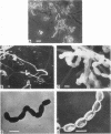
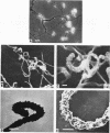
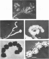
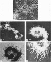
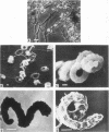
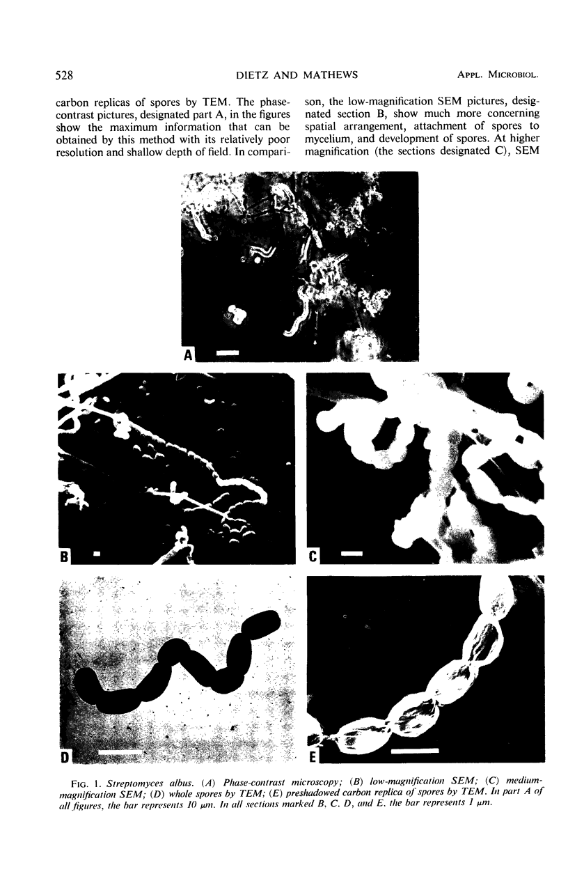
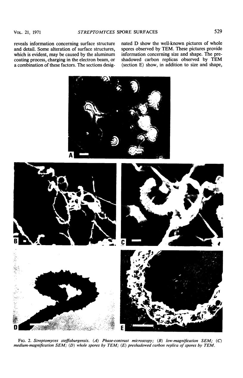
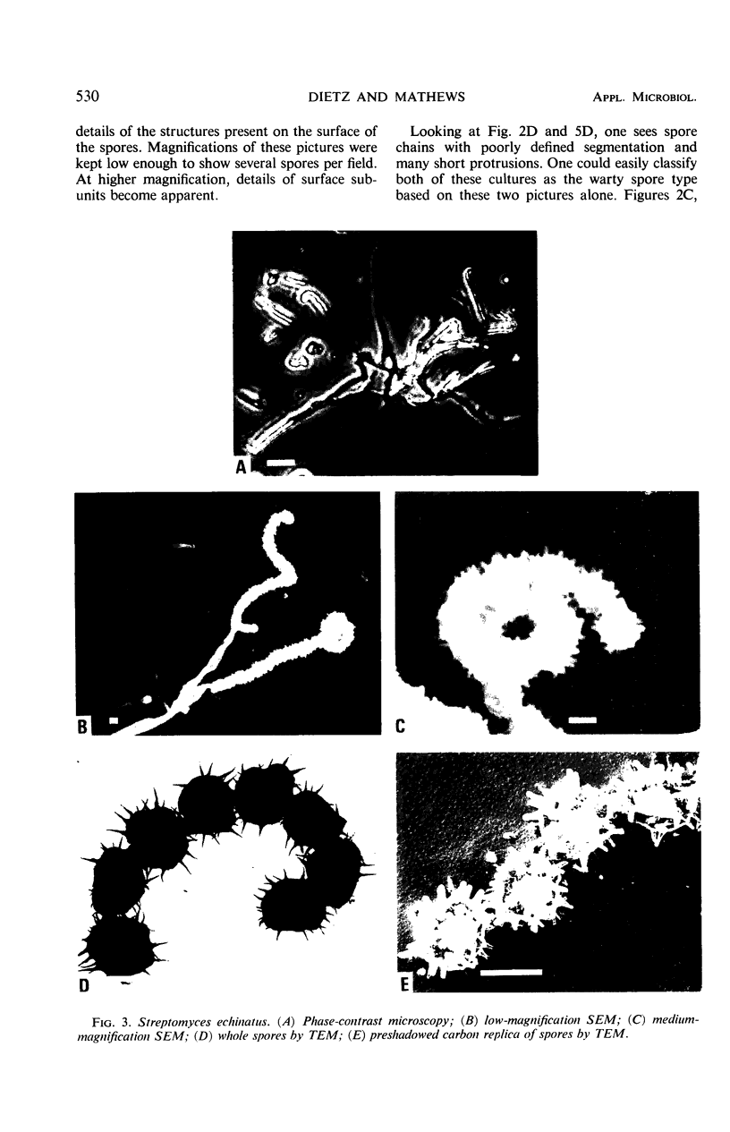
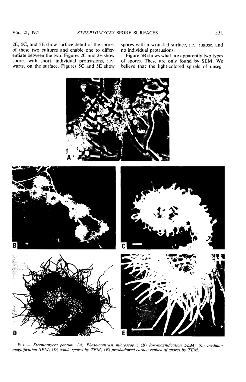
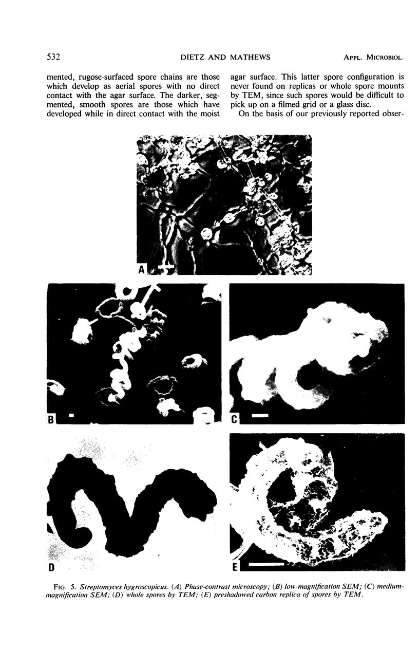
Streptomyces spores surfaces have been classified into five groups, smooth, warty, spiny, hairy, and rugose, by examination of carbon replicas of spores with the transmission electron microscope and by direct examination of spores with the scanning electron microscope.
Full text
PDF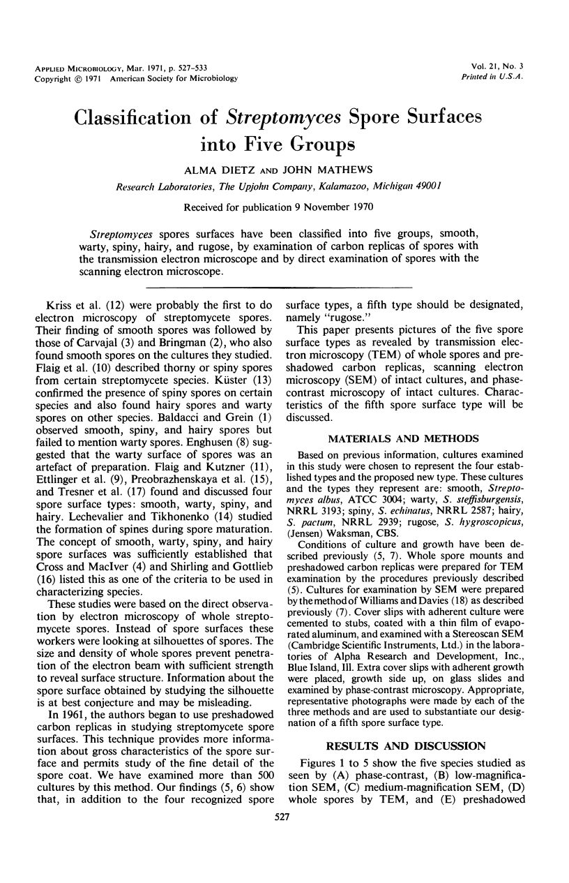
Images in this article
Selected References
These references are in PubMed. This may not be the complete list of references from this article.
- DIETZ A., MATHEWS J. Taxonomy by carbon replication. I. An examination of Streptomyces hygroscopicus. Appl Microbiol. 1962 May;10:258–263. doi: 10.1128/am.10.3.258-263.1962. [DOI] [PMC free article] [PubMed] [Google Scholar]
- Dietz A., Mathews J. Scanning electron microscopy of selected members of the Streptomyces hygroscopicus group. Appl Microbiol. 1969 Oct;18(4):694–696. doi: 10.1128/am.18.4.694-696.1969. [DOI] [PMC free article] [PubMed] [Google Scholar]
- Dietz A., Mathews J. Taxonomy by carbon replication. II. Examination of eight additional cultures of Streptomyces hygroscopicus. Appl Microbiol. 1968 Jun;16(6):935–941. doi: 10.1128/am.16.6.935-941.1968. [DOI] [PMC free article] [PubMed] [Google Scholar]
- ENGHUSEN H. Elekronenoptische Darstellungen von Streptomyceten-Sporen und-Hällen. Arch Mikrobiol. 1955;21(3):329–334. [PubMed] [Google Scholar]
- LESHEVAL'E Kh A., TIKHONENKO A. S. [Effect of the conditions of nutrition on the structure of the surface of actinomycete spores]. Mikrobiologiia. 1960 Jan-Feb;29:43–50. [PubMed] [Google Scholar]
- TRESNER H. D., DAVIES M. C., BACKUS E. J. Electron microscopy of Streptomyces spore morphology and its role in species differentiation. J Bacteriol. 1961 Jan;81:70–80. doi: 10.1128/jb.81.1.70-80.1961. [DOI] [PMC free article] [PubMed] [Google Scholar]
- Williams S. T., Davies F. L. Use of scanning electron microscope for the examination of actinomycetes. J Gen Microbiol. 1967 Aug;48(2):171–177. doi: 10.1099/00221287-48-2-171. [DOI] [PubMed] [Google Scholar]


