Abstract
This study was conducted to compare lipid components of sebum from persons from three ethnic backgrounds—Caucasian, African American and Northern Asian. Men and women with no acne in two age groups (18‒25 y and 35‒45 y) were recruited. Skin surface hydration (SkiCon 200EX and NovaMeter), barrier function (Delfin VapoMeter), high-resolution clinical imaging, self-assessments and two pairs of sebutapes on the forehead that extracted the lipids on the surface of their skin were used. Significant differences (p < 0.05) in skin hydration between African Americans and Caucasians in both age groups were noted, with the order from highest to lowest absolute values: African American > Northern Asian > Caucasian. Transepidermal water loss (TEWL) measurements demonstrated that African Americans and Caucasians were significantly different (p < 0.05), with the trend being the inverse of the hydration trend—Caucasian > Northern Asian > African American, which would indicate better barrier function for African Americans with a lower TEWL. African American women had more total lipid production than Northern Asian or Caucasian women. When analyzing the three lipid classes (free fatty acids, triglycerides and wax esters), the trend became significant (p < 0.05) in the wax ester fraction when directly comparing African Americans with Caucasians. Additionally, six lipids were identified in the wax ester fractions that were significantly different in quantity (p < 0.05) between African Americans and Caucasians. These results identified significant differences in sebaceous lipid profiles across ethnic groups and determined that the differences correlated with skin barrier function.
Keywords: age, ethnic, fatty acid, lipid, sebum, skin, squalene
Introduction
Previous studies have indicated that different skin types have different levels of skin hydration1 and barrier function2 based on bioinstrumental measures of conductance and transepidermal water loss (TEWL). Another study has indicated that skin hydration and TEWL are age-dependent but linked to ethnic skin type.3,4 However, another major component of skin that has been less investigated due to its complexity, but still contributes significantly to hydration and barrier function, are the sebaceous lipids.5 To investigate this topic further, in vivo biochemical experiments on the skin from different ethnic populations living in the same environment are needed.
Several previous studies have focused on dissecting the relationship between sebum output and the pathophysiology of acne. Two recent studies showed a correlation between higher sebum output and acne development.6,7 Most of the published studies assess the total sebum output by instrumental analysis (e.g., the sebumeter), self-evaluation or by using a sebum absorbing adhesive tape (Sebutape), which is a specially designed tape that has been proven to be reproducible and convenient to estimate sebaceous gland output.8 Additionally, Sebutape is used for collecting sebum for further quantitation of its components.8 To our knowledge no study thus far has measured individual sebaceous lipids in search of specific sebaceous lipids that could be uniquely associated with ethnic groups or would be correlated with instrumental parameters. This is likely due to the cumbersome and lengthy tasks associated with the individual extraction,7,8 separation and quantitation of lipids and their corresponding subclasses. To date, a few studies have reported on the correlation of sebaceous lipids and acne status; however, these studies were conducted in diseased (acne) skin and not in healthy skin.7,9,10
Elevated sebum excretion is involved in the pathophysiology of acne,11-13 as body sites that are rich in sebaceous glands are where acne lesions are typically manifested. High levels of sebum are associated with acne in adolescence and may offer a possible benefit by lubricating the skin, contributing to a better skin barrier as well as better skin hydration. Because acne is unique to humans it has been suggested that the unique sebaceous lipids could be associated with this human-specific disease. The accumulation of squalene and the presence of unique fatty acids and waxes are unique manifestations of sebum.14,15 Our study aimed to identify possible lipid components with sebum that could correlate with ethnic skin hydration and barrier and might be associated with age or ethnicity.
In the current study we conducted a series of in vivo assessments on the skin of women from three different ethnic populations living in the same environment (i.e., same locale). This study was conducted to determine what, if any, differences were quantifiable in skin lipid content across age and ethnic skin type composition between females aged 18–25 and 35–45 y with no acne or other chronic skin diseases.
Results
Extracted lipids from each Sebutape were subjected initially to thin layer chromatography and three major lipid classes were separated: free fatty acids, triglycerides and wax and cholesterol esters. Subsequently, each lipid class was extracted separately from the silica plate and subjected to saponification and derivatization to be prepared for individual fatty acid analysis. This was performed by gas chromatography with fluorescence ionization detector (GC-FID), as previously described.7 We were able to estimate the individual fatty acid population of each of the lipid classes listed above.
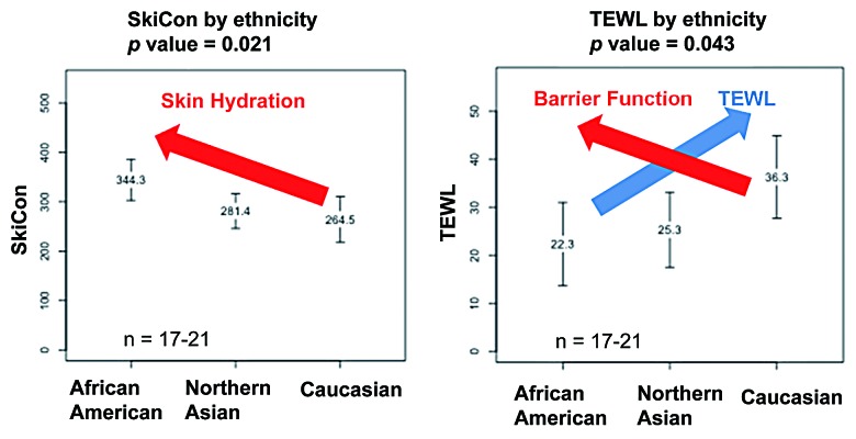
The initial pilot study involved male and females from three ethnic groups (n = 17‒21). Skin hydration and TEWL were assessed. Results showed significant differences (p < 0.05) in skin hydration between African Americans and Caucasians—with the order of African-American > Northern Asian > Caucasian (Fig. 1). The results for TEWL demonstrated that African Americans and Caucasians were again significantly different (p < 0.05), with the trend being the inverse of the hydration trend—Caucasian > Northern Asian > African American, which would indicate a better barrier function for African Americans with a lower TEWL (Fig. 1).
Figure 1. Correlation of hydration and TEWL values among three ethnic groups of males and females aged 20‒45 y. Hydration and TEWL values were grouped per the total gender and age combined ethnic population examined. Higher SkiCon were value associated with greater hydration (red arrow, left panel). Greater TEWL (blue arrow, right panel) is associated with worse barrier function (red arrow, right panel). Data are shown as mean ± standard deviation. Abbreviation: TEWL, transepidermal water loss.
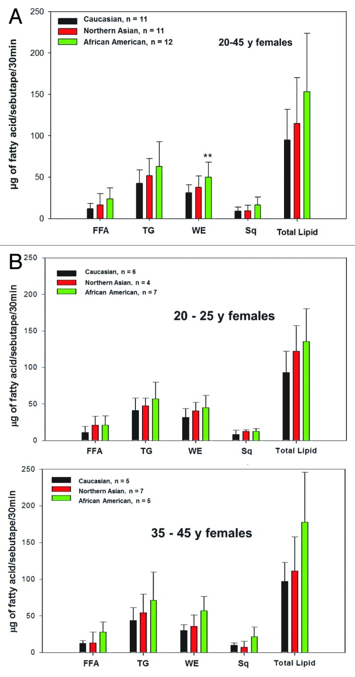
Consequently, Sebutape and lipid analysis were performed. A smaller cohort group due to the labor-intensive methodology was chosen, including females 20–25 and 35–45 y (n = 6–8) for all ethnic groups. Figure 2 summarizes the quantitative results in micrograms of lipid and the comparative analysis of the sebaceous lipid classes among the three different ethnic groups, total (top panel), 18‒25 y (middle panel) and 35‒45 y (bottom panel). It is apparent that the total amount of lipid is higher in the African American groups (150‒170 ug of fatty acids/Sebutape/30 min) than in the Caucasian Americans (90‒100 ug of fatty acids/Sebutape/30 min) regardless of the age ranges. The sebum levels of Asian Americans (110‒130 ug of fatty acids/Sebutape/30 min) lies between the two other ethnic groups. The greatest difference and with a statistical significant difference (p < 0.05) among the ethnic groups was observed in the wax ester fraction that is a sebum-specific lipid class. African American females in total had higher levels of wax esters (62 ug of fatty acid/Sebutape/30 min) than Caucasian American females (40 ug of fatty acid/Sebutape/30 min) (Fig. 2, top panel).

Figure 2. Quantitative lipid analysis of females segmented by age and ethnic group. Results were expressed as micrograms of fatty acids and were grouped per individual class of lipid. FFA, TG, WE and squalane were graphed as well as total lipid, which was expressed as the sum of all of the aforementioned classes (A) for the total group tested and (B) per age group. Data are shown as mean ± standard deviation. **p < 0.05. Abbreviations: FFA, free fatty acids; TG, triglycerides; Sq, squalene; WE, wax esters.
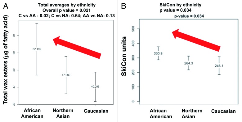
Because the overall wax esters fraction analysis demonstrated a significant difference—African American females had higher production than the other two ethnic groups—we looked at the individual lipid classes and we correlated the lipid analysis data to the SkiCon values acquired from the same subjects. As illustrated by the results (Fig. 3), there was a strong pattern/trend between the level of the wax ester fraction in sebum with SkiCon values that indicate conductivity and water content.
Figure 3. Correlation of wax ester fraction and hydration values among three ethnic groups of females aged 20‒45 y. Wax ester results and hydration values were grouped by combining females from all ethnic groups and analyzed by one-way analysis of variance and subsequent t-tests. Trends are shown by red arrows. Data are shown as mean ± standard deviation.
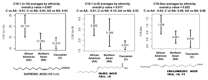
The wax ester fraction was further analyzed and six fatty acids were identified that were significantly different in quantity (p < 0.05) between African and Caucasian Americans (Table 1). Some are synthesized naturally in skin, as the 14:0, 16:1, n10 and 18:1, n9; some are acquired exclusively from diet, as the iso-18:2, whereas others may be a product of bacterial metabolism, as the 15:0 or 17:1 fatty acids, which would indicate flora differences on the surface of the skin between these ethnic groups. Three of these fatty acids were at higher levels than 1 µg/per Sebutape/30 min (Fig. 4), whereas the rest were at less significant quantities.
Table 1. Significant differences in wax ester fraction fatty acids among three ethnic groups.
| Lipid |
p-value 3-way |
Caucasian vs African American |
Caucasian vs Northern Asian |
African American vs. Northern Asian |
|---|---|---|---|---|
| C14:0 |
0.027 |
0.020 |
0.470 |
0.270 |
| C15:0 |
0.025 |
0.030 |
0.070 |
0.940 |
| C16:1 (n-10) |
0.007 |
0.010 |
0.930 |
0.030 |
| C17:1 |
0.045 |
0.050 |
0.940 |
0.120 |
| C18:1 (n-9) |
0.017 |
0.010 |
0.150 |
0.530 |
| C18:2 iso | 0.022 | 0.030 | 0.950 | 0.060 |
Results were grouped by combining females from all ethnic groups and analyzed by one way analysis of variance and subsequent t-tests. Differences between groups were determined to be statistically significant if p < 0.05.

Figure 4. Correlation of the most predominant fatty acids from the wax ester fraction among three ethnic groups of females aged 20‒45 y. Statistically significant differences in major sebum fatty acids were observed only in the wax ester fraction (25‒30% of sebum). Data are expressed as µg fatty acid/sebutape/30min. Results were grouped by combining data from women in all ethnic groups and analyzed by one way analysis of variance and subsequent t-tests. Data are shown as mean ± standard deviation.
Discussion
In this study we analyzed sebum, hydration and TEWL of subjects from different ethnic and age groups. African American women secreted larger amounts of sebum than the Caucasian women. The class of lipids that was significantly higher in African American women was wax esters. The only mammalian cells that synthesize wax esters are the sebaceous cells; therefore this class of lipids serves as a marker for sebaceous differentiation. One point of uniqueness in human sebum is that this non-polar lipid class accumulates in unusually high levels (~25%). Waxes are long, highly hydrophobic molecules in nature that act as a barrier against excessive hydration or dehydration. Waxes could potentially alter the rheological properties of sebum, as it is one of the most non-polar molecules in sebum as well as in nature. Because waxes may serve as a lipid marker for sebaceous activity, they could be far more upregulated than the rest of the lipids in cases of oily skin.
We also performed the comparative analysis of the lipid classes in females from three different ethnic groups in conjunction with other instrumental parameters such as hydration/conductivity and TEWL. In this study, besides demonstrating that African American women have higher amounts of wax esters and sebum than Caucasian American women, we coupled these results with skin hydration and barrier function measures as well as instrumental measures. These instrumental measures were highly correlated with the total lipid content and wax measures.
This is the first report that links an individual class of lipids, the wax esters, to a better barrier and higher hydration among three ethnic groups. In fact, the notion that sebum may have a moisturizing or conditioning effect is highly supported by these higher wax ester levels that may act as another layer of protection that enhances the skin’s barrier. Moreover, we demonstrate that both elevated sebum and higher hydration may contribute to a better skin barrier that is manifested with lower TEWL. Indeed, evidence in the literature indicates that African Americans have better barrier or oilier skin.7 Qualitative analysis was also performed but did not reveal any noteworthy differences among the ethnic groups.
Significant differences in six fatty acids in the wax ester fraction among the ethnic groups were identified. The most prevalent fatty acid in sebum, sapienic acid (16:1, n10), is significantly higher in African Americans and correlated with the higher sebum output in that ethnic group. Also higher in the African American group are oleic acid (18:1, n9) and myristic acid (14:0), precursor of palmitic acid (16:0). Sapienic acid is the most abundant fatty acid in sebum; therefore it is normal to be at higher levels in individuals with higher sebum output. The same argument can apply to the second most abundant monounsaturated fatty acid, oleic acid, a known adipogenic fatty acid.16,17 The most intriguing differences found are two fatty acids that are in lower levels but do not belong to the mammalian fatty acid metabolism, as they have a chain with an odd number of carbons, 15:0 and 17:1. It is well known that Propionibacterium acnes colonize within the philosebaceous hair canal, and they could contribute significantly in the lipid output; however, the higher presence of these fatty acids in the wax esters of African American females may indicate that (1) some bacterial fatty acids could get incorporated into sebaceous wax synthesis and (2) different ethnic groups could potentially have differences in the skin microflora. The last intriguing result is that we identified significant differences at the level of an isomer of linoleic acid, which is simply a dietary fatty acid. Although our analytical method could not give us the exact location of the two double bonds, it is certain that the observed differences could account for different nutritional habits of the different ethnic groups. This result is preliminary and requires more sophisticated instrumentation for more concrete results.
It would be of interest to perform a blind analysis of this marker in larger cohorts and to retrospectively identify high sebum-producer individuals to correlate with differential wax ester secretion. Better analytical techniques would help to increase our understanding of waxes and their precursor, plus their role in the induction or the maintenance of the skin’s conditioning state and barrier function. The field of “sebum-omics” is open for many new discoveries and is constantly enhanced by advances in analytical chemistry.
Consequent steps could be to repeat this study in another season to determine the impact the climate/environment has on skin hydration, barrier function and lipid production among the three ethnic groups.
Materials and Methods
Study design
Acne-free subjects of both males and females with no serious dermatological issues from Skillman, NJ, area were recruited (after signing an informed consent form) from two age groups (18‒25 and 35‒45) and three ethnic origins, Caucasian, African American and Northern Asian. Each subject underwent a 30 min acclimation period in a temperature- and humidity-controlled room and then was subjected to the following tests on their facial skin: surface hydration measurements with SkiCon 200EX (IBS Co.); Nova Dermal Phase Meter (Nova DPM); barrier function measurements (VapoMeter; Delfin Technologies, Ltd.); multi-modal high resolution digital clinical imaging; self-assessments; and skin surface lipid sampling with two pairs of Sebutape Adhesive Patches (CuDerm Corporation) on the forehead. Sebutapes were sent to an external lab (SCRI, Scotland), where the lipid classes were extracted via thin layer chromatography (TLC) and subsamples underwent mass spectroscopy and GC-FID. Photos with regular, polarized and fluorescent lights were obtained from each subject.
Skin surface lipid sampling and quantification
Two pairs of Sebutapes were applied for 30 min (to the forehead areas above both eyebrows) after degreasing with a cotton swab soaked in 70% isopropanol alcohol. Sebutapes were subsequently extracted by using the Folch method.18 The extracted organic layer was dried under a gentle stream of nitrogen at 40°C. The dried samples were stored under nitrogen at -20°C until processed for TLC and GC-FID analysis according to a method established by Mylnefield Lipid Analysis.19
In brief, the sample (50 mL) was spotted on a glass TLC plate, which was developed with 80:20:2 isohexane/diethyl ether/formic acid until the solvent front is a short distance from the top. The plate was air-dried and subsequently sprayed with 0.01% primuline to visualize the FFA, TAG and CE/WE bands under UV light. Each marked band was removed from the glass TLC plate and was placed into a test tube, where 1 mL toluene and 2 mL 1% sulphuric acid in methanol were added. The tubes were capped and left overnight at 50°C. Afterwards 5 mL of 5% NaCl solution was added and the samples were extracted with 2 × 2mL isohexane. Extracts were transferred to fresh test tubes that were shaken with 3 mL of 2% potassium hydrogen carbonate and then passed through a prewashed (3 mL isohexane) sodium sulfate column and also subsequently post washed with 2 mL of isohexane. The solvent was removed by nitrogen and isohexane + BHT (70 μL) before transferring them to a GC vial and injected with 5 μL of the sample. A C17:0 was used as an internal standard on a Cp-Wax 52CB (0.25 mm × 25 min × 0.2 μm) column, flow rate 1 mL/min. The GC was Agilent 6890 and the GC was performed using the method published by William Christie.19
Data analysis
Overall differences between ethnicity means were tested using one-way analysis of variance models. Pairwise comparisons of ethnicities were tested using Tukey’s test to adjust for multiple comparisons. Differences of means between males and females within an ethnic group were tested using t-tests. Considering the small sample sizes, significant results were verified via nonparametric tests (Kruskal-Wallis test for overall difference between ethnicities and Wilcoxon rank sum test for pairs).
Acknowledgments
We would like to thank Dr Michael Southall for fruitful discussions and critical reading of this manuscript.
Glossary
Abbreviations:
- FFA
free fatty acids
- GC-FID
gas chromatography-flame ionization detector
- TEWL
transepidermal water loss
- TG
triglycerides
- TLC
thin layer chromatography
- WE
wax esters
Disclosure of Potential Conflicts of Interest
All authors were employees of Johnson and Johnson Consumer Companies, Inc., Skillman, NJ and had no conflict of interest.
Footnotes
Previously published online: www.landesbioscience.com/journals/dermatoendocrinology/article/25366
References
- 1.Diridollou S, de Rigal J, Querleux B, Leroy F, Holloway Barbosa V. Comparative study of the hydration of the stratum corneum between four ethnic groups: influence of age. Int J Dermatol. 2007;46(Suppl 1):11–4. doi: 10.1111/j.1365-4632.2007.03455.x. [DOI] [PubMed] [Google Scholar]
- 2.Wilson D, Berardesca E, Maibach HI. In vitro transepidermal water loss: differences between black and white human skin. Br J Dermatol. 1988;119:647–52. doi: 10.1111/j.1365-2133.1988.tb03478.x. [DOI] [PubMed] [Google Scholar]
- 3.Rogers J, Harding C, Mayo A, Banks J, Rawlings A. Stratum corneum lipids: the effect of ageing and the seasons. Arch Dermatol Res. 1996;288:765–70. doi: 10.1007/BF02505294. [DOI] [PubMed] [Google Scholar]
- 4.Rawlings AV. Ethnic skin types: are there differences in skin structure and function? Int J Cosmet Sci. 2006;28:79–93. doi: 10.1111/j.1467-2494.2006.00302.x. [DOI] [PubMed] [Google Scholar]
- 5.Yamamoto A, Serizawa S, Ito M, Sato Y. Effect of aging on sebaceous gland activity and on the fatty acid composition of wax esters. J Invest Dermatol. 1987;89:507–12. doi: 10.1111/1523-1747.ep12461009. [DOI] [PubMed] [Google Scholar]
- 6.Mourelatos K, Eady EA, Cunliffe WJ, Clark SM, Cove JH. Temporal changes in sebum excretion and propionibacterial colonization in preadolescent children with and without acne. Br J Dermatol. 2007;156:22–31. doi: 10.1111/j.1365-2133.2006.07517.x. [DOI] [PubMed] [Google Scholar]
- 7.Pappas A, Johnsen S, Liu JC, Eisinger M. Sebum analysis of individuals with and without acne. Dermatoendocrinol. 2009;1:157–61. doi: 10.4161/derm.1.3.8473. [DOI] [PMC free article] [PubMed] [Google Scholar]
- 8.Nordstrom KM, Schmus HG, McGinley KJ, Leyden JJ. Measurement of sebum output using a lipid absorbent tape. J Invest Dermatol. 1986;87:260–3. doi: 10.1111/1523-1747.ep12696640. [DOI] [PubMed] [Google Scholar]
- 9.Perisho K, Wertz PW, Madison KC, Stewart ME, Downing DT. Fatty acids of acylceramides from comedones and from the skin surface of acne patients and control subjects. J Invest Dermatol. 1988;90:350–3. doi: 10.1111/1523-1747.ep12456327. [DOI] [PubMed] [Google Scholar]
- 10.Morello AM, Downing DT, Strauss JS. Octadecadienoic acids in the skin surface lipids of acne patients and normal subjects. J Invest Dermatol. 1976;66:319–23. doi: 10.1111/1523-1747.ep12482300. [DOI] [PubMed] [Google Scholar]
- 11.Zouboulis CC. Acne and sebaceous gland function. Clin Dermatol. 2004;22:360–6. doi: 10.1016/j.clindermatol.2004.03.004. [DOI] [PubMed] [Google Scholar]
- 12.Cunliffe WJ. Acne. London, England: Martin Dunitz, 1989. [Google Scholar]
- 13.Thiboutot D. Regulation of human sebaceous glands. J Invest Dermatol. 2004;123:1–12. doi: 10.1111/j.1523-1747.2004.t01-2-.x. [DOI] [PubMed] [Google Scholar]
- 14.Nicolaides N. Skin lipids: their biochemical uniqueness. Science. 1974;186:19–26. doi: 10.1126/science.186.4158.19. [DOI] [PubMed] [Google Scholar]
- 15.Nicolaides N, Ansari MN. Fatty acids of unusual double-bond positions and chain lengths found in rat skin surface lipids. Lipids. 1968;3:403–10. doi: 10.1007/BF02531278. [DOI] [PubMed] [Google Scholar]
- 16.Ntambi JM, Miyazaki M. Regulation of stearoyl-CoA desaturases and role in metabolism. Prog Lipid Res. 2004;43:91–104. doi: 10.1016/S0163-7827(03)00039-0. [DOI] [PubMed] [Google Scholar]
- 17.Miyazaki M, Man WC, Ntambi JM. Targeted disruption of stearoyl-CoA desaturase1 gene in mice causes atrophy of sebaceous and meibomian glands and depletion of wax esters in the eyelid. J Nutr. 2001;131:2260–8. doi: 10.1093/jn/131.9.2260. [DOI] [PubMed] [Google Scholar]
- 18.Folch J, Lees M, Sloane Stanley GH. A simple method for the isolation and purification of total lipides from animal tissues. J Biol Chem. 1957;226:497–509. [PubMed] [Google Scholar]
- 19.Christie WW. Gas chromatography and lipids: a practical guide. Dundee,Scotland: The Oily Press Ltd, 1994:68. [Google Scholar]


