Summary
Our thesis is that thin filament function can only be fully understood and muscle regulation then elucidated if atomic structures of the thin filament are available to reveal the positions of tropomyosin on actin in all physiological states. After all, it is tropomyosin influenced by troponin that regulates myosin-crossbridge cycling on actin and therefore controls contraction in all muscles. In addition, we maintain that a complete appreciation of thin filament activation also requires that the mechanical properties of tropomyosin itself are recognized and then related to the effect of myosin-association on actin. Taking the Gestalt-binding of tropomyosin into account, coupled with our electron microscopy structures and computational chemistry, we propose a comprehensive mechanism for tropomyosin regulatory movement over the actin filament surface that explains the cooperative muscle activation process. In fact, well-known point mutations of critical amino acids on the actin-tropomyosin binding interface disrupt Gestalt-binding and are associated with a number of inherited myopathies. Moreover, dysregulation of tropomyosin may also be a factor that interferes with the gatekeeping operation of non-muscle tropomyosin in the controlling interactions of a wide variety of cellular actin-binding proteins. The clinical relevance of Gestalt-binding is discussed in articles by the Marston and the Gunning groups in this special journal issue devoted to the impact of tropomyosin on biological systems.
Keywords: Actin, muscle regulation, myosin, troponin, tropomyosin
Introduction
All muscles require an on-off switching mechanism to regulate actin-myosin interactions; otherwise they would be permanently contracted and of little use. The requisite control systems must adapt with precision to changes in physiological and myopathic stimuli. Indeed, force development in all muscles is controlled by changes in sarcoplasmic Ca2+ levels, and, for example in skeletal and cardiac muscle, Ca2+ alters the conformational arrangements of tropomyosin and troponin on thin filaments, leading to relaxation at low and contraction at high Ca2+ concentrations (reviewed in Gordon et al. 2000).
Coiled-coil tropomyosin binds to seven successive actin subunits on F-actin and the 40 nm long molecule is linked end-to-end to form a polymeric cable that runs continuously along muscle thin filaments (reviewed in Brown and Cohen 2005). The troponin complex, linked to each tropomyosin molecule, couples Ca2+-concentration changes to the movement of tropomyosin over the surface of actin (ibid.). In turn, tropomyosin’s location on actin controls whether the myosin-binding patch on the actin surface is exposed or not and thereby regulates the myosin-crossbridge cycling on actin that powers contraction (Haselgrove 1972; Huxley 1972; Parry and Squire 1972; Lehman et al. 1994; Vibert et al. 1997; Lehman and Craig 2008).
Previous structural work on isolated and reconstituted thin filaments showed that at low Ca2+ levels, tropomyosin is constrained by troponin to sterically obstruct access to myosin-binding sites on actin, thus producing the blocked B-state of the thin filament, which in whole tissue causes muscle relaxation (Lehman et al. 1994; Vibert et al. 1997; Lehman and Craig 2008). Conversely, full switching-on of troponin-tropomyosin regulated filaments involves tropomyosin movement exposing the myosin-binding sites, allowing myosin-crossbridge associations to occur and consequently muscle contraction to result. The “opening” of myosin-binding sites on thin filaments is a two-step process. It first takes Ca2+-binding to troponin to release the troponin-constraint, and tropomyosin then moves to a “C-state” position, partially exposing the myosin-binding sites (Vibert et al. 1997). The binding of small numbers of myosin-heads on actin then presumably shifts semi-rigid tropomyosin further to a fully active open “M-state” position of the filament, resulting in full access to all myosin-binding sites (Vibert et al. 1997; Craig and Lehman 2001). These actions of tropomyosin are entirely consistent with the steric mechanism of regulation first formulated by Hanson and Lowy (1964) and by Huxley (1972). Our subsequent structural work (Lehman et al. 1994, 2000; Vibert et al. 1997; Xu et al. 1999; Craig and Lehman 2001; Poole et al. 2006) and the biochemistry of the Geeves and Lehrer groups (McKillop and Geeves, 1993; Lehrer and Geeves, 1998; Geeves and Lehrer, 2002) validated this proposed sequential mechanism, which evolved into a 3-state model of muscle regulation (i.e. Blocked → Closed → Open functional states, which no doubt parallel the B → C → M structural transitions.) These two sets of terms are frequently applied interchangeably, yet some care should be observed in their use, as they describe characteristics that are measured differently (in one case biochemically, in the other structurally) and which therefore are not necessarily identical (see Table 1 for nomenclature).
Table 1.
Nomenclature used to define structural and functional thin filament states.
| Filament Preparation | Structural State | Functional State |
|---|---|---|
| Actin-Tropomyosin (no troponin) | A | Closed |
| Thin Filaments – low Ca2+ (actin-tropomyosin-troponin) | B | Blocked |
| Thin Filaments – high Ca2+ (actin-tropomyosin-troponin) | C | Closed |
| Thin Filaments + rigor bonded S1 (no ATP, S1 + actin-tropomyosin or actin-tropomyosin-troponin) | M | Open |
In the following discussion, we review our current understanding of the mechanical properties of the tropomyosin molecule as well as its regulatory movements over the surface of the actin filament. We then put forward our view on the structural role played by myosin in the activation process.
Gestalt-binding is fundamental to tropomyosin behavior
The binding of individual tropomyosin molecules to F-actin is extraordinarily weak [Ka ~ 2–5 × 10³ M−1, (Wegner, 1979)]. Thus, contrary to the observation, micromolar protein concentrations typically used for in vitro work should lead to little tropomyosin-binding to actin filaments. The fact that tropomyosin binds to F-actin readily and saturates filaments in standard buffers (20 – 150 mM KCl (or NaCl) and 1 – 3 mM MgCl2 at neutral pH) (Eaton et al. 1975) is accounted for by the tendency of tropomyosin to readily polymerize end-to-end on the F-actin substrate. Thus once the polymer is seeded, this linkage leads to a collective apparent affinity for actin of ~ Kan, where n is the number of tandem tropomyosin molecules linked together (about 25 in striated muscle filaments), while in vitro an apparent Ka > 106 M−1 is measured (Wegner, 1979). These binding characteristics are well-suited to tropomyosin function on thin filaments: viz. at a local level, the intrinsic weak binding to actin allows tropomyosin position to be easily perturbed by troponin, myosin-S1 or other actin-binding proteins, while globally the polymerized tropomyosin cable remains effectively and strongly bound to the thin filament.
Given the low Ka of a single unpolymerized molecule of tropomyosin for F-actin and the molecule’s elongated 40 nm shape, Holmes and Lehman (2008) argued that induced-fitting mechanisms during thin filament assembly are improbable. Instead, they proposed that tropomyosin must be “preshaped” to the contours of the F-actin helix in order to bind effectively. We found that this indeed is the case (Li et al. 2010a, b), showing that on average the 3D trajectories of tropomyosin matched the helix of the actin filament very well with an associated small but anisotropic variance, indicating that tropomyosin is preshaped with an overall design to bind effectively to F-actin. At the same time, Li et al. (2010a, 2012) resolved longstanding controversies regarding the flexibility of tropomyosin by quantifying the tropomyosin stiffness both in hundreds of EM images of isolated tropomyosin molecules and in tens of thousands of snapshots taken during MD simulations. This analysis showed that tropomyosin is semi-rigid with a persistence length about 12 times its own length (Li et al. 2010a, 2010b, 2012; Sousa et al. 2010). Thus local troponin- and myosin-induced shifts of semi-rigid tropomyosin on the actin filament can be expected to be transmitted distally with limited decrement in azimuthal displacement, as is needed to achieve the observed cooperativity of myosin-binding to the actin-tropomyosin filament.
The above studies supported the view proposed by Holmes and Lehman (2008) that the form-function relationship or Gestalt of the tropomyosin strand emerges from the preshaping of the molecule coupled with the inherent tendency to polymerize by end-to-end association. The term Gestaltbindung or Gestalt-binding was coined to additionally convey the notion that the assembly and organization of tropomyosin on thin filaments cannot be easily understood if examination of tropomyosin interactions is restricted to discrete molecular sites and then strict lock-and-key binding mechanisms invoked. Recent studies describing tropomyosin behavior tend to support this view. For example, experimentally induced mutation of tropomyosin, replacing an alanine cluster along the molecule’s coiled-coil hydrophobic stripe with larger hydrophobic residues (A74L/A78V/A81L), disrupts tropomyosin binding on F-actin (Singh and DeGregori, 2003, 2006). The effect not only is associated with subtle changes in local molecular bending in the surrounds of the affected residues, but more importantly with adjustments in global curvature, as much of the tropomyosin molecule becomes straightened even at significant distances from the mutation sites, thereby losing some of its pre-shaped average conformation (Li et al. 2010a). Likewise, cardiomyopathy-linked tropomyosin mutations E180G and D175N not only cause local increases in tropomyosin flexibility but also unexpected changes in flexibility (and susceptibility to proteolysis) at a considerable distance from the mutation (Ly and Lehrer 2012; Li et al. 2012). Thus, treating polymeric tropomyosin as part of an unbroken complex cooperative system and not simply a sum of its parts is an appropriate and necessary approach to understanding thin filament regulation. Indeed, in order to further understand the concerted cooperative transitions taking place on thin filaments, the behavior of thin filament components has been modeled by treating overlapping tropomyosins as continuous flexible chains or other representations (e.g. see Smith et al. 2003; Smith and Geeves 2003; Mijailovich et al. 2012, 2010; Geeves et al. 2011, Loong et al. 2012). However, presently the only effective experimental approach available to study the global structural mechanics of large macromolecular systems like thin filaments at high resolution is cryo-electron microscopy (cryo-EM) (often correlated with the results of fiber diffraction studies). Docking crystal structures of thin filament components into the three-dimensional EM maps is additionally useful to provide near-atomic detail, and further computational chemistry yields insights that are not accessible by current experimental approaches.
Troponin-induced tropomyosin movement
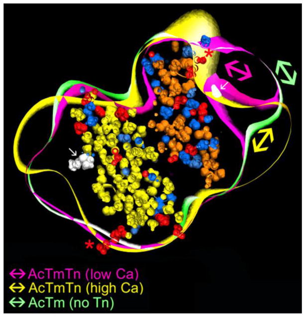
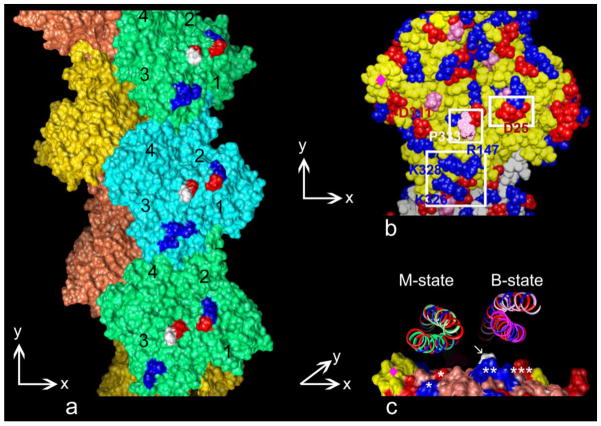
Reconstruction of low-Ca2+-treated thin filaments shows that the polymeric tropomyosin cable is constrained by troponin in the B-state position over the outer domain of actin (Fig. 1). Here, tropomyosin interacts with actin subdomain 1 and bridges over the smaller subdomain 2 to contact successive actin subunits along their helical path (Lehman et al. 1994; Vibert et al. 1997; Lehman and Craig 2008). Ca2+-binding by troponin results in azimuthal tropomyosin movement toward subdomains 3 and 4 across a minor ridge located at the inner edge of subdomain 1 (formed by residues Lys326, Lys328, Arg147 and Pro333, see Fig. 2). Thus tropomyosin in the Ca2+-induced C-state is located largely at positions over subdomains 2 and 4, and now, as mentioned above, exposes a good deal of the myosin-binding site on actin (ibid.). In contrast, tropomyosin in troponin-free filaments (here called the “A-state” for “Apo”-actin-tropomyosin) is disordered azimuthally and does not strictly occupy one or the other structural state, although on average its location on actin is closer to the blocking position rather than to the C-state position (Fig. 1)(Lehman et al. 2009, Li et al. 2011). Even though the A- and B-state positions of tropomyosin are close to each other, troponin-free A-state filaments respond functionally to myosin-binding as if in the closed-state, since there is no troponin-imposed blockage (McKillop and Geeves 1993; Geeves and Lehrer 2002, see Table 1).
Figure 1.
Control of tropomyosin position by troponin and Ca2+. Cross-sections of three superposed reconstructions showing the EM envelopes of reconstituted thin filaments (viewed from the pointed end of actin) consisting of troponin-free F-actin-tropomyosin (green), as well as troponin-decorated F-actin-tropomyosin maintained at low-Ca2+ (magenta) or treated with high-Ca2+ concentrations (yellow). Two azimuthally related actin subunits (Oda et al. 2009) (yellow, brown) were fitted within the reconstructions for reference, with acidic residues (red), basic residues (blue), and Pro333 (white, arrows) highlighted. Double-sided arrows indicate tropomyosin positions on one side of the filament. Note the good positional discrimination between high- and low-Ca2+ positions of tropomyosin on the inner and outer domains of actin for the troponin-regulated filaments, but the broader positioning of tropomyosin on the troponin-free filament. Also note, the position of actin residue Asp25 (asterisk), which is both a binding site for tropomyosin and a structural barrier restricting troponin-induced tropomyosin movement. Tropomyosin reconstructions were rendered with the program Chimera (Pettersen et al. 2004) based on data in Lehman et al. (2009) and Li et al. (2011).
Figure 2.
Surface residues on the actin interface specify tropomyosin positioning. (a) An actin filament model showing residues defining the axial path over which tropomyosin runs on troponin-free actin (actin subdomain numbers marked, acidic (red) and basic (blue) amino acids that interact with tropomyosin are highlighted, Pro333 colored white (actin’s pointed end facing up)). (b) Enlargement of a single actin subunit viewed face-on and in (c) viewed end-on (from actin’s barbed end with only the top surface of the actin shown). Charged residues (Lys326, Lys328, Arg147, and Asp25) project from actin (Oda et al. 2009) and are poised to interact with tropomyosin (Li et al. 2011); in (c) the 326/328/147 residue cluster is indicated by a double asterisk, Asp25 by a triple asterisk and Pro333 by a white arrow. Tropomyosin (ribbon representations) shown in the B-state position lies over these residues, but is well-removed from them when it is in the M-state position, where interactions with residues Asp311 and Lys315 are likely (single asterisks in (c)). Note that the edges of the relatively flat interface over which tropomyosin moves is demarcated on one side by Asp25 on actin subdomain 1 and on the other by a cluster of amino acids (Thr229 to Leu236) on subdomain 4 (magenta diamond). Figures rendered using the program Chimera (Pettersen et al. 2004) based on data in Li et al. (2011) and Behrmann et al. (2012); (a,b) are modifications of Figure 2f and 2j in Li et al. (2011) with permission. The y-axis markers indicate the filament (and actin subunit) pointed end direction and the x-axis indicates the azimuthal direction traversed by tropomyosin from the M-state to the B-state across the relatively flat front facing interface of actin.
The decreased azimuthal definition of tropomyosin noted on troponin-free filaments suggests that the precise positioning of tropomyosin is troponin-dependent (Lehman et al. 2009). By decreasing azimuthal instability and biasing tropomyosin to respective B- or C-state positions, troponin appears to display a dual-structural function: in relaxed muscles at low-Ca2+, troponin operates as inhibitor of actin-myosin interaction by pinning tropomyosin over the myosin-binding site on the thin filament, while in activated muscles at high-Ca2+, it acts as a promoter of actin-myosin contact by facilitating the B- to C-state tropomyosin transition and thus increasing the probability of myosin-binding.
The actin “playing field”
Docking atomic models of actin and tropomyosin into EM reconstructions shows that the regulatory movement of tropomyosin occurs over a fairly flat actin “playing field” that is delimited by structural landmarks on the edges of the inner and outer actin domains (Fig. 2b)(Li et al. 2011; Behrmann et al. 2012). Residues Ala22-Pro27 bulging out from actin subdomain 1 form one boundary of the flat interface, likely to restrict troponin-induced tropomyosin movement outward, while residues Thr229-Leu236 projecting from actin subdomain 4 probably limit troponin- or myosin-induced tropomyosin movement inward on the opposite side of the interface (Fig. 2c)(ibid.).
Actin residues Lys326, Lys328, and Arg147 as well as Asp25, Arg28 and Glu334 form two broad clusters of charged amino acids projecting from the surface of actin subdomain 1 (highlighted in Fig. 2a). In the A- and B-states, these clusters interact with specific residues on each tropomyosin pseudo-repeat, and therefore each successive actin subunit along F-actin is matched to tropomyosin (Li et al. 2011). Here, roughly 30 electrostatic interactions define the actin-tropomyosin binding (Li et al. 2011; Orzechowski et al. 2012). As expected, mutation of many of the residues interferes with actin-tropomyosin binding (Barua et al. 2012, 2013). The azimuthal edges of tropomyosin locate between residues Asp25 and Arg28, part of the subdomain 1 bulge, and Pro333, a residue at high radius on actin that demarcates the boundary between subdomains 1 and 3 (Fig. 2b,c). In order for tropomyosin to move to the C-state position, it must slide over Pro333. Molecular modeling indicates that this is accomplished without notable clashes occurring since residues on tropomyosin abutting Pro333 do not contain bulky side chains (Dominguez, 2011). Once in the C-state, tropomyosin is still likely to interact electrostatically with actin residues Lys326 and Lys328 and approach residues Asp311 and Lys315 on actin subdomain 3. However, the C-state transition appears to eliminate tropomyosin interaction with actin residues Arg147, Asp25 and Arg28.
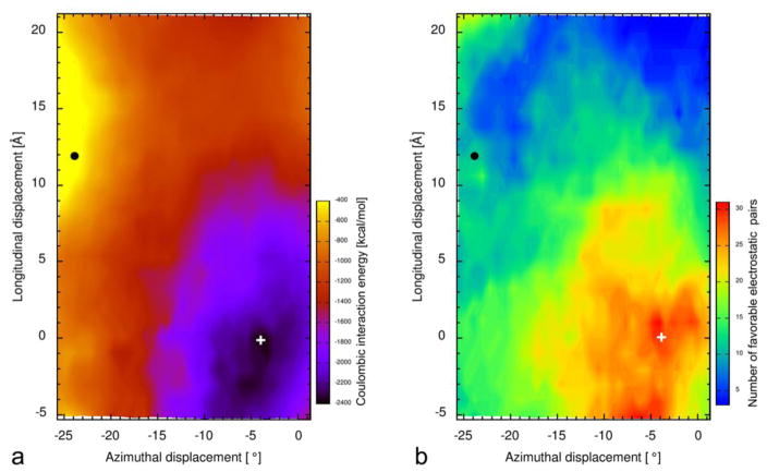
An electrostatic (Coulombic) energy landscape for locations of tropomyosin on troponin-free actin (corresponding to the A-state) identifies an energy minimum very close to the position of tropomyosin in the B-state filament configuration (Fig. 3, Table 2). The energy landscape between B- and C-states is quite flat (Fig. 3a), suggesting that easily induced troponin-dependent repositioning of tropomyosin is inherent to the control mechanism. In marked contrast, the myosin-induced M-state “open” position of tropomyosin is well removed from the B-/C-state energy basin at an unfavorable peak on the energy landscape. Indeed, the region of actin covered by tropomyosin in the M-state has far fewer charged amino acids ready to pair together with opposing charges on tropomyosin. In this region on actin, only isolated residues Asp311 and Lys315 are likely to interact with tropomyosin electrostatically (Table 3). Although tropomyosin – actin interaction appears to be considerably weakened in the M-state, tropomyosin does not unravel from the actin filament because now myosin-tropomyosin interactions (described below) trap the tightly wrapped tropomyosin cable on actin (Behrmann et al. 2012).
Figure 3.
Maps of the interaction energy landscape between tropomyosin and F-actin. (a) Electrostatic Coulombic energy values were computed for a single tropomyosin placed super-helically on F-actin (Oda et al. 2009) at different axial and azimuthal positions (Orzechowski et al. 2012), as done previously for a similar but narrower search around the A-state position (Li et al. 2011). (a) The Coulombic landscape has a broad basin (blue) surrounding the energy minimum (indicated by +) (dark blue area) located close to the B-state position found by EM (0,0 position on the map). In contrast, the Coulombic energy maximum (indicated by ●) (yellow area) is close to the open M-state position of tropomyosin (Behrmann et al. 2012). Note: there is only one obvious energy minimum, and not three separate and discrete minima, which might represent B-, C- and M-state positions for tropomyosin on F-actin (cf. Lehman et al. 2000). (b) Number of oppositely charged pairs of residues on actin and tropomyosin making favorable electrostatic interactions at close range (< 5 Å) for each grid location in (a) were tabulated; the same conformers used in the map in (a) were examined in (b). Note the favorable basin in (a) corresponds to the region with greatest number of complementary charged residue pairs in (b).
To compute these maps, actin-tropomyosin structures were first generated by sliding an atomic model of tropomyosin (Li et al. 2011) in longitudinal and azimuthal directions over actin in ±1 Å × 0.75° (~0.6 Å) increments to create a grid of tropomyosin locations. Intermediate positions were derived by interpolating/extrapolating between two actin-tropomyosin reference structures (+ and ●, mentioned above), then subjecting surface residues of F-actin and tropomyosin to an energy minimization. This removes steric clashes without significant changes in position of tropomyosin on F-actin. This procedure differs from that in Li et al. (2011), where only tropomyosin was minimized and F-actin was fixed, resulting in a small (~4°) azimuthal difference in the global minimum between the two determinations. All calculations used the CHARMM27 force-field (MacKerell et al. 1998, 2004; Brooks et al. 2009). Solvation was implemented according to the Generalized Born model and the GBSW protocol (Im et al. 2003) at 150 mM salt concentration.
Table 2.
Energetics of the actin-tropomyosin interaction on myosin-free F-actin.
Respective electrostatic and total energy terms were calculated based on the published actin-tropomyosin structures for which we have atomic coordinates. Note that electrostatic interaction energy (calculated as in Fig. 3) between actin and tropomyosin in the Li et al. (2011) reference position (i.e. very close to the B-state) is considerably lower (more favorable) that that for the Behrman et al. (2012) M-state structure. Computation of total energy (Brooks et al. 2009), here including solvation and Van der Waals energy terms, also indicates that M-state tropomyosin binding to myosin-free F-actin is not energetically favorable.
| Configuration Tropomyosin on F-actin | Electrostatic Interaction Energy1 (kcal) | Relative Total Energy (Solvation, Van der Waals, Electrostatic)2 (kcal) |
|---|---|---|
| Blocking B-Position (Li et al., 2011) | −2190 | −36.0 |
| Open M-Position (Behrmann et al., 2012 | −306 | 0 |
| Isolated Actin + Tropomyosin3 (Separated from each other) | 0 | −15.2 |
electrostatic energy measured for unsolvated actin-tropomyosin
actin-tropomyosin solvated
Note that measurement of total energy suggests that, in the absence of myosin, tropomyosin dissociated from F-actin is favored over that bound in the M-state configuration.
Table 3.
Electrostatic interactions between tropomyosin and actin and/or myosin in the M-state. The pseudo-atomic model of the actin-tropomyosin-myosin complex (Behrmann et al. 2012) was evaluated to identify acidic and basic residues on actin and myosin, which are in close proximity to oppositely charged ones along tropomyosin’s seven repeat periods.
| Tropomyosin pseudo- repeat period | Tropomyosin residues in close proximity to actin and myosin | ||
|---|---|---|---|
|
| |||
| actin Asp311 and myosin Glu286 | actin Lys315 and myosin Arg288 | myosin Glu286 | |
| 1 | - | Asp-20 | Arg-21 |
| 2 | Lys-59 | Glu-62 | - |
| 3 | - | Asp-100 | Arg-101 |
| 4 | Lys-136 | Glu-139 | Lys-140 |
| 5 | Arg-178 | Glu-181 | Arg-182 |
| 6 | Lys-217 | Asp-219 | Lys-220 |
| 7 | - | Asp-258 | - |
Myosin is a “game changer”
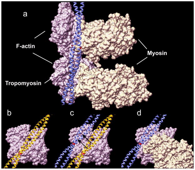
Strong binding of myosin on actin displaces tropomyosin from B- or C-state positions, thus overriding relatively weak local actin-tropomyosin associations (Vibert et al. 1997; Behrmann et al. 2012) (Fig. 4a). The semi-rigid tropomyosin slides between B- and M-state positions over the relatively flat actin interface, without any major twisting or rolling (Behrmann et al. 2012), confirming earlier predictions based on the mechanical properties of tropomyosin (Li et al. 2010a). Flexible fitting routines used to fit high resolution crystal structures into cryo-EM densities (Behrmann et al. 2012) indicate that no obvious tropomyosin pseudo-rotation occurs during tropomyosin sliding (Fig. 4b–d), although pseudo-rotation would be expected if azimuthal rolling takes place, as is sometimes envisioned.
Figure 4.
Tropomyosin movement from B- to M-state. (a) The pseudo-atomic model of the actin-tropomyosin-myosin complex, obtained by fitting crystal structures into the cryo-EM density map of Behrmann et al. (2012), reveals that tropomyosin in the M-state fits snugly into a groove formed by actin and myosin. (b) Tropomyosin in the B-state (Li et al. 2011) (c) rotates slightly and shifts by ~23 Å to reach the M-state position (d), where tropomyosin is wedged (locked) between the myosin head and a bulge on actin subdomain 4 (Behrmann et al. 2012). Actin subunits are displayed in lavender, myosin heads in pale beige, and tropomyosin in the B- and M-states in yellow and blue respectively; tropomyosin residue R181 locations are highlighted in red.
In marked contrast to the A-, B- and C-state organization of tropomyosin on the thin filament, tropomyosin in the M-state now is physically restricted to a narrow structural well formed by the myosin crossbridge head bound to actin on one side and the Thr229-Leu236 actin bulge on the other (ibid.) (Figs. 2c, 4a). Thus, in the M-state configuration of the thin filament, little azimuthal oscillation of the tropomyosin is expected in the presence of myosin.
Unlike results of the B-state conformational mapping, where 30 potential salt bridges between tropomyosin and actin are thought to result in a relatively strong interaction (Li et al., 2011; Orzechowski et al., 2012), Behrmann et al. (2012) found that tropomyosin in the M-state is stabilized by only ~11 electrostatic interactions with actin (Table 3). However, tropomyosin additionally interacts with myosin, resulting in a total of 16 possible salt bridges between tropomyosin, actin and myosin.
Interestingly, in the M-state, every tropomyosin pseudo-repeat shows a glutamate or aspartate residue associating with Lys315 on actin and Arg288 on myosin. In some but not all pseudo-repeats, arginine or lysine residues at neighboring positions strengthen this interaction by forming additional salt bridges with myosin Glu286 and actin Asp311. Hence, the resulting motif K(R)XXD(E)K(R) is only complete at pseudo-repeats 4, 5 and 6, making these positions likely to be favored target zones for myosin. It should be noted that the troponin core domain is thought to localize over tropomyosin pseudo-repeats 4 and 5 (Mudalige et al., 2009; Mudalige and Lehrer, 2010), possibly favoring additional myosin-thin filament linkages of physiological significance (cf. Perz-Edwards et al., 2011).
Conclusion: Tropomyosin’s Gestalt-binding and inherent semi-rigidity determine cooperative transitions between thin filament on- and off-states
As discussed above, at low-Ca2+, tropomyosin is pinned-down on thin filaments by troponin to block myosin-binding sites, crossbridge-cycling and contraction, while at high-Ca2+, troponin and myosin in effect push tropomyosin across the surface of actin to unmask these sites and initiate contraction. We also indicated that energy-landscape measurements display an electrostatic energy minimum for tropomyosin on actin-subunits, which is established by ~30 favorable electrostatic interactions localizing tropomyosin near its blocking-site on filaments (Li et al., 2011; Orzechowski et al., 2012). In that state, the inherent semi-flexibility of tropomyosin is likely to be attenuated, as tropomyosin is weakly “glued” onto its actin substrate. This interaction will necessarily dampen tropomyosin azimuthal “vibration” and also the propensity for spontaneous tropomyosin switching to the M-state positions. Indeed, comparisons of Molecular Dynamics simulations of free- and actin-bound tropomyosin (Li et al., 2012) indicate a decrease in both overall and local flexibility once tropomyosin is linked to actin (Table 4).
Table 4.
Local and global flexibility of isolated and actin-bound tropomyosin.
Flexibility determined from MD simulations of either isolated tropomyosin molecules or tropomyosin when bound to F-actin (Li et al., 2010a, b; Li et al., 2012). measures flexibility as the mean deviation angle from the time-averaged position between the two ends of a given tropomyosin segment (ibid.), here either over the entire tropomyosin molecule or locally over 9 residues centered on a specific amino acid. The individual residues quantified are examples of charged amino acids in tropomyosin pseudo-repeats 5, 4 and 3 that interact closely with actin surface residues. Error of corresponding half-data sets reported.
| Region examined | Residues measured | δ - Isolated Tropomyosin Fluctuation angle (°) | δ – Actin-Bound Tropomyosin Fluctuation angle (°) |
|---|---|---|---|
| Full length tropomyosin | Met 1 – Ile 284 | 22.0 ± 1.91 | 9.9 ± 0.11 |
| Repeat Period 5 | Glu 181 | 4.6 ± 0.2 | 3.9 ± 0.4 |
| Repeat Period 5 | Arg 167 | 5.3 ± 0.7 | 4.0 ± 0.4 |
| Repeat Period 4 | Glu 139 | 4.4 ± 0.2 | 3.5 ± 0.4 |
| Repeat Period 4 | Lys 128 | 5.3 ± 0.8 | 4.4 ± 0.4 |
| Repeat Period 4 | Lys 125 | 6.0 ± 1.0 | 4.6 ± 0.4 |
| Repeat Period 3 | Glu 104 | 4.5 ± 0.2 | 3.6 ± 0.4 |
| Repeat Period 3 | Arg 90 | 5.0 ± 0.6 | 4.2 ± 0.4 |
Data from Li et al. 2012.
Following Ca2+-activation and release of troponin-based pinning of tropomyosin on actin, strong actin-myosin binding and myosin-induced movement of tropomyosin to the M-state position on actin breaks the “electrostatic glue” between actin and tropomyosin discussed above. Given that atomic structures for transitions between B- and M-end states are not available, it is difficult to predict the interplay required between initial myosin-head binding, myosin-cleft closure (needed for strong actin-myosin association) and the repositioning of tropomyosin (needed for cleft closure; see Poole et al. 2006). Whatever the case may be, the new M-state interactions now formed between tropomyosin and actin-subunits are relatively weakly coupled, which presumably is compensated by tropomyosin-myosin interaction along the tropomyosin cable (Behrmann et al. 2012). In this sense, M-state tropomyosin becomes a myosin-binding protein as well as an actin-binding partner.
We propose that filament mechanics following a tropomyosin shift to the myosin-induced open M-state must reflect the low number of complementary electrostatic interactions possible between tropomyosin and F-actin (Orzechowski et al., 2012). Hence, as myosin begins to populate actin filaments, and locally displaces tropomyosin from its favored binding patches on actin, the semi-rigidity of tropomyosin will induce movement of adjacent stretches of the tropomyosin cable. As this process proceeds and tropomyosin separates and moves from the “gluing” region of actin, tropomyosin then will assume more of the mechanical Gestalt-characteristics of the unbound, ~500 nm persistence-length molecule. Thus it follows that, at the onset of myosin-induced movement of tropomyosin when most tropomyosin is still largely in B- or C-state configuration, local myosin-induced repositioning of tropomyosin will propagate because here semi-rigid tropomyosin, now largely unfettered by actin interactions, can move as an undampened unit to the open position on neighboring actin subunits. We regard this process as the basis of the cooperative activation of actin-myosin interaction during contraction. Given the very modest local energy gain derived from a single myosin-tropomyosin interaction, the cooperativity only operates in this scheme because the first myosin bound to actin must “do the heavy lifting of tropomyosin” to the M-state position, and the total energy gain of the event is, of course, coupled to strong actin-myosin binding. The cooperative propagated movement of tropomyosin, beyond the point of myosin-thin filament contact, can be noted for at least 120 nm (i.e. over ~3 successive tropomyosin molecules) (Vibert et al., 1997) and is particularly important in intact muscle, since, once activated, crossbridge binding to thin filaments is asynchronous and hence myosin heads do not saturate actin subunits. In this activation scheme, increasing levels of Ca2+, Ca2+-binding to troponin and B- to C-state filament transitions have a permissive effect on the myosin interactions with actin, but it is myosin itself that is the agent which dominates cooperative thin filament activation.
In the reverse process following detachment of myosin heads from F-actin during muscle relaxation, semi-rigid tropomyosin would be expected to move away from the now energetically unfavorable M-state position and revert back to its energy minimum site, facilitating the relaxation. The possibility that tropomyosin rapidly “snaps-back” to low energy positions, as myosin dissociates during muscle relaxation, is consistent with results of time-resolved fiber diffraction studies on intact muscles (Perz-Edwards et al., 2011).
We previously described how Gestalt-binding of tropomyosin is required for the assembly of the actin-tropomyosin filament. Here we have illustrated how the activation of the thin filament takes advantage of the semi-rigidity and Gestalt of tropomyosin to manipulate form-function relationships fundamental to muscle regulation.
Acknowledgments
This work was supported by grants from the NIH (R37-HL036153 to W.L. and P01-HL086655 to W.L. and Kathleen G. Morgan), the Deutsche Forschungsgemeinschaft (RA1781/1-1) and the Max Planck Gesellschaft (to S.R.).
Contributor Information
William Lehman, Email: wlehman@bu.edu, Department of Physiology and Biophysics, Boston University School of Medicine, 72 East Concord Street, Boston, MA 02118 USA, Tel. (617)638-4397, Fax (617)638-4273.
Marek Orzechowski, Department of Physiology and Biophysics, Boston University School of Medicine, Boston, MA 02118 USA.
Xiaochuan Edward Li, Department of Physiology and Biophysics, Boston University School of Medicine, Boston, MA 02118 USA.
Stefan Fischer, Computational Biochemistry Group, IWR, University of Heidelberg, Heidelberg D69120 Germany.
Stefan Raunser, Molecular Electron Microscopy Laboratory, Max Planck Institute of Molecular Physiology, Dortmund D44227 Germany.
References
- Barua B, Fagnant PM, Winkelmann DA, Trybus KM, Hitchcock-Degregori SE. A periodic pattern of evolutionarily-conserved basic and acidic residues constitutes the binding interface of actin- tropomyosin. J Biol Chem. 2013 doi: 10.1074/jbc.M113.451161. (in press) [DOI] [PMC free article] [PubMed] [Google Scholar]
- Barua B, Winkelmann DA, White HD, Hitchcock-DeGregori SE. Regulation of actin-myosin interaction by conserved periodic sites of tropomyosin. Proc Natl Acad Sci USA. 2012;109:18425–18430. doi: 10.1073/pnas.1212754109. [DOI] [PMC free article] [PubMed] [Google Scholar]
- Behrmann E, Müller M, Penczek PA, Mannherz HG, Manstein DJ, Raunser S. Structure of the rigor actin-tropomyosin-myosin complex. Cell. 2012;150:327–338. doi: 10.1016/j.cell.2012.05.037. [DOI] [PMC free article] [PubMed] [Google Scholar]
- Brooks BR, Brooks CL, Mackerell AD, Jr, Nilsson L, Petrella RJ, et al. CHARMM: The Biomolecular Simulation Program. J Comput Chem. 2009;30:1545–1614. doi: 10.1002/jcc.21287. [DOI] [PMC free article] [PubMed] [Google Scholar]
- Brown JH, Cohen C. Regulation of muscle contraction by tropomyosin and troponin: how structure illuminates function. Adv Protein Chem. 2005;71:121–159. doi: 10.1016/S0065-3233(04)71004-9. [DOI] [PubMed] [Google Scholar]
- Craig R, Lehman W. Crossbridge and tropomyosin positions observed in native, interacting thick and thin filaments. J Mol Biol. 2001;311:1027–1036. doi: 10.1006/jmbi.2001.4897. [DOI] [PubMed] [Google Scholar]
- Dominguez R. Tropomyosin: the gatekeeper’s view of the actin filament revealed. Biophys J (2011) 2011;100:797–798. doi: 10.1016/j.bpj.2011.01.018. [DOI] [PMC free article] [PubMed] [Google Scholar]
- Eaton BL, Kominz DR, Eisenberg E. Correlation between the inhibition of the acto-heavy meromyosin ATPase and the binding of tropomyosin to F-actin: effects of Mg2+, KCl, troponin I, and troponin C. Biochemistry. 1975;14:2718–2725. doi: 10.1021/bi00683a025. [DOI] [PubMed] [Google Scholar]
- Geeves M, Griffiths H, Mijailovich S, Smith D. Cooperative Ca2+-dependent regulation of the rate of myosin binding to actin: solution data and the tropomyosin chain model. Biophys J. 2011;100:2679–2687. doi: 10.1016/j.bpj.2011.04.020. [DOI] [PMC free article] [PubMed] [Google Scholar]
- Geeves MA, Lehrer SS. Modeling thin filament cooperativity. Biophys J. 2002;82:1677–1681. doi: 10.1016/S0006-3495(02)75518-5. [DOI] [PMC free article] [PubMed] [Google Scholar]
- Gordon AM, Homsher E, Regnier M. Regulation of contraction in striated muscle. Physiol Rev. 2000;80:853–924. doi: 10.1152/physrev.2000.80.2.853. [DOI] [PubMed] [Google Scholar]
- Hanson J, Lowy J. The structure of actin filaments and the origin of the axial periodicity in the I-substance of vertebrate striated muscle. Proc Royal Soc Lond B. 1964;160:449–460. doi: 10.1098/rspb.1964.0055. [DOI] [PubMed] [Google Scholar]
- Haselgrove JC. X-ray evidence for a conformational change in actin-containing filaments of vertebrate striated muscle. Cold Spring Harbor Symp Quant Biol. 1972;37:341–352. [Google Scholar]
- Holmes KC, Lehman W. Gestalt-binding of tropomyosin to actin filaments. J Muscle Res Cell Motility. 2008;29:213–219. doi: 10.1007/s10974-008-9157-6. [DOI] [PubMed] [Google Scholar]
- Huxley HE. Structural changes in actin and myosin-containing filaments during contraction. Cold Spring Harbor Symp Quant Biol. 1972;37:361–376. [Google Scholar]
- Im W, Lee MS, Brooks CL. Generalized Born Model with a simple smoothing function. J Comput Chem. 2003;24:1691–1702. doi: 10.1002/jcc.10321. [DOI] [PubMed] [Google Scholar]
- Lehman W, Gali ska-Rakoczy A, Hatch V, Tobacman LS, Craig R. Structural basis for the activation of muscle contraction by troponin and tropomyosin. J Mol Biol. 2009;388:673–681. doi: 10.1016/j.jmb.2009.03.060. [DOI] [PMC free article] [PubMed] [Google Scholar]
- Lehman W, Craig R. Tropomyosin and the steric mechanism of muscle regulation. Adv Exp Med Biol. 2008;644:95–109. doi: 10.1007/978-0-387-85766-4_8. [DOI] [PubMed] [Google Scholar]
- Lehman W, Craig R, Vibert P. Ca2+-induced tropomyosin movement in Limulus thin filaments revealed by three-dimensional reconstruction. Nature. 1994;368:65–67. doi: 10.1038/368065a0. [DOI] [PubMed] [Google Scholar]
- Lehman W, Hatch V, Korman V, Rosol M, Thomas L, Maytum R, Geeves MA, Van Eyk JE, Tobacman LS, Craig R. Tropomyosin and actin isoforms modulate the localization of tropomyosin strands on actin filaments. J Mol Biol. 2000;302:593–606. doi: 10.1006/jmbi.2000.4080. [DOI] [PubMed] [Google Scholar]
- Lehrer SS, Geeves MA. The muscle thin filament as a classical cooperative/allosteric regulatory system. J Mol Biol. 1998;277:1081–1089. doi: 10.1006/jmbi.1998.1654. [DOI] [PubMed] [Google Scholar]
- Li XE, Holmes KC, Lehman W, Jung H-S, Fischer S. The shape and flexibility of tropomyosin coiled-coils: Implications for actin filament assembly and regulation. J Mol Biol. 2010a;395:327–399. doi: 10.1016/j.jmb.2009.10.060. [DOI] [PubMed] [Google Scholar]
- Li XE, Lehman W, Fischer S. The relationship between curvature, flexibility and persistence length in the tropomyosin coiled-coil. J Struct Biol. 2010b;107:313–318. doi: 10.1016/j.jsb.2010.01.016. [DOI] [PMC free article] [PubMed] [Google Scholar]
- Li XE, Suphamungmee W, Janco M, Geeves MA, Marston SB, Fischer S, Lehman W. The flexibility of two tropomyosin mutants, D175N and E180G, that cause hypertrophic cardiomyopathy. Biochem Biophys Res Commun. 2012;424:493–496. doi: 10.1016/j.bbrc.2012.06.141. [DOI] [PMC free article] [PubMed] [Google Scholar]
- Li XE, Tobacman LS, Mun JY, Craig R, Fischer S, Lehman W. Tropomyosin position on F-actin revealed by EM reconstruction and computational chemistry. Biophys J. 2011;100:1005–1013. doi: 10.1016/j.bpj.2010.12.3697. [DOI] [PMC free article] [PubMed] [Google Scholar]
- Loong CK, Badr MA, Chase PB. Tropomyosin flexural rigidity and single Ca(2+) regulatory unit dynamics: implications for cooperative regulation of cardiac muscle contraction and cardiomyocyte hypertrophy. Front Physiol. 2012;3:article 80, 1–10. doi: 10.3389/fphys.2012.00080. [DOI] [PMC free article] [PubMed] [Google Scholar]
- Ly S, Lehrer SS. Long-range effects of familial hypertrophic cardiomyopathy mutations E180G and D175N on the properties of tropomyosin. Biochemistry. 2012;51:6413–6420. doi: 10.1021/bi3006835. [DOI] [PMC free article] [PubMed] [Google Scholar]
- MacKerell AD, Bashford D, Dunbrack RL, Evanseck JD, Field MJ, et al. All-atom empirical potential for molecular modeling and dynamics studies of proteins. J Phys Chem B. 1998;102:3586–3616. doi: 10.1021/jp973084f. [DOI] [PubMed] [Google Scholar]
- Mackerell AD, Feig M, Brooks CL. Extending the treatment of backbone energetics in protein force fields: Limitations of gas-phase quantum mechanics in reproducing protein conformational distributions in molecular dynamics simulations. J Comput Chem. 2004;25:400–1415. doi: 10.1002/jcc.20065. [DOI] [PubMed] [Google Scholar]
- McKillop DF, Geeves MA. Regulation of the interaction between actin and myosin subfragment-1: Evidence for three states of the thin filament. Biophys J. 1993;65:693–701. doi: 10.1016/S0006-3495(93)81110-X. [DOI] [PMC free article] [PubMed] [Google Scholar]
- Mijailovich SM, Kayser-Herold O, Li X, Griffiths H, Geeves MA. Cooperative regulation of myosin-S1 binding to actin filaments by a continuous flexible Tm-Tn chain. Eur Biophys J. 2012;41:1015–1032. doi: 10.1007/s00249-012-0859-8. [DOI] [PMC free article] [PubMed] [Google Scholar]
- Mijailovich SM, Li X, Griffiths RH, Geeves MA. Resolution and uniqueness of estimated parameters of a model of thin filament regulation in solution. Comput Biol Chem. 2010;34:19–33. doi: 10.1016/j.compbiolchem.2009.11.002. [DOI] [PMC free article] [PubMed] [Google Scholar]
- Mudalige WA, Lehrer SS. What region of tropomyosin interacts with the N- terminal half of troponin T? Biophys J. 2010;98:351a. [Google Scholar]
- Mudalige WA, Tao TC, Lehrer SS. Ca2+-dependent photocrosslinking of tropomyosin residue 146 to residues 157–163 in the C-terminal domain of troponin I in reconstituted skeletal muscle thin filaments. J Mol Biol. 2009;389:575–583. doi: 10.1016/j.jmb.2009.04.027. [DOI] [PMC free article] [PubMed] [Google Scholar]
- Oda T, Iwasa M, Aihara T, Maéda Y, Narita A. The nature of the globular- to fibrous-actin transition. Nature. 2009;457:441–445. doi: 10.1038/nature07685. [DOI] [PubMed] [Google Scholar]
- Orzechowski M, Fischer S, Lehman W. Structural simulations of troponin-regulated tropomyo-sin movement on F-actin. Biophys J. 2011;100:586a–587a. [Google Scholar]
- Parry DAD, Squire JM. Structural role of tropomyosin in muscle regulation: analysis of the X-ray diffraction patterns from relaxed and contracting muscles. J Mol Biol. 1973;75:33–55. doi: 10.1016/0022-2836(73)90527-5. [DOI] [PubMed] [Google Scholar]
- Pettersen EF, Goddard TD, Huang CC, Couch GS, Greenblatt DM, Meng EC, Ferrin TE. UCSF Chimera - A visualization system for exploratory research and analysis. J Comput Chem. 2004;25:1605–1612. doi: 10.1002/jcc.20084. [DOI] [PubMed] [Google Scholar]
- Poole KJ, Lorenz M, Evans G, Rosenbaum G, Pirani A, Tobacman LS, Lehman W, Holmes KC. A comparison of muscle thin filament models obtained from electron microscopy reconstructions and low-angle X-ray fibre diagrams from non- overlap muscle. J Struct Biol. 2006;155:273–284. doi: 10.1016/j.jsb.2006.02.020. [DOI] [PubMed] [Google Scholar]
- Perz-Edwards RJ, Irving TC, Baumann BA, Gore D, Hutchinson DC, et al. X-ray diffraction evidence for myosin-troponin connections and tropomyosin movement during stretch activation of insect flight muscle. Proc Natl Acad Sci USA. 2011;108:120–125. doi: 10.1073/pnas.1014599107. [DOI] [PMC free article] [PubMed] [Google Scholar]
- Singh A, Hitchcock-DeGregori SE. Local destabilization of the tropomyosin coiled coil gives the molecular flexibility required for actin binding. Biochemistry. 2003;42:14114–14121. doi: 10.1021/bi0348462. [DOI] [PubMed] [Google Scholar]
- Singh A, Hitchcock-DeGregori SE. Dual requirement for flexibility and specificity for binding of the coiled-coil tropomyosin to its target, actin. Structure. 2006;14:43–50. doi: 10.1016/j.str.2005.09.016. [DOI] [PubMed] [Google Scholar]
- Smith DA, Geeves MA. Cooperative regulation of myosin-actin interactions by a continuous flexible chain II: actin-tropomyosin-troponin and regulation by calcium. Biophys J. 2003;84:3168–3180. doi: 10.1016/S0006-3495(03)70041-1. [DOI] [PMC free article] [PubMed] [Google Scholar]
- Smith DA, Maytum R, Geeves MA. Cooperative regulation of myosin-actin interactions by a continuous flexible chain I: actin-tropomyosin systems. Biophys J. 2003;84:3155–3167. doi: 10.1016/S0006-3495(03)70040-X. [DOI] [PMC free article] [PubMed] [Google Scholar]
- Sousa D, Cammarato A, Jang K, Graceffa P, Tobacman LS, Li XE, Lehman W. Electron microscopy and persistence length analysis of semi-rigid smooth muscle tropomyosin strands. Biophys J. 2010;99:1–7. doi: 10.1016/j.bpj.2010.05.004. [DOI] [PMC free article] [PubMed] [Google Scholar]
- Vibert P, Craig R, Lehman W. Steric-model for activation of muscle thin filaments. J Mol Biol. 1997;266:8–14. doi: 10.1006/jmbi.1996.0800. [DOI] [PubMed] [Google Scholar]
- Wegner A. The interaction of alpha, alpha- and alpha, beta-tropomyosin with actin filaments. FEBS Lett. 1980;119:245–248. doi: 10.1016/0014-5793(80)80263-8. [DOI] [PubMed] [Google Scholar]
- Xu C, Craig R, Tobacman L, Horowitz R, Lehman W. Tropomyosin positions in regulated thin filaments revealed by cryo-electron microscopy. J Mol Biol. 1999;77:985–992. doi: 10.1016/S0006-3495(99)76949-3. [DOI] [PMC free article] [PubMed] [Google Scholar]


