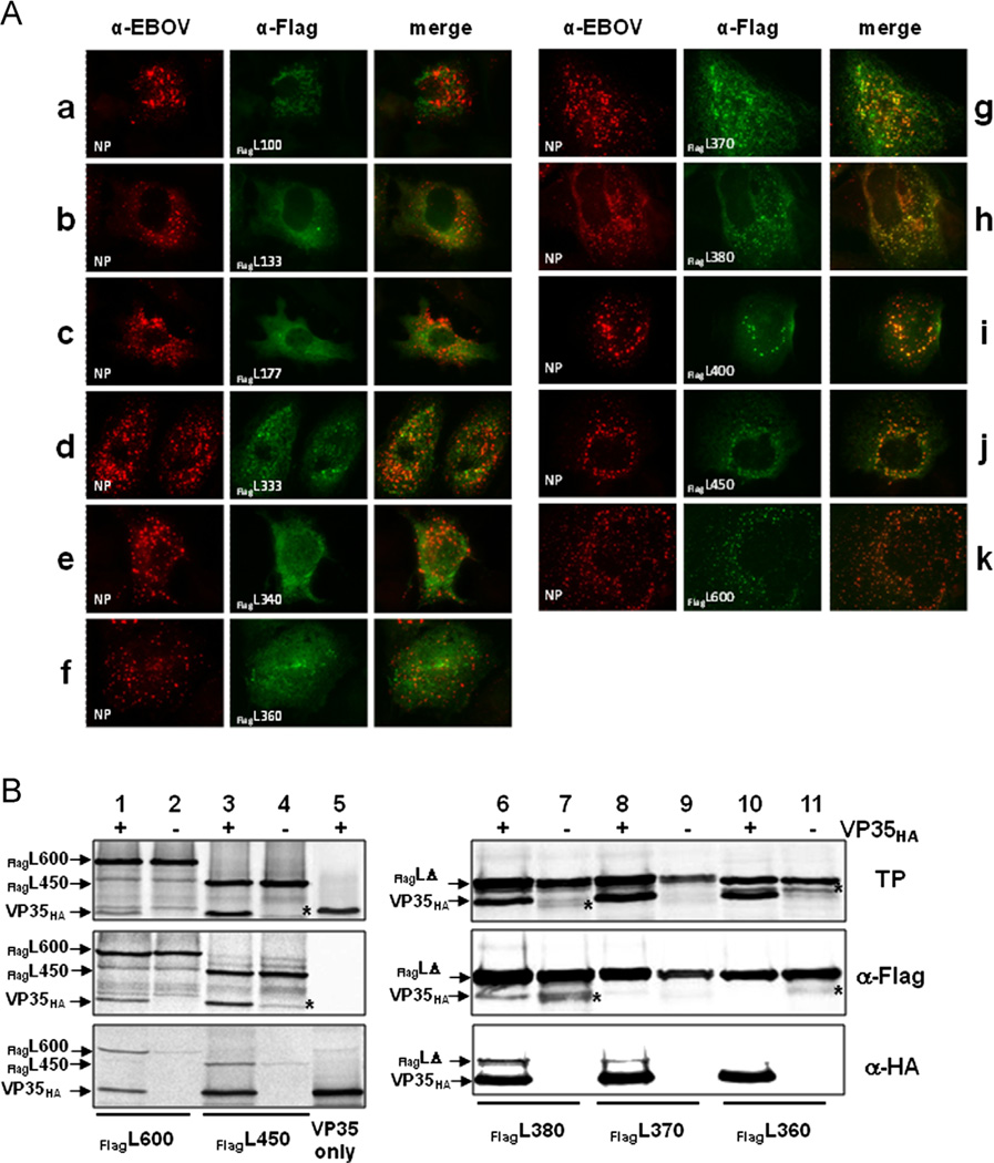
Fig. 3.
Interaction of C-terminal L deletion mutants with VP35. (A) Interaction of FlagL deletion mutants with VP35 was analyzed by IFA as described in Fig. 1. NP is stained red, FlagL mutants green. (B) The interaction of VP35HA and FlagL was confirmed by CoIP after in vitro translation of the proteins as described in Fig. 1. The positions of FlagL fragments 600 and 450 are indicated on the left; smaller FlagL fragments that were very similar in size (380, 370, and 360) are indicated by FlagLΔ. Lane 5 shows expression and precipitation of VP35HA only. The asterisk (*) in lanes 4, 7 and 11 indicates a FlagL fragment that runs close to the size of VP35HA. TP, in vitro translation products. Experiments were performed at least three times with similar outcome and representative images are shown.

