Abstract
Objective
Delirium affects 60– 80% of ventilated patients and is associated with worse clinical outcomes including death. Unfortunately there are limited data regarding the prevalence and risk factors of delirium in critically ill burn patients. The objectives of this study were to evaluate the prevalence of delirium in ventilated burn patients, using validated instruments, and to identify its risk factors.
Methods
Adult ventilated burn patients at 2 tertiary centers were prospectively evaluated for delirium using the Confusion Assessment Method in the ICU for 30 days or until ICU discharge. Patients with neurological injuries, severe dementia and those not expected to survive > 24 hours, were excluded. Markov logistic regression was used to identify risk factors of delirium, adjusting for clinically relevant covariates.
Results
The 82 ventilated burn patients had a median (interquartile range) age of 48 (38, 62), APACHE II scores 27 (21, 30) and percent burns of 20 (7, 32). Prevalence of delirium was 77% with a median duration of 3 (1, 6) days. Exposure to benzodiazepines was an independent risk factor for the development of delirium [Odds Ratio 6.8, (confidence interval 3.1, 15), p <0.001) while exposure to intravenous opiates [0.5 (0.4, 0.6), p <0.001] and methadone [0.7 (0.5, 9), p=0.02] were both associated with a lower risk of delirium.
Conclusion
Delirium occurred atleast once in approximately 80% of ventilated burn patients. Exposure to benzodiazepines was an independent risk factor for delirium while opiates and methadone reduced the risk of developing delirium, possibly through reduction of pain in these patients.
Keywords: burn critically ill, delirium, benzodiazepines, risk factors, mechanically ventilated
INTRODUCTION
Delirium is a form of acute brain organ dysfunction defined as a disturbance of consciousness that is accompanied by inattention, perceptual disturbances, and a fluctuation of mental status.1 With the recent validation of delirium monitoring instruments such as the Confusion Assessment Method in the Intensive Care Unit (CAM-ICU)2,3 and the Intensive Care Delirium Screening Checklist (ICDSC),4 it has become possible for non-psychiatrists to evaluate patients in the ICU for delirium even if mechanically ventilated (MV).5 Cohort studies using these instruments have shown that delirium occurs in up to 80% of MV patients,3,6,7 is frequently under-diagnosed if monitoring instruments are not used,8 and is independently associated with worse clinical outcomes, higher costs, and increased mortality.7,9–13 Furthermore, delirium is associated with significant cognitive impairment and increased disability after hospital discharge.14–16
While the prevalence and risk factors for delirium have previously been studied in the medical, surgical, and trauma ICU populations,6,7,13,17 available data in the burn population is lacking. According to the American Burn Association (www.ameriburn.org), there are approximately 500,000 burn injuries per year that receive medical treatment, with 40,000 hospitalizations, 25,000 referrals to specialized burn centers, and 4,000 deaths. Burn patients often experience longer periods of mechanical ventilation and ICU care, making this a population at risk for developing delirium and its associated complications. Much of the available literature on cognitive disturbances in burn patients is either in the form of case reports18 or studies that focused on post-traumatic stress disorder (PTSD) and depression in burn survivors.19 In a case series looking specifically at delirium, Andreasen et. al.20 reported that 31% of the 32 enrolled patients developed delirium. While the number of risk factors assessed in this study was limited, the authors found that seven of the ten delirious patients had a total body surface area burn of greater that 30%. In addition, pre-existing psychological factors were found to be risk factors for delirium.20
The risk factors for delirium appear to be different, based on the severity of illness (hospitalized versus critically-ill) and type of ICU,6,7,13,17,21 and as such may be different for critically-ill burn patients. Additionally, while sedatives, and in particular benzodiazepines,6,13,17 have been associated with delirium in the medical, surgical, and trauma ICU patients, there are conflicting data on the risk of delirium with opiates used to treat pain, depending on the patient population studied.6,22,23 Burn patients frequently receive greater amounts of sedative and analgesic medications, given their profoundly painful injuries, and the need for frequent operative and extensive non-operative burn care. It is unknown if this practice puts these patients at additional risk for delirium, or if appropriate analgesic management is actually protective.
The sparse available data in burn patients are with the use of standard psychiatric evaluation techniques for evaluating delirium, which is impractical for routine monitoring of these patients, due to the nature of their injuries and the fact that the majority of the sickest patients will be mechanically ventilated and non-verbal. In addition, the basic goals of these studies were not to study prevalence and specific risk factors for delirium, which are questions that must be answered before pharmacological or psychological interventions may be considered. The objective of this prospective, observational trial was therefore to study the prevalence of delirium in MV burn ICU patients, using the validated CAM-ICU delirium monitoring instrument, and to identify potentially modifiable risk factors.
METHODS
Patients
The institutional review boards at Vanderbilt Medical Center and Maricopa Medical Center approved this investigation with a waiver of consent due to the non-interventional/non-invasive nature of the study and the use of a de-identified dataset. Enrollment criteria included all patients 18 years or older, admitted to the Burn ICU at Vanderbilt Medical Center or the Arizona Burn Center, and required mechanical ventilation (MV) for greater than 24 hours. Patients were excluded who had (1) significant baseline neurological diseases, anoxic brain injury or intracranial neurotrauma that would confound the evaluation of delirium, (2) inability to understand English, (3) significant hearing loss, and (4) moribund patients not expected to survive > 24 hours and (5) admitted to the Burn ICU due to unavailability of other appropriate ICU beds.
Study procedures
Baseline demographics as well as information pertaining to known pre-morbid risk factors for delirium were collected at time of enrollment.6,7,13,17,21 All aspects of patient management with regards to treatment of burns, sepsis and sedation were per existing treatment protocols at the respective ICUs. Sedation was prescribed based on the Society of Critical Care Medicine practice guidelines, titrated to target Richmond Agitation-Sedation Scale (RASS)24,25 scores deemed appropriate by the treating team; daily wake up trials were not standard of care in these ICUs at the time of the study, but were performed per the discretion of the treating team.
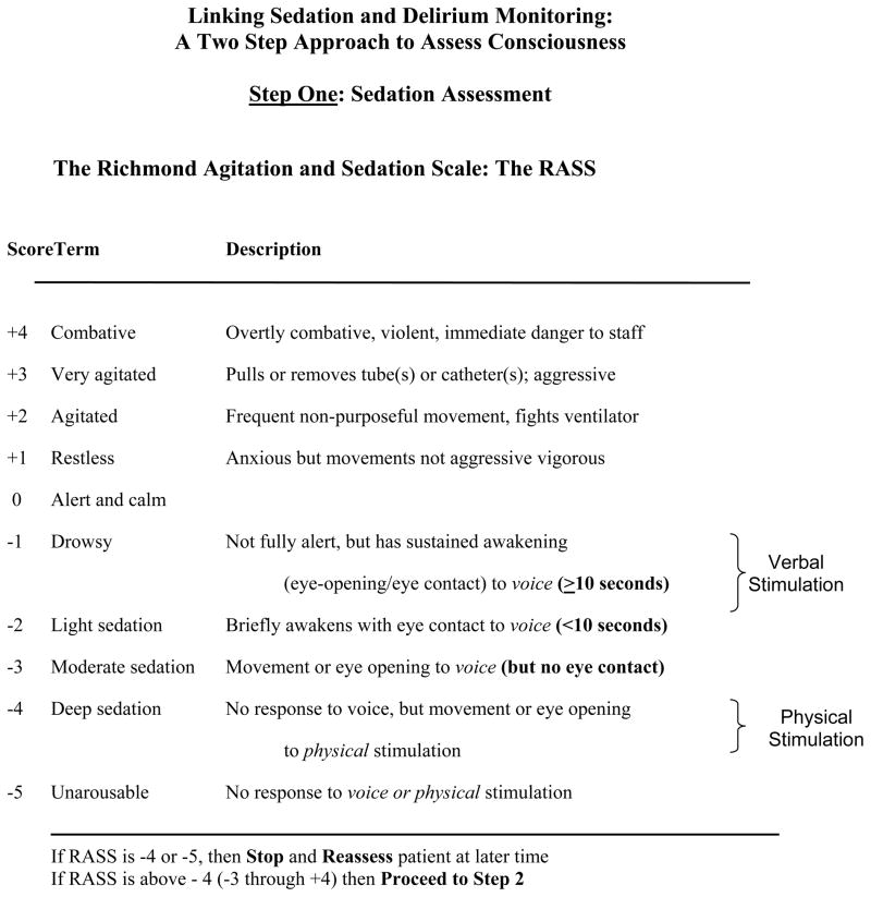
Level of arousal was measured using the RASS24,25 scale, a 10 point scale ranging from + 4 to −5 with a score of 0 denoting a calm and alert patient. Positive RASS scores denote positive or aggressive symptomology ranging from +1 (mild restlessness) to + 4 (dangerous agitation). The negative RASS scores apply to decreased levels of consciousness and differentiate between response to verbal commands (RASS score −1 to −3) and physical stimuli (RASS score −4 and −5).
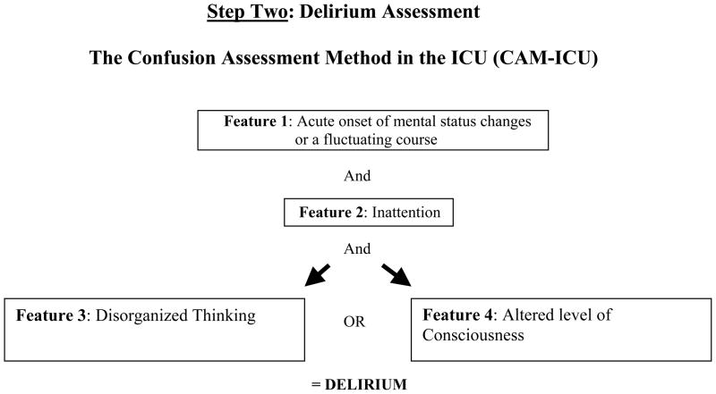
Patients were evaluated once daily for delirium using the validated CAM-ICU 2,3 if they were responsive to verbal commands (a RASS score of −3 or lighter level of sedation) for a maximum of 30 days or until ICU discharge. The CAM-ICU comprises four features which assess the following: acute change or fluctuation in mental status (Feature 1), inattention (Feature 2), disorganized thinking (Feature 3), and an altered level of consciousness (Feature 4). Each day the patient was categorized as being either in a coma, normal, or delirious based on previously published standardized definitions.2,3 Coma was defined as a RASS score of −4 or −5 in which case the CAM-ICU is not assessed due to lack of any response to any verbal stimulation. Normal was defined as RASS scores −3 and above and CAM-ICU negative. To be diagnosed as delirious, one needed to have a RASS score of −3 or higher (lighter level of sedation), and be CAM-ICU positive [i.e. with an acute change or fluctuation in mental status (Feature 1), accompanied by inattention (Feature 2) and either disorganized thinking (Feature 3) or an altered level of consciousness (Feature 4)] (Figure 1)
Figure 1. Richmond Agitation- Sedation Scale (RASS) and the Confusion Assessment Method of the ICU (CAM-ICU).
This sedation scale and delirium instrument can be used together as a two-step approach to assess consciousness and diagnose delirium. Patients are considered to have delirium, if they have RASS scores of −3 and above and are CAM-ICU positive by having Features 1 and 2, and either 3 or 4 positive. With permission from Dr. E. Wesley Ely from www.icudelirium.org
We chose to evaluate and label our delirium assessment as prevalent delirium instead of incident delirium (the first positive CAM-ICU assessment following a period of normal mental status) because it was difficult to obtain a reliable assessment of a patients’ pre-enrollment delirium status prior to ICU admission. Therefore prevalent delirium was defined as a positive CAM-ICU assessment during the first non-comatose mental status evaluation. We believe most of our patients were not delirious prior to their ICU admission, and that the new or incident delirium rates would be the same as the prevalent delirium rates. We further classified patients into the motoric subtypes of delirium, based on the criteria used by Peterson et al.26: Hyperactive, indicating that the patient only had positive RASS scores (+1 to + 4) associated with every CAM-ICU positive assessment; hypoactive, indicating that the patient only had RASS scores between 0 and −3 associated with every CAM-ICU positive assessment; and mixed, indicating that the patient had both positive and negative RASS scores associated with CAM-ICU positive assessments during the study.
Daily laboratory data, as ordered by the treating ICU team, were collected to gather information related to metabolic disturbances that may contribute to delirium. Sedatives and analgesic medications were prescribed by physicians according to institutional protocols adapted from the guidelines of the Society of Critical Care Medicine.27 The medications were titrated by the bedside nurses to achieve a target sedation level determined by the treating team using the RASS score 24,25 and for pain using a modification of the behavioral pain scale.28 Total 24-hour doses of sedative (lorazepam, midazolam, diazepam and ketamine) and analgesic medications (fentanyl, morphine, and methadone), including those administered for burn wound care, were collected while in the ICU.
Definitions of clinical injury severity and critical illness scoring systems
Several clinical injury severity and critical illness scoring systems were used. The Acute Physiology and Chronic Health Evaluation (APACHE) II 29,30 is a severity of illness scoring system, and these data were calculated using the most abnormal parameters within (before or after) 24 hours of institution of mechanical ventilation. APACHE II scores range from 0 (best) to 71 (worst).29,30 The Sequential Organ Failure Assessment (SOFA)31 is an organ failure scoring system that was calculated using the most abnormal parameters within (before or after) 24 hours of institution of mechanical ventilation. SOFA scores range from 0 (best) to 24 (worst).31 In addition, given the unique nature of the injuries to patients in the burn ICU, specific information with regards to percentage burn, depth of burn, and mechanism of injury was obtained from the medical record.
Statistical Analysis
Patients’ baseline demographic and clinical variables are presented using medians and interquartile range (IQRs) for continuous variables and proportions for categorical variables. In order to identify temporal association between potential risk factors for delirium, a Markov logistic regression model32 was used with an outcome of delirium (vs. normal mental status) on a given day and predetermined covariates, including the previous day’s mental status. In this study, our Markov models included the following 6 (3 by 2) transitions: from normal, delirious, or comatose during the previous 24 hours to either normal or delirious status presently. Generalized estimating equation (GEE) methods33 were used to account for correlations within patients among multiple days in the ICU. Covariates used in the model included the previous day’s mental status, doses of benzodiazepines (midazolam, lorazepam and diazepam), intravenous opiates (fentanyl and morphine), methadone, and a summary value for baseline variables. In order to avoid overfitting the model, principal component analysis was used as a data reduction technique to calculate a single value incorporating age, acute physiology component of the APACHE, history of alcohol and substance abuse, burn percentage, and presence of an inhalation injury. Benzodiazepines exposure was expressed as midazolam equivalents, defined as follows: 0.4 mg lorazepam = 1 mg midazolam; 2 mg diazepam = 1 mg midazolam.34 Similarly, all intravenous opiate exposure was expressed as fentanyl equivalents defined as follows: 10 mg morphine = 150 micrograms of fentanyl. For all analyses, R-software version 2.10 (www.r-project.org) was used. Two-sided 5% significance level was used for all statistical inferences. Odds ratios are presented for continuous covariates using the 25th and 75th percentiles as comparators; this approach is more clinically meaningful than the traditional one-unit increase approach. All drug doses were originally allowed to have a nonlinear relationship with the outcome, although drugs for which any association was clearly linear (p for nonlinearity > 0.20) had this nonlinear term removed.
RESULTS
We screened 198 patients, of which we enrolled 82 patients. Of the 116 patients excluded, majority (85) were because they were non-burn patients admitted due to lack of ICU beds elsewhere, 25 were <18 years of age, 2 patients had severe anoxic injury precluding delirium assessment and 4 patients were moribund and not expected to survive 24 hours. Demographic data of the cohort are presented in Table 1. The enrolled patients were a relatively younger ICU population compared to previous delirium studies with median (interquartile) age of 48 (38, 62). Further, these patients exhibited a high severity of illness, as reflected by their median APACHE II score of 27 (21, 30) and burn percentage of 20 (7, 32), both representative of the typical burn demographics seen in tertiary referral centers. More than two thirds of the patients had thermal injuries, and about half of the population had a concomitant inhalation injury. Of the co-morbid risk factors of delirium evaluated, about a third of our patients (27/82) had a history of alcohol or substance abuse, but rates of depression (12/82), previous cognitive impairment (2/82) and visual/hearing impairments (3/82) were low.
Table 1.
Patient Characteristics
| Variable* | N=82 |
|---|---|
| Age in years | 48 (38, 62) |
| Men, n (%) | 54 (66%) |
| Race, n (%) | |
| White | 71(87%) |
| Black | 7 (8 %) |
| Native American | 3 (4%) |
| Asian | 1 (1%) |
| APACHE II score † | 27 (21, 30) |
| SOFA score ‡ | 6 (5, 7) |
| Alcohol or substance abuse, n (%) | 27 (30%) |
| Depression | 12 (15%) |
| Burn injury description | |
| Burn percentage | 20 (7, 32) |
| Burn Depth, n (%) | |
| Full thickness | 31 (38%) |
| Partial thickness | 41 (50%) |
| Superficial | 10 (12%) |
| Burn type, n (%) | |
| Flame/Flash | 63 (77%) |
| Electrical | 6 (7%) |
| Scald | 3 (4%) |
| Chemical | 10 (12%) |
| Inhalation injury, n (%) | 45 (55%) |
| Sedative and antipsychotic use | |
| Benzodiazepine, (%) | 100% |
| Benzodiazepine, days | 15 (5, 24) |
| Intravenous opiates, % | 100% |
| Intravenous opiates, days | 13 (6, 22) |
| Propofol, % | 33% |
| Propofol, days | 3 (1, 5) |
| Dexmedetomidine, % | 11% |
| Dexmedetomidine, days | 3 (2, 7) |
| Haloperidol, % | 30% |
| Haloperidol, days | 3 (2, 6) |
| Atypical antipsychotics, % | 11% |
| Atypical antipsychotics, days | 1 (1, 7) |
Median (Interquartile range) unless specified otherwise
APACHE II - Acute Physiology and Chronic Health Evaluation II
SOFA - Sequential Organ Failure Assessment
Table 2 shows important clinical outcomes of our cohort. Approximately 80% of our patients had at least one episode of delirium, with the majority (71%) experiencing pure hypoactive delirium (negative symptomology such as lethargy and inattention), 22% experiencing mixed delirium (i.e. showing symptoms consistent with hypoactive and hyperactive delirium), and only 6% experiencing solely the pure hyperactive (positive or aggressive symptomology) motoric subtype. The median duration of delirium was 3 (1, 6) days.
Table 2.
Patient brain dysfunction outcomes
| Variable* | N=82 |
|---|---|
| Ever had delirium, n (%) | 63 (77%) |
| Hyperactive delirium only, n (%) | 4 (6%) |
| Hypoactive delirium only, n (%) | 45 (71%) |
| Mixed delirium | 14 (22%) |
| Comatose during entire 30-days follow up, n (%) | 12 (15%) |
| Days of delirium | 3 (1, 6) |
| Days of coma | 8 (2, 15) |
| Days on mechanical ventilation | 14.5 (5.5, 25) |
| Died during 30-day study follow up, n (%) | 10 (13%) |
Median (Interquartile range) unless specified otherwise
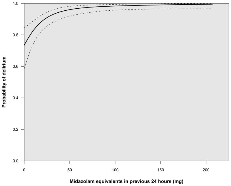
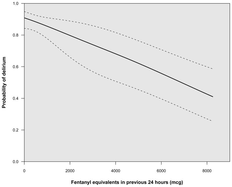
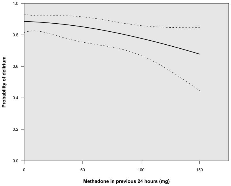
In the multivariable analyses, adjusting for previous cognitive status and clinically relevant covariates at baseline, benzodiazepine exposure (calculated as midazolam equivalents) was a strong independent predictor of developing delirium [odds ratio (OR) 6.8 (CI 3.1, 15), p < 0.001] (Table 3 and Figure 2). Intravenous opiate exposure (fentanyl equivalents) [OR 0.5 (CI 0.4, 0.6), p < 0.001] (Figure 3) as well as exposure to methadone [OR 0.7 (CI 0.5, 0.9), p = 0.02] (Figure 4) reduced the likelihood of development of delirium in this burn population. The combined effect of age, percentage of body surface area with burn injury, inhalation injury, history of substance abuse and severity of illness had no significant association with development of delirium (p=0.19). As in all our previous studies, (6, 16) abnormal mental status on a given day was a strong predictor of development of delirium the following day (p for overall effect < 0.001). Patients who were delirious at the previous assessment had about 27 times the odds of being delirious the following day compared to those with a normal assessment; those who were comatose at the previous assessment had 45 times the odds of being delirious than those with a normal assessment.
Table 3.
Risk Factors for Delirium
| Variable* | Odds Ratio | P Value** |
|---|---|---|
| Mental status on previous day | 0.001 | |
| Delirious vs. normal | 26.5 (11.9, 58.9) | |
| Comatose vs. normal | 44.9 (19, 105.8) | |
| Benzodiazepine dose (midazolam equivalents) in previous 24 hours † | 6.8 (3.1, 15) | <0.001 |
| Opiate dose (fentanyl equivalents) in previous 24 hours ‡ | 0.5 (0.4, 0.6) | <0.001 |
| Methadone dose in previous 24 hours | 0.7 (0.5, 0.9) | 0.02 |
| Baseline variable component*** | 0.6 (0.3, 1.0) | 0.19 |
Odds ratios are presented using the 25th and 75th percentile values as comparators. For example, patients receiving 60 mg of benzodiazepines (expressed as midazolam equivalents) in the previous 24 hours had 6.8 times higher odds of delirium the following day (vs. a normal assessment) than those receiving 5 mg of benzodiazepines.
P-values are calculated using group added last tests for the overall effect of each variable, including all related terms. For example, the p-value for mental status on the previous day includes model terms for delirium vs. normal status as well as for coma vs. normal status; the p-value for benzodiazepines includes both the linear and nonlinear terms.
For the purpose of this study, all benzodiazepines were converted to midazolam equivalents, such that 1 mg of midazolam was equal to 0.4 mg of lorazepam and 2 mg of diazepam.
For the purpose of this study, all intravenous opioids administered were converted to fentanyl equivalents, such that 10 mg of morphine was equal to 150 micrograms of fentanyl.
In order to conserve power, principal component analysis was used as a data reduction technique to calculate a single value incorporating age, acute physiology component of the APACHE, history of alcohol/substance abuse, burn percentage, and presence of an inhalation injury.
Figure 2. Benzodiazepines (midazolam equivalents) and the probability of developing delirium.
The probability of developing delirium increased with the dose of benzodiazepines administered in the previous 24 h. For the purpose of this study, all benzodiazepines were converted to midazolam equivalents, such that 1 mg of midazolam was equal to 0.4 mg of lorazepam and 2 mg of diazepam. This incremental risk of developing delirium was large at low doses and plateaued at around 50 mg/day of midazolam equivalent benzodiazepine exposure.
Figure 3. Opioid exposure (fentanyl equivalents) and the probability of developing delirium.
The probability of developing delirium decreased with the dose of opiates administered in the previous 24 h. For the purpose of this study, all intravenous opioids administered were converted to fentanyl equivalents, such that 10 mg of morphine was equal to 150 micrograms of fentanyl.
Figure 4. Methadone and the probability of developing delirium.
The probability of developing delirium decreased with increasing dose of methadone administered in the previous 24 h.
Organ and metabolic dysfunctions such as liver and renal dysfunction, hypoglycemia and hyponatremia, as well as outcomes in patients with and without any episodes of delirium are shown in Table 4.
Table 4.
Outcomes Based on Ever vs. Never Delirium*
| Variable** | Never Delirium*** N=7 | Ever Delirium N=63 |
|---|---|---|
| Liver dysfunction (bilirubin ≥2), n (%) | 0 (0%) | 12 (19%) |
| Liver dysfunction (bilirubin ≥2), days | 0 (0, 0) | 1 (1, 4) |
| Renal dysfunction (creatinine ≥2), n (%) | 0 (0%) | 8 (13%) |
| Renal dysfunction (creatinine ≥2), days | 0 (0, 0) | 2 (1, 3) |
| Hyponatremia (sodium <130 meq/l), n (%) | 0 (0%) | 10 (16%) |
| Hyponatremia (sodium <130 meq/l), days | 0 (0, 0) | 1 (1, 3) |
| Hypoglycemia (glucose <40 g/dl), n (%) | 0 (0%) | 2 (3%) |
| Hypoglycemia (glucose <40 g/dl), days | 0 (0, 0) | 1 (1,1) |
| ICU length of stay, days | 2 (2, 4) | 19 (14, 30) |
| Mechanical ventilator days | 1 (1, 1) | 16 (10, 26) |
| Died during 30-day study follow up, n (%) | 0 (0%) | 3 (5%) |
This table displays descriptive statistics according to whether delirium was diagnosed at any time during the study. Univariate comparisons were not performed because such comparisons can be significantly biased by confounders and immortal time bias. Multivariable analyses were not performed due to limited sample size (i.e., only seven patients were never delirious).
Median (Interquartile ranges) unless specified otherwise
Twelve patients were persistently comatose before dying; these patients are excluded since they were not eligible for delirium assessment, nor did they have any days of “normal mental status.”
DISCUSSION
Our study is the first prospective, observational trial that has elucidated the prevalence and risk factors for delirium in MV burn ICU patients using monitoring instruments validated for non-verbal patients. Several important findings need to be emphasized. First, we found that nearly eight out of ten patients developed delirium and that this brain organ dysfunction lasted for a median of three days. Second, and perhaps the most important finding, is that higher doses of benzodiazepines were independently associated higher odds of development of delirium. Conversely, higher doses of intravenous opiates used to treat pain reduced the odds of delirium development by half. Additionally, patients transitioned from intravenous opiates to methadone also had lower odds of development of delirium. Third, as seen in the medical and surgical patients,26,35 hyperactive delirium was the least frequent motoric subtype in burn ICU patients. Thus, unless routine monitoring is instituted with the CAM-ICU or the ICDSC, majority of the cases of delirium will be missed. This is due to the fact that healthcare teams are more attuned to noticing the patient with hyperactive delirium with positive symptomology (i.e., agitation) than the hypoactive delirium that manifests negative symptomology, such as inattention and depressed level of consciousness. This has important clinical ramifications, given that hypoactive delirium has been shown to be associated with worse outcomes than hyperactive delirium.36
Our data are consistent with recent studies of medical, surgical and trauma cohorts in which lorazepam and midazolam were found to be independent risk factors for delirium.6,7,13,17 Similarly, the prevalence rates and subtypes of delirium were in line with those reported earlier in non-burn patients.7,26,35 A unique finding in this study was that intravenous opiate administration (fentanyl and morphine) reduced the odds of development of delirium by half. Similarly, patients on methadone also experienced a lower risk of delirium. This supports the hypothesis that adequate pain control, so vital in caring for critically-ill burn ICU patients, reduces the risk of development of brain organ dysfunction. This is in line with a previous investigation in hip surgical patients, where adequate pain control with morphine reduced the likelihood of developing delirium,23 and with our previous work6 in trauma patients where morphine appeared to have a protective effect in the development of delirium. However, this contradicts many other studies in which opiates administered intravenously or via an epidural catheter were associated with increased risk of delirium.6,7,21,23,37 It is possible that the risk of delirium could be opioid specific and also dependent on the indication of use. Meperidine has almost universally been shown to be associated with delirium,21,23,37 while data on fentanyl and morphine have been mixed.6,7,21,23,37 Additionally, sedative and analgesic practices vary in ICUs around the world,38 with some physicians opting to use opiates for the “double effect” of analgesia and sedation thereby reducing the need for benzodiazepines or propofol. It is likely that when fentanyl or morphine are used for achieving deeper levels of sedation and are administered at high doses continuously, they are risk factors for delirium.6,7 On the other hand, if these analgesics are utilized judiciously for pain control23 or in cases where likelihood of pain is high (trauma and burn patients), they are less likely to be risk factors for delirium or may even be protective6 as was demonstrated in this study.
The mechanism or prognostic implications of delirium caused by the different psychoactive medications remains unknown and are not answered by the current study. Benzodiazepines and propofol have high affinity for the gamma-amino butyric acid (GABA)-receptor in the central nervous system,39 potentially leading to alterations in the levels of numerous neurotransmitters believed to be deliriogenic.40 Altering sedation paradigms with the alpha2 agonist, dexmedetomidine, has been shown in two randomized controlled trials41,42 to improve delirium outcomes when compared to benzodiazepines, though it is not known if the properties of dexmedetomidine per se or the ability to reduce benzodiazepine burden, contributed to the improved outcomes.41,42 While it should be emphasized that sedative and analgesic medications have a very important role in patient comfort, healthcare professionals must also strive to achieve the right balance of sedative and analgesic administration through greater focus on reducing unnecessary use. Instituting daily interruption of sedatives and analgesics and protocolizing their delivery with sedation and analgesic goals have both been shown to improve patients’ outcomes and need to be implemented to avoid overzealous use of these medications.43–45
Several limitations of this investigation warrant consideration. First, we evaluated for delirium once daily and measured sedative and analgesic exposure in the intervening 24 hours to study the effect of these medications in any change in cognitive status. However, it is possible that the sedative and analgesic medications were given as a consequence of delirium experienced during these 24 hours. In order to completely remove this bias, one would need to assess patient’s cognitive status more frequently (every 2–4 hours) and measure sedative and analgesic exposure between these periods. Given that this is extremely difficult task to accomplish in a research setting due to resource and time constraints, our next best alternative was to use Markov transitions in our modeling, which at least allowed examination of possible temporal associations within 24 hour periods. Despite their limitations, these methods are still much more rigorous than previously published non-ICU databases or burn ICU studies addressing this topic. Second, this study was not designed or powered to determine the role of delirium in leading to adverse outcomes such as length of stay, cost, or mortality. This remains unknown in the burn patient population, though the strong relationship between delirium and these outcomes has been repeatedly demonstrated in many other studies of critically-ill patients in medical, surgical, geriatric, orthopedic, and other patient populations.7,10–12,14,46,47 Third, our sample size, though larger than any previous cohort that has evaluated delirium in burn patients, limited our ability to study individual benzodiazepines, opiates, propofol and antipsycotics. Though our model incorporated numerous covariates that were deemed relevant a priori, we had to use principal component analysis as a data reduction technique to calculate a single value incorporating age, acute physiology component of the APACHE, alcohol and substance abuse, burn percentage, and presence of an inhalation injury. Additionally, we were unable to investigate the role of hypoxemia as risk factors for delirium, though 90% of our patient days were spent at oxygen saturations of 90% or higher, and half of them were at 95% or higher, making it unlikely that these would have contributed significantly towards the delirium. Additionally hypoxemia has yet to be shown to be associated with delirium in critically ill patients.
This study has delineated the magnitude of brain dysfunction in burn patients and has provided insight into risk factors that can potentially be modified. The high percentage of hypoactive delirium seen in our study will hopefully provide an impetus for routine delirium monitoring in burn patients, since delirium diagnosis in these patients would be missed without use of validated instruments. Future studies would need to assess if incorporating sedation and delirium protocols, including daily wake up trials, or using non-pharmacological and pharmacological interventions (antipsychotic medications, alpha2 agonists, analgesia based sedation etc) impact delirium rates and improve patient outcomes.
CONCLUSIONS
In the current study, we found delirium to be present in 77% of mechanically ventilated burn ICU patients. We used Markov logistic regression modeling to show that there was an independent association between higher doses of benzodiazepines and developing delirium the next day, while higher doses of opiates appeared to be strongly protective. Thus, the sedative and analgesic medications that are routinely employed have both therapeutic and hazardous ramifications for our patients and must be dosed very carefully with a specific aim to treat patient activity and pain adequately. Considering that delirium is a predictor of death, prolonged cognitive impairment, and higher cost of care, interventional studies should be conducted to determine whether alternative management strategies (or specific choices of sedative/analgesic agents) are associated with reductions in delirium and other short and long-term clinical outcomes in the burn population.
Acknowledgments
Funding/Support: Dr. Pandharipande is the recipient of a VA Career Development Award (Clinical Science Research and Development Service), and was funded by the ASCCA-FAER-Abbott Physician Scientist Award and Vanderbilt Physician Scientist Development Award during the conduct of the study. Dr. Ely is supported by the VA Clinical Science Research and Development Service (VA Merit Review Award) and the National Institutes of Health (AG0727201).
Footnotes
Potential financial conflict of interest:
Dr. Pandharipande has received research grant and honoraria from Hospira Inc. Ms. Pun has received honoraria from Hospira Inc. Dr. Ely has received research grant and honoraria from Hospira, Inc, Pfizer, Eli Lilly, GSK, and a research grant from Aspect Medical Systems. The other authors report no financial disclosures.
Reference List
- 1.American Psychiatric Association. Diagnostic and statistical manual of mental disorders. 4. Washington, DC: American Psychiatric Association; 2000. text revision. [Google Scholar]
- 2.Ely EW, Truman B, May L, et al. Validation of the CAM-ICU for delirium assessment in mechanically ventilated patients. J Am Geriatr Soc. 2001;49:S2. [Google Scholar]
- 3.Ely EW, Inouye SK, Bernard GR, et al. Delirium in mechanically ventilated patients: validity and reliability of the confusion assessment method for the intensive care unit (CAM-ICU) JAMA. 2001;286:2703–2710. doi: 10.1001/jama.286.21.2703. [DOI] [PubMed] [Google Scholar]
- 4.Bergeron N, Dubois MJ, Dumont M, et al. Intensive Care Delirium Screening Checklist: evaluation of a new screening tool. Intensive Care Med. 2001;27:859–864. doi: 10.1007/s001340100909. [DOI] [PubMed] [Google Scholar]
- 5.Pun BT, Gordon SM, Peterson JF, et al. Large-scale implementation of sedation and delirium monitoring in the intensive care unit: A report from two medical centers. Crit Care Med. 2005;33:1199–1205. doi: 10.1097/01.ccm.0000166867.78320.ac. [DOI] [PubMed] [Google Scholar]
- 6.Pandharipande P, Cotton B, Shintani A, et al. Prevalence and Risk Factors for Development of Delirium in Surgical and Trauma Intensive Care Unit Patients. Journal of Trauma. 2008;65:34–41. doi: 10.1097/TA.0b013e31814b2c4d. [DOI] [PMC free article] [PubMed] [Google Scholar]
- 7.Lat I, McMillian W, Taylor S, et al. The impact of delirium on clinical outcomes in mechanically ventilated surgical and trauma patients. Crit Care Med. 2009;37:1898–1905. doi: 10.1097/CCM.0b013e31819ffe38. [DOI] [PubMed] [Google Scholar]
- 8.Ely EW, Siegel MD, Inouye SK. Delirium in the intensive care unit: an under-recognized syndrome of organ dysfunction. Semin Respir Crit Care Med. 2001;22:115–126. doi: 10.1055/s-2001-13826. [DOI] [PubMed] [Google Scholar]
- 9.Ely EW, Gautam S, Margolin R, et al. The impact of delirium in the intensive care unit on hospital length of stay. Intensive Care Med. 2001;27:1892–1900. doi: 10.1007/s00134-001-1132-2. [DOI] [PMC free article] [PubMed] [Google Scholar]
- 10.Ely EW, Shintani A, Truman B, et al. Delirium as a predictor of mortality in mechanically ventilated patients in the intensive care unit. JAMA. 2004;291:1753–1762. doi: 10.1001/jama.291.14.1753. [DOI] [PubMed] [Google Scholar]
- 11.Lin SM, Liu CY, Wang CH, et al. The impact of delirium on the survival of mechanically ventilated patients. Crit Care Med. 2004;32:2254–2259. doi: 10.1097/01.ccm.0000145587.16421.bb. [DOI] [PubMed] [Google Scholar]
- 12.Milbrandt EB, Deppen S, Harrison PL, et al. Costs associated with delirium in mechanically ventilated patients. Crit Care Med. 2004;32:955–962. doi: 10.1097/01.ccm.0000119429.16055.92. [DOI] [PubMed] [Google Scholar]
- 13.Ouimet S, Kavanagh BP, Gottfried SB, et al. Incidence, risk factors and consequences of ICU delirium. Intensive Care Med. 2007;33:66–73. doi: 10.1007/s00134-006-0399-8. [DOI] [PubMed] [Google Scholar]
- 14.Jackson JC, Gordon SM, Hart RP, et al. The association between delirium and cognitive decline: a review of the empirical literature. Neuropsychol Rev. 2004;14:87–98. doi: 10.1023/b:nerv.0000028080.39602.17. [DOI] [PubMed] [Google Scholar]
- 15.Jackson JC, Gordon SM, Girard TD, et al. Delirium as a risk factor for long term cognitive impairment in mechanically ventilated ICU survivors. Am J Respir Crit Care Med. 2007;175:A22. [Google Scholar]
- 16.Rahkonen T, Eloniemi-Sulkava U, Halonen P, et al. Delirium in the non-demented oldest old in the general population: risk factors and prognosis. Int J Geriatr Psychiatry. 2001;16:415–421. doi: 10.1002/gps.356. [DOI] [PubMed] [Google Scholar]
- 17.Pandharipande P, Shintani A, Peterson J, et al. Lorazepam is an independent risk factor for transitioning to delirium in intensive care unit patients. Anesthesiology. 2006;104:21–26. doi: 10.1097/00000542-200601000-00005. [DOI] [PubMed] [Google Scholar]
- 18.Stanford GK, Pine RH. Postburn delirium associated with use of intravenous lorazepam. J Burn Care Rehabil. 1988;9:160–161. doi: 10.1097/00004630-198803000-00006. [DOI] [PubMed] [Google Scholar]
- 19.Blank K, Perry S. Relationship of psychological processes during delirium to outcome. Am J Psychiatry. 1984;141:843–847. doi: 10.1176/ajp.141.7.843. [DOI] [PubMed] [Google Scholar]
- 20.Andreasen NJ. Neuropsychiatric complications in burn patients. Int J Psychiatry Med. 1974;5:161–171. doi: 10.2190/FATV-K0DW-12M7-J9BH. [DOI] [PubMed] [Google Scholar]
- 21.Dubois MJ, Bergeron N, Dumont M, et al. Delirium in an intensive care unit: a study of risk factors. Intensive Care Med. 2001;27:1297–1304. doi: 10.1007/s001340101017. [DOI] [PubMed] [Google Scholar]
- 22.Marcantonio ER, Goldman L, Orav EJ, et al. The association of intraoperative factors with the development of postoperative delirium. Am J Med. 1998;105:380–384. doi: 10.1016/s0002-9343(98)00292-7. [DOI] [PubMed] [Google Scholar]
- 23.Morrison RS, Magaziner J, Gilbert M, et al. Relationship between pain and opioid analgesics on the development of delirium following hip fracture. J Gerontol A Biol Sci Med Sci. 2003;58:76–81. doi: 10.1093/gerona/58.1.m76. [DOI] [PubMed] [Google Scholar]
- 24.Sessler CN, Gosnell MS, Grap MJ, et al. The Richmond Agitation-Sedation Scale: validity and reliability in adult intensive care unit patients. Am J Respir Crit Care Med. 2002;166:1338–1344. doi: 10.1164/rccm.2107138. [DOI] [PubMed] [Google Scholar]
- 25.Ely EW, Truman B, Shintani A, et al. Monitoring sedation status over time in ICU patients: reliability and validity of the Richmond Agitation-Sedation Scale (RASS) JAMA. 2003;289:2983–2991. doi: 10.1001/jama.289.22.2983. [DOI] [PubMed] [Google Scholar]
- 26.Peterson JF, Pun BT, Dittus RS, et al. Delirium and its motoric subtypes: a study of 614 critically ill patients. J Am Geriatr Soc. 2006;54:479–484. doi: 10.1111/j.1532-5415.2005.00621.x. [DOI] [PubMed] [Google Scholar]
- 27.Jacobi J, Fraser GL, Coursin DB, et al. Clinical practice guidelines for the sustained use of sedatives and analgesics in the critically ill adult. Crit Care Med. 2002;30:119–141. doi: 10.1097/00003246-200201000-00020. [DOI] [PubMed] [Google Scholar]
- 28.Payen JF, Bru O, Bosson JL, et al. Assessing pain in critically ill sedated patients by using a behavioral pain scale. Crit Care Med. 2001;29:2258–2263. doi: 10.1097/00003246-200112000-00004. [DOI] [PubMed] [Google Scholar]
- 29.Knaus WA, Draper EA, Wagner DP, et al. APACHE II: a severity of disease classification system. Crit Care Med. 1985;13:818–829. [PubMed] [Google Scholar]
- 30.Knaus WA, Wagner DP, Draper EA, et al. The APACHE III prognostic system. Risk prediction of hospital mortality for critically ill hospitalized adults. Chest. 1991;100:1619–1636. doi: 10.1378/chest.100.6.1619. [DOI] [PubMed] [Google Scholar]
- 31.Vincent JL, Mendonca Ad, Cantraine F, et al. Use of the SOFA score to assess the incidence of organ dysfunction/failure in intensive care units: Results of a multicenter, prospective study. Crit Care Med. 1998;26:1793–1800. doi: 10.1097/00003246-199811000-00016. [DOI] [PubMed] [Google Scholar]
- 32.Diggle PJ, Liang KY, Zeger SL. Analysis of longitudinal data. Oxford: Clarendon Press; 1994. [Google Scholar]
- 33.Zeger SL, Liang KY. Longitudinal data analysis for discrete and continuous outcomes. Biometrics. 1986;42:121–130. [PubMed] [Google Scholar]
- 34.Dundee JW, McGowan WA, Lilburn JK, et al. Comparison of the actions of diazepam and lorazepam. Br J Anaesth. 1979;51:439–446. doi: 10.1093/bja/51.5.439. [DOI] [PubMed] [Google Scholar]
- 35.Pandharipande P, Cotton BA, Shintani A, et al. Motoric subtypes of delirium in mechanically ventilated surgical and trauma intensive care unit patients. Intensive Care Med. 2007;33:1726–1731. doi: 10.1007/s00134-007-0687-y. [DOI] [PubMed] [Google Scholar]
- 36.Meagher DJ, Trzepacz PT. Motoric subtypes of delirium. Semin Clin Neuropsychiatry. 2000;5:75–85. doi: 10.153/SCNP00500075. [DOI] [PubMed] [Google Scholar]
- 37.Marcantonio ER, Juarez G, Goldman L, et al. The relationship of postoperative delirium with psychoactive medications. JAMA. 1994;272:1518–1522. [PubMed] [Google Scholar]
- 38.Soliman HM, Melot C, Vincent JL. Sedative and analgesic practice in the intensive care unit: the results of a European survey. Br J Anaesth. 2001;87:186–192. doi: 10.1093/bja/87.2.186. [DOI] [PubMed] [Google Scholar]
- 39.Mihic SJ, Harris RA. GABA and the GABAA receptor. Alcohol Health Res World. 1997;21:127–131. [PMC free article] [PubMed] [Google Scholar]
- 40.Van Der Mast RC. Pathophysiology of delirium. J Geriatr Psychiatry Neurol. 1998;11:138–145. doi: 10.1177/089198879801100304. [DOI] [PubMed] [Google Scholar]
- 41.Pandharipande PP, Pun BT, Herr DL, et al. Effect of sedation with dexmedetomidine vs lorazepam on acute brain dysfunction in mechanically ventilated patients: the MENDS randomized controlled trial. JAMA. 2007;298:2644–2653. doi: 10.1001/jama.298.22.2644. [DOI] [PubMed] [Google Scholar]
- 42.Riker RR, Shehabi Y, Bokesch PM, et al. Dexmedetomidine vs midazolam for sedation of critically ill patients: a randomized trial. JAMA. 2009;301:489–499. doi: 10.1001/jama.2009.56. [DOI] [PubMed] [Google Scholar]
- 43.Kollef MH, Levy NT, Ahrens TS, et al. The use of continuous i.v. sedation is associated with prolongation of mechanical ventilation. Chest. 1998;114:541–548. doi: 10.1378/chest.114.2.541. [DOI] [PubMed] [Google Scholar]
- 44.Brook AD, Ahrens TS, Schaiff R, et al. Effect of a nursing-implemented sedation protocol on the duration of mechanical ventilation. Crit Care Med. 1999;27:2609–2615. doi: 10.1097/00003246-199912000-00001. [DOI] [PubMed] [Google Scholar]
- 45.Kress JP, Pohlman AS, O’Connor MF, et al. Daily interruption of sedative infusions in critically ill patients undergoing mechanical ventilation. N Engl J Med. 2000;342:1471–1477. doi: 10.1056/NEJM200005183422002. [DOI] [PubMed] [Google Scholar]
- 46.Inouye SK, Rushing JT, Foreman MD, et al. Does delirium contribute to poor hospital outcomes? a three-site epidemiologic study. J Gen Intern Med. 1998;13:234–242. doi: 10.1046/j.1525-1497.1998.00073.x. [DOI] [PMC free article] [PubMed] [Google Scholar]
- 47.McCusker J, Cole M, Abrahamowicz M, et al. Delirium predicts 12-month mortality. Arch Intern Med. 2002;162:457–463. doi: 10.1001/archinte.162.4.457. [DOI] [PubMed] [Google Scholar]


