Abstract
Gap junction channels composed of connexin43 (Cx43) are essential for normal heart formation and function. We studied the potential role of the Wnt family of secreted polypeptides as regulators of Cx43 expression and gap junction channel function in dissociated myocytes and intact hearts. Neonatal rat cardiomyocytes responded to Li+, which mimics Wnt signaling, by accumulating the effector protein β-catenin and by inducing Cx43 mRNA and protein markedly. Induction of Cx43 expression was also observed in cardiomyocytes cocultured with Rat-2 fibroblasts or N2A neuroblastoma cells programmed to secrete bioactive Wnt-1. By transfecting a Cx43 promoter-reporter gene construct into cardiomyocytes, we demonstrated that the inductive effect of Wnt signaling was transcriptionally mediated. Enhanced expression of Cx43 increased cardiomyocyte cell coupling, as determined by Lucifer Yellow dye transfer and by calcium wave propagation. Conversely, in a transgenic cardiomyopathic mouse model that exhibits ventricular arrhythmias and gap junctional remodeling, β-catenin and Cx43 expression were downregulated concordantly. In response to Wnt signaling, the accumulating Cx43 colocalized with β-catenin in the junctional membrane; moreover, forced expression of Cx43 in cardiomyocytes reduced the transactivation potential of β-catenin. These findings demonstrate that Wnt signaling is an important modulator of Cx43-dependent intercellular coupling in the heart, and they support the hypothesis that dysregulated signaling contributes to altered impulse propagation and arrhythmia in the myopathic heart.
Introduction
Gap junctions are composed of arrays of intercellular channels at the interface of cells (1–3). By regulating the direct exchange of ions and small molecules between cells, gap junction channels have been implicated in a diverse assortment of biologic processes including cellular differentiation and development, metabolic homeostasis, and electrotonic coupling of excitable tissues. In mammals, gap junction channel proteins are encoded by connexins, a multigene family with as many as 15 distinct isoforms in the mouse. Many tissues express a number of connexin isoforms, with complex and overlapping temporal and spatial profiles. Moreover, different isoforms may aggregate to form complex heteromeric or heterotypic channels, thereby creating even greater potential for functional diversity.
In the rodent heart, at least 3 connexin proteins are expressed at readily detectable levels (4, 5). Cx43 is by far the most abundant cardiovascular gap junction channel protein, found in both atrial and ventricular gap junctions, but relatively excluded from pacemaking cells and those forming the specialized His-Purkinje conduction system. Cx40 is preferentially localized in atrial gap junctions, as well as regions of the specialized conduction system. Cx45 expression patterns are less well defined, but the most recent studies suggest that it too may be localized to the specialized conduction system (6).
Increasing theoretical and experimental evidence suggests that downregulation and/or spatial redistribution of gap junction channel proteins, a process referred to as gap junction remodeling, may contribute to the high incidence of arrhythmias associated with various forms of heart disease. The remodeling process has been observed in patients with ischemic and hypertrophic cardiomyopathies (7, 8) and also in several experimental animal myopathies associated with reentrant ventricular tachycardia, including a canine infarct model and a transgenic mouse model in which the diphtheria toxin A gene is conditionally expressed in cardiomyocytes (9, 10). Genetic studies also suggest an essential role for gap junction channel proteins in maintaining normal cardiac conduction properties. Homozygous loss of function of Cx40 results in atrial conduction slowing (11), whereas heterozygous Cx43-deficient mice show a similar effect in ventricular tissue (12).
Complete loss of Cx43 expression in the mouse leads to a number of developmental consequences, including obstruction of the right ventricular outflow tract and perinatal death (13). Although this congenital cardiac defect was unexpected and its occurrence remains unexplained, recent studies support the hypothesis that loss of function of Cx43 in cells of neural crest origin may account for this phenotype (14).
One potential regulator of gap junction expression and function that may have important implications for developmental processes as well as for normal function in adult stages is the Wnt family of genes. Wnt genes encode a large family of secreted polypeptides that can influence cell-cell communication, both during embryonic development and in adult life (15). Wnt proteins are thought to act via the Frizzled class of cell-surface proteins. Receptor activation leads to inhibition of glycogen synthase kinase 3β (GSK3β), resulting in stabilization and accumulation of nonphosphorylated β-catenin within the cytosolic compartment. Increased abundance of this pool is associated with entry of β-catenin into the nucleus, where the protein complexes with Tcf transcription factors and modulates the expression of specific target genes (16). β-Catenin also is associated with the plasma membrane, where it acts as a component of the cell-cell adhesive junction. Wnt-1 has been shown to enhance gap junctional communication between ventral cells of the developing Xenopus embryo (17, 18). In addition, ectopic expression of Wnt-1 in the limb mesenchyme of transgenic mice increases the local accumulation of Cx43 transcripts (19). The mechanisms leading to these effects are uncertain, but recent studies in rat pheochromocytoma (PC12) cells suggest that the Cx43 gene may be a downstream target of Wnt-1 signaling (20). Interestingly, the gene product of wingless (wg), the Drosophila Wnt homologue, has been shown to be crucial for normal heart development, where it appears to be absolutely required for specifying the heart progenitors (21, 22). Although the role of Wnt signaling in mammalian cardiovascular development and disease is less well characterized, recent studies have described preferential expression of individual Wnt and Frizzled-related genes in the cardiovasculature (23–25). Moreover, experimental attenuation of Wnt-1 expression in the mouse embryo by antisense methodology leads to cardiomegaly (26), and recent human genetic studies have found that the frizzled gene Fzd9 is deleted in Williams-Beuren syndrome (27).
Despite these diverse observations suggesting an important association between Wnt signaling and normal heart formation and function, the relationship between Wnt proteins and Cx43 expression in the mammalian heart has not, to our knowledge, been previously investigated. Accordingly, we embarked upon this study to determine whether Wnt proteins regulate Cx43 expression and function in cardiomyocytes.
Methods
Neonatal rat ventricular myocyte culture.
Primary cultures of 2- to 3-day-old neonatal rat ventricular myocytes were prepared as reported previously (28). Briefly, neonatal ventricles were carefully excised from atria and great vessels, minced into about 1-mm3 pieces, and dissociated in a pancreatin solution at 37°C. Cells were resuspended in heart medium (HM) consisting of Hanks’ solution supplemented with 10% FCS, MEM amino acids and MEM essential amino acids, MEM Vitamine, hypoxanthine, and penicillin/streptomycin. After preplating to remove fibroblasts, the remaining cell suspension was plated onto variously sized primary culture dishes at a density of 105 cells/cm2. The culture medium was changed after overnight incubation; at 36 hours, the medium was replaced with 2% FCS-containing HM plus various additives as indicated in specific experiments. Cell culture components were obtained from GIBCO BRL (Gaithersburg, Maryland, USA), and, unless otherwise indicated, other chemicals and additives, including LiCl and dibutyryl cyclic AMP (db-cAMP), were obtained from Sigma Chemical Co. (St. Louis, Missouri, USA).
Transfection and luciferase assay.
Freshly isolated ventricular myocytes were transiently transfected by electroporation (29), with mixtures of plasmid DNA as detailed in the individual figure legends. Plasmids used in these studies included a rat Cx43 promoter-luciferase reporter construct (30), the pTOPFLASH wild-type Tcf-luciferase reporter construct, and the pFOPFLASH mutant Tcf-luciferase reporter construct (31); wild-type rat β-catenin expression vector or its parental plasmid (32); a c-myc epitope–tagged human Cx43 expression vector (33) or parental plasmid and CMV-driven β-galactosidase expression vector (CLONTECH Laboratories Inc., Palo Alto, California, USA). For each transfection, 10 × 106 myocytes were suspended in 0.4 mL PBS containing 0.1% glucose and plasmid DNA and electroporated in a 1-mL cuvette at 280V and 250 μF with a Gene Pulser (Bio-Rad Laboratories Inc., Hercules, California, USA) with a total of 12 μg of plasmid DNA. After 5 minutes at room temperature, 0.4 mL of PBS-glucose was added to the cell mixture. Cells were plated at a density of 2.5 × 10⁶/cm2 in 10% FCS-HM. The culture medium was changed after 12 hours of incubation, and cells were harvested 48 hours later for determination of luciferase and β-galactosidase activities, using a Bio-orbit 1251 luminometer (Wallace Inc., Gaithersburg, Maryland, USA). For each well, luciferase activity was normalized to β-galactosidase activity.
Cell lines and coculture methodology.
N2A neuroblastoma cells were maintained in 10% FCS in DMEM, cultured to 80% confluence, and subsequently infected with MV7, a parental retroviral vector conferring G418 resistance (34), or MV Wnt-1, which expresses Wnt-1, as described previously (35). Infected cells were selected in the presence of 400 μg/mL G418, and surviving cells were maintained as pools of drug-resistant clones. For coculture experiments, the pooled clones were switched from 10 % FCS in DMEM to HM 2 days before coculturing. Rat ventricular myocyte cultures were prepared as already described here. Thirty-six hours after plating, the transduced N2A cells were seeded atop the myocytes. Parental Rat-2 fibroblasts or those stably transduced with a retroviral vector encoding Wnt-1, have been described previously (35). Rat-2 fibroblasts transduced with a Wnt-4 expression construct were similarly prepared. For transmembrane coculture experiment, myocytes were seeded on the upper side of polycarbonate membranes of transwell inserts (24 mm; Corning-Costar Corp., Cambridge, Massachusetts, USA) at a density of 105 cells/cm2 and maintained in HM. The culture medium was changed 12 hours later. At 36 hours, fibroblasts (parental Rat-2, Rat-2 Wnt-1, Rat-2 Wnt-4) were applied onto the lower side of the membrane at a density of 5 × 10⁴ cells/cm2 and maintained in HM until harvesting, typically 48 hours later. All cultured cells were maintained at 37°C in an atmosphere of 5% CO2.
Northern blotting.
Total RNA from cardiomyocytes were prepared using Trizol reagent (GIBCO BRL) according to the manufacturer’s protocol. Samples of RNA were fractionated with formaldehyde/agarose electrophoresis and transferred onto nitrocellulose membrane. Blots were hybridized with a rat 32P-labeled Cx43 probe (clone G2A; kindly provided by E. Beyer, University of Chicago, Illinois, USA), prepared with the PrimeIt Kit (Stratagene, La Jolla, California, USA) by standard techniques. A GAPDH probe was used to normalize loading. The washed membranes were exposed to x-ray film at –75°C, and the autoradiograms were quantified using NIH Image software (National Institutes of Health, Bethesda, Maryland, USA).
Immunoblotting.
Cultured myocytes were washed twice with ice-cold PBS and subsequently lysed in 2× Laemmli buffer. Myocytes from transmembrane inserts were harvested after carefully removing the cocultured fibroblasts from the lower side of membrane with a rubber policeman. For studies of murine hearts, ventricular tissue was harvested from transgenic and age-matched nontransgenic controls at 2 months of age and solubilized by sonication in 1× Laemmli buffer. The viscosity of all samples was reduced by multiple passages of the lysate through a 25-gauge needle. Equal amounts of protein were separated by 10% SDS-PAGE and transferred to nitrocellulose membranes. The equivalency of protein loading was verified by staining the membranes with Ponceau Red (Sigma Chemical Co.). Membranes were then incubated for 30 minutes at room temperature in blocking buffer (20 mm Tris-HCl [pH 7.4], 150 mM NaCl, 0.05% Tween-20, and 5% nonfat dry milk [TBST]). Cx43 was detected using a rabbit polyclonal antibody raised against the COOH terminus of Cx43 (10), which was diluted 1:5,000 in blocking buffer and incubated at room temperature for 2 hours. β-Catenin was detected by incubating the same membrane with a 1:1,000 dilution of a mouse monoclonal anti–β-catenin antibody (Transduction Laboratories, Lexington, Kentucky, USA) overnight at 4°C. Actin was detected with an mAb against actin at 1:2,000 dilution (Boehringer Mannheim, Indianapolis, Indiana, USA). Blots were subsequently incubated with a 1:5,000 dilution of horseradish-conjugated protein A or goat anti-mouse IgG (Bio-Rad Laboratories) in TBST at room temperature for 1 hour. After further washing, the blots were incubated for 1 minute in enhanced chemilluminescence (ECL) detection reagents (Amersham Pharmacia Biotech, Piscataway, New Jersey, USA) and exposed to x-ray film.
Cell fractionation.
To determine the subcellular localization of β-catenin, cell fractionation was performed essentially as described previously (36). In brief, cells were washed twice in ice-cold PBS and collected in physiological buffer (PB) (10 mM Tris-HCl [pH 7.4], 5mM EDTA, 2 mM DTT, 1 mM PMSF, 2 μg/mL leupeptin, 2 μg/mL pepstatin A, 140 mM NaCl; 800 μL per 100 mm dish). The cells were homogenized by 30 strokes with a Dounce homogenizer, and the lysates were clarified by centrifugation at 500 g at 4°C for 10 minutes. The crude total cellular lysates were then centrifuged at 200,000 g for 90 minutes at 4°C. The supernatants from these spins were designated as the cytosolic fractions. The pellets were resuspended in PBS with 1% Triton (150 μL per 100 mm dish), and the solubilized proteins were designated as the membrane fractions. Protein concentrations in each fraction were determined by a commercial protein assay kit (Bio-Rad Laboratories). Laemmli buffer was added to each of the fractions for analysis by SDS-PAGE.
Immunoprecipitation.
Myocytes were lysed in 1% Triton X-100 in PB on ice for 30 minutes with agitation and clarified by brief centrifugation at 1,500 g. Rabbit polyclonal anti–Cx43 antisera or mouse monoclonal anti–β-catenin antibody were added to lysates containing 75 μg of total protein and incubated at 4°C overnight with rocking. A total of 60 μL protein A or protein G-agarose (Boehringer Mannheim) was then added, and the incubation was continued for an additional 4 hours. The agarose beads were washed 5 times with 1% Triton X-100 and 0.1% SDS in PBS, and the bound proteins were solubilized by 2× Laemmli buffer and subjected to SDS-PAGE and immunoblotting, as already described here.
Confocal microscopy.
Cells were cultured on glass coverslips coated with cell-Tak (Becton Dickinson Labware, Bedford, Massachusetts, USA). After washing with PBS, cells were fixed with 50% methanol/50% acetone at room temperature for 2 minutes and then permeabilized with 1% Triton X-100 in PBS for 15 minutes at room temperature. After blocking by 0.4% Triton X-100 PBS with 10% goat serum at room temperature for 30 minutes, the cells were incubated in the same buffer with rabbit polyclonal anti-Cx43 antiserum (1:1,000) and mouse monoclonal anti–β-catenin IgG (1:250) (Transduction Laboratories) concurrently at 4°C overnight. After washing with 0.4% Triton X-100 in PBS, the cells were then incubated with both FITC-conjugated goat anti-rabbit IgG (1:1,000) and Texas Red-conjugated goat anti-mouse IgG (1:1,000) (Jackson ImmunoResearch Laboratories Inc., West Grove, Pennsylvania, USA) at room temperature for 1 hour. Cells were examined using a Leica TCS-SP (ultraviolet [UV]) confocal laser scanning microscope (Heidelberg, Germany) equipped with a 4-channel spectrophotometer scan head and 4 lasers (Ar-UV, Argon, Krypton, and HeNe). For these studies, the Argon (488 nm), Krypton (568 nm) lasers were used to simultaneously illuminate the cells, and a 100× 1.4 N.A. objective lens was used for imaging. The spectrophotometer windows in each channel were set such that no signal from the FITC channel crossed into the Texas Red channel and vice versa (i.e., the left edge of the Texas Red window was set such that no signal appeared when only the 488-nm line was used, and the right edge of the FITC window was set such that no signal appeared when only the 568-nm line was used). The pinhole size was adjusted such that resultant “optical sections” were approximately 0.25- to 0.3-μm thick. Serial optical sections (in the x-y plane) were collected at 0.25-μm intervals from the top to the bottom of the cells. The sensitivity of the photomultiplier detectors was set such that the intensity levels of the output signal in the plane of maximum fluorescence intensity for the brightest sample was distributed in a linear fashion (via a glow over/under lookup table) over 256 gray levels (with the dimmest pixel, black = 0 and the brightest pixel, white = 255). For all other samples and control slides (in which the primary antibody was omitted), the detector settings were maintained at the same values. The FITC and Texas Red channels were pseudocolorized, and the individual images were merged to view the extent of colocalization (Photoshop, Adobe Systems).
Lucifer yellow dye transfer.
To assess dye coupling, individual myocytes were injected with Lucifer Yellow (4% wt/vol in 150 mM LiCl) through microelectrodes (20 MΩ if filled with KCl) using overcompensation of the negative capacitance circuit on an electrometer (World Precision Instruments, Sarasota, Florida, USA) or with the passage of 0.1 nA hyperpolarizing pulses (900 milliseconds; 0.5 Hz) until the cell glowed brightly (< 1 minute). Cells were viewed on a Nikon diaphot microscope equipped with fluorescence illumination and FITC filters and photographed on Kodak TMAX 400 film at 2 minutes after injection using a constant exposure time of 30 seconds.
Intracellular calcium measurements.
Myocytes plated on glass-bottomed microwells were loaded with Indo-1-AM (10 mM; Molecular Probes Inc., Eugene, Oregon) at 37°C for 45 minutes, after which they were rinsed and used for confocal microscopy. Intracellular Ca2+ was measured in loaded myocytes bathed in Tyrode’s solution (pH 7.3). The ratio of Indo-1 fluorescence intensity emitted at 2 wavelengths (390–440 nm and > 440 nm) was imaged using UV laser excitation at 351 nm. Ratio images were continuously acquired at 1–4 Hz after background and shading correction using a Nikon real-time confocal microscope (RCM 8000) with UV large pinhole and Nikon 40× water immersion objective (N.A. 1.15; working distance 0.2 mm). Indo-1 fluorescence ratio images were continuously acquired before and 1–2 minutes after the induction of intercellular calcium waves. The ratiometric images were saved on an optical magnetic disk recorder as the average of 16 or 32 frames and then played back for measurement. Changes in calcium levels (number of pixels per area) within the regions of interest (circular spots with radii of 6.41 mm, containing about 200 pixels) were averaged and then used for analysis. Calcium waves between cultures of Indo-1AM loaded myocytes were evoked by brief mechanical stimulation of the cells in the confocal field (171 × 128 μm) using a glass pipette with 1-2 μm outer diameter.
Statistics.
Autoradiographic signals were quantified using NIH Image software. For each treatment, the mean and standard deviation were calculated, and differences between treatments were analyzed by t test or, for those experiments with multiple groups, by ANOVA, and were further assessed post hoc by the Fisher protected least significant difference test. Values of P < 0.05 were considered statistically significant.
Results
Wnt-1 increases expression of Cx43 in cardiomyocytes.
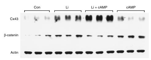
Our initial experiments were designed to examine the consequences of GSK3β inhibition in cardiomyocytes, thereby mimicking the consequences of Wnt signaling. We therefore treated rat neonatal myocytes with lithium, in the form of LiCl, which has been shown previously to inhibit GSK3β (37, 38). Western blot analysis of cardiac myocytes treated with 20 mM LiCl for 24 hours showed more than a 3-fold increase of Cx43 protein, compared with untreated control cells, as shown in Figure 1 (3.4 fold ± 0.1; P < 0.05). The inductive effect of LiCl was substantially greater than that observed with db-cAMP (1 mM) (1.5 fold ± 0.5), previously shown to be one of the more potent inducers of Cx43 expression in cardiac myocytes (39). Treatment with an equal concentration of NaCl was without effect, arguing against a nonspecific effect related to osmolarity (not shown). The combination of LiCl and db-cAMP treatment appeared synergistic, with Cx43 expression increasing more than the additive effect of either treatment alone (5.8 fold ± 0.17; P < 0.05, compared with control).
Figure 1.
Regulation of Cx43 expression by lithium treatment. (a) Whole-cell lysates from control myocytes (Con) or those treated for 24 hours with either 20 mM LiCl (Li), 1 mM db-cAMP (cAMP) or LiCl plus db-cAMP were subjected to Western blotting. The same blots were sequentially probed by anti–Cx43 antibody (upper) and anti–β-catenin antibody (middle). Each treatment was performed in triplicate, and equivalency of loading was verified with an antibody against actin (lower).
We next probed the same membranes with an anti–β-catenin mAb. LiCl treatment led to a significant increase in whole-cell β-catenin protein levels (3.0 fold ± 0.44; P < 0.05), as shown in Figure 1, implying that GSK3β and downstream components of the Wnt signaling pathway are present in cardiomyocytes and comprise a functional signaling mechanism. Treatment with db-cAMP also led to a significant increase in β-catenin levels (3.8 fold ± 0.30; P < 0.05), suggesting its mechanism of action also might converge through GSK3β or otherwise stabilize β-catenin. Interestingly, despite the marked elevation in Cx43 levels in myocytes treated with a combination of LiCl and db-cAMP, β-catenin levels were significantly lower than those in myocytes treated with each compound individually (1.9 fold ± 0.19 for combined treatment versus 3.0–3.8-fold for individual treatments; P < 0.05), suggesting a possible inhibitory effect of excess Cx43 on the accumulation of β-catenin.
We next examined the biologic effects of Wnt proteins themselves, inasmuch as Li+ treatment at such relatively high concentrations may have additional effects besides inhibition of GSK3β. Owing to the well-established difficulty in purifying biologically active Wnt proteins (15), we instead used a coculture strategy, whereby cardiomyocytes were plated with cell lines programmed to secrete Wnt proteins.
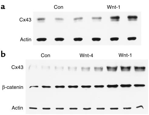
We initially used N2A cells, which express no endogenous Cx43, as previously demonstrated and confirmed here (data not shown). N2A cells were transduced with either a parental retrovirus with no insert, or one expressing Wnt-1. After several weeks of G418 selection, pools of surviving clones from each infection were cocultured with cardiomyocytes. Compared with N2A cells infected with the parental vector, coculture of cardiomyocytes with Wnt-1–expressing N2A cells resulted in a significant induction of Cx43 protein (2.4 fold ± 0.50; P < 0.05; Figure 2a).
Figure 2.
Wnt-1 increases Cx43 expression. (a) Whole-cell lysates from myocytes cocultured for 48 hours with N2A cells transduced with retroviral vector alone (Con) or those expressing Wnt-1 were subjected to Western blotting with anti-Cx43 antibody. (b) Whole-cell lysates from myocytes cocultured on opposite sides of polycarbonated membranes with fibroblasts transduced with retroviral vector alone (Con), or those expressing Wnt-4 or Wnt-1. The membrane was sequentially probed with anti-Cx43 and anti–β-catenin antibodies. Each treatment was performed in triplicate and equivalency of loading was verified with an antibody against actin.
To confirm and extend these findings, we also examined whether a second cell type programmed to secrete biologically active Wnt proteins could regulate expression of Cx43 in cocultured cardiac myocytes. Accordingly, Rat-2 fibroblasts were transduced with either the parental vector or with retroviral constructs encoding Wnt-1 or Wnt-4. Inasmuch as Rat-2 cells express endogenous Cx43 (data not shown), for these experiments we cocultured myocytes and the transduced fibroblasts on opposite sides of polycarbonate membranes (0.4-μΜ pore), enabling intercellular signaling but preventing mixing of cells. In this manner, effects on Cx43 expression could be assessed in the myocytes alone. Compared with myocytes cocultured with control cells for 48 hours, myocytes cocultured with Wnt-1 cells showed nearly a 5-fold increase in the abundance of Cx43 protein (4.9 ± 0.14, P < 0.05), as shown in Figure 2b. In contrast, myocytes cocultured with Wnt-4 transduced fibroblasts showed a significantly weaker effect on Cx43 levels (2.1 ± 0.33; P < 0.05), suggesting a specific inductive effect of Wnt-1. Consistent with these data, myocytes cocultured with Wnt-1–expressing fibroblasts show almost a 3-fold increase in β-catenin levels (2.8 fold ± 0.14 versus control; P < 0.05), whereas levels in myocytes exposed to Wnt-4–transduced cells were not significantly different from controls (P = 0.22), as illustrated in Figure 2b.
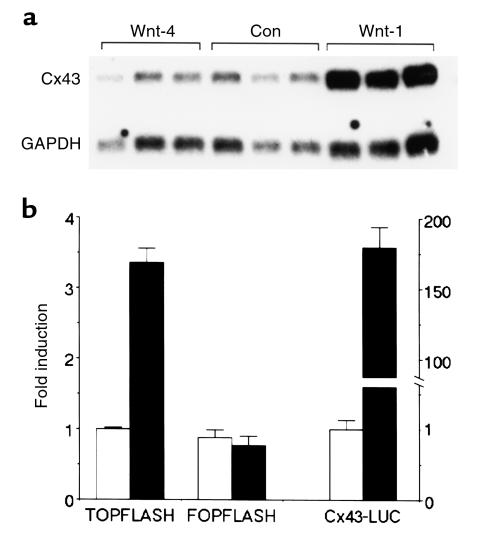
To examine whether the increase in Cx43 protein level was mediated by enhanced transcription, we next examined Cx43 mRNA levels in myocytes cocultured with fibroblasts expressing Wnt-1 or Wnt-4. As illustrated in Figure 3a, Wnt-1, but not Wnt-4, led to a substantial accumulation of Cx43 transcripts in cardiomyocytes (4.2 fold ± 0.80, P < 0.05; 0.69 fold ± 0.10, P = NS, respectively, versus control). These results are consistent with the presence of consensus T-cell factor (Tcf) binding sites in the rat Cx43 gene (20) and indicate that the upregulation of Cx43 is likely to result, at least in part, through nuclear translocation of β-catenin, binding to Tcf-family members and subsequent transcriptional transactivation.
Figure 3.
Wnt-1 increases Cx43 transcription. (a) Northern blot of total RNA isolated from myocytes cocultured with Rat-2 fibroblasts transduced with retroviral vector alone (Con) or those expressing Wnt-1 or Wnt-4. The membrane was sequentially probed for Cx43 and for GAPDH to verify equivalency of loading. (b) Reporter gene analysis. Neonatal myocytes were transfected with 2 μg of pTOPFLASH, pFOPFLASH, or Cx43-LUC reporter genes, 9.8 μg of either a β-catenin expression plasmid (filled squares) or its parental vector (open squares), and 0.2 μg of a β-galactosidase expression plasmid to normalize transfection efficiency.
To confirm that activation of Wnt-1 signaling directly induced Cx43 transcription, we examined the effects of forced expression of β-catenin on Cx43 promoter activity in neonatal cardiomyocytes. We first tested the response of the well-characterized pTOPFLASH and pFOPFLASH plasmids, which harbor concatemerized wild-type or mutant Tcf-binding sites, respectively, upstream of a luciferase reporter gene. As shown in Figure 3b, compared with vector alone, forced expression of β-catenin in cardiomyocytes increased the activity of the pTOPFLASH reporter gene significantly (3.37 ± 0.19 fold; P < 0.05), whereas the mutant pFOPFLASH reporter was insensitive. The Cx43 reporter construct was markedly sensitive to β-catenin expression. Compared with vector alone, luciferase activity in cells cotransfected with the β-catenin expression plasmid rose nearly 200-fold (178 ± 14 fold; P < 0.05) compared with vector alone. These results strongly suggest that the increase in Cx43 mRNA abundance in response to Wnt-1 signaling is due, at least in part, to enhanced transcription.
Wnt-signaling augments intercellular coupling.
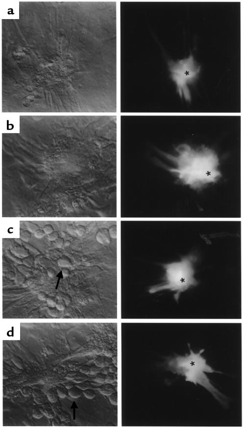
We next asked whether the induction of Cx43 by Wnt signaling resulted in a functional effect on cell coupling. We approached this question using 2 measures of gap junction–mediated intercellular communication. First, we tested the extent of intercellular transfer of the gap junction permeant fluorescent dye Lucifer Yellow, inasmuch as gap junction channels composed of Cx43 are highly permeable to Lucifer Yellow. As shown in Figure 4, the number of visibly fluorescent neighbor cells after injection into a centrally situated myocyte increased significantly after treatment with Li+, on average increasing from 6.0 ± 0.3 to 9.1 ± 0.6 (P < 0.5). A similar effect was seen in cardiomyocytes cocultured with Wnt-1–expressing N2A cells, where the number of coupled cells increased from 5.3 ± 0.3 to 8.6 ± 0.6 (P < 0.05). Not only did the extent of dye transfer increase with Wnt-1 signaling, but the spontaneous beat rate of myocytes also was affected. Whereas control myocytes on average contracted at 76.3 ± 1.3 beats per minute (bpm), the beat frequency in those treated with lithium increased significantly to 87.7 ± 1.9 bpm (P < 0.05). Moreover, compared with myocytes cocultured with N2A cells transduced with vector alone, those cocultured with N2A cells programmed to secrete Wnt-1 increased their beat frequency markedly (83.0 ± 0.6 versus 121 ± 0.3, respectively; P < 0.05).
Figure 4.
Dye coupling in control, Li-treated, and Wnt-1–exposed cardiomyocytes. In each panel, phase contrast micrographs are to the left and fluorescence micrographs (using FITC excitation and emission filters) are shown to the right. (a) Untreated cardiac myocytes. (b) Cardiac myocytes treated for 24 hours with 20 mM LiCl. (c) Cardiac myocytes cocultured with parental N2A cells. (d) Cardiac myocytes cocultured with Wnt-1–expressing N2A cells. Photographs taken at 1 minute after dye injection. N2A cells are indicated by arrows, and injected cardiac myocytes are indicated by asterisks. Note that LiCl and Wnt-1 exposure significantly increases dye coupling among cardiac myocytes (differences in transfer efficacies, which are highly significant for both LiCl and Wnt-1 treatments, are presented in the text).
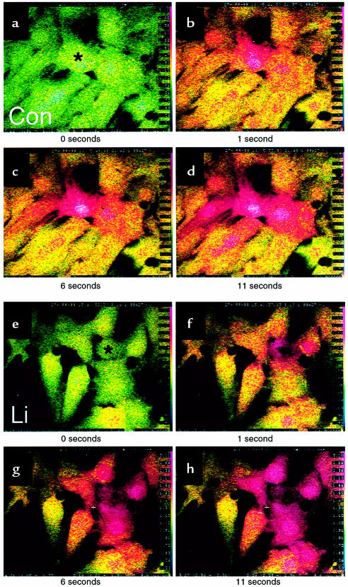
We also examined an additional measure of gap junction channel function, assessing the velocity and extent of slow calcium wave propagation evoked by mechanical stimulation. As illustrated in Figure 5, Li+ treatment increased both the velocity of intercellular calcium waves and also the number of cells recruited into the response. Velocity of Ca2+ waves in Li+-treated cells was 23 ± 2 μm/s, compared with 13 ± 2 μm/s in untreated cells (n = 9 and 10 experiments, respectively; P < 0.001); the percentage of cells recruited into the Ca2+ waves was 91 ± 5 in Li+-treated cultures and 59 ± 8 in controls (P = 0.005). Peak elevations in intracellular Ca2+ in responding cells did not differ between the groups.
Figure 5.
Slow Ca2+ wave propagation in normal (a–d) and LiCl-treated (e–h) cardiac myocytes. Ratio images shown are averages of 16 frames acquired at 32 frames per sec using the Nikon RCM800 real-time confocal microscope with UV excitation; pseudocoloring uses the rainbow representation, where low (∼50–100 nm) Ca2+ is represented as blue to green, and highest Ca2+ (> 2 μM) is red. Intracellular Ca2+ levels are illustrated before stimulation (0), within 1 second after mechanically stimulating the cell indicated by the asterisk, and at 6-second and 11-second intervals thereafter, showing the propagation of a slow (∼10–20 μm/s) Ca2+ wave whose velocity is more rapid in the LiCl-treated (Li) than in the untreated control (Con) myocytes. Values for the numbers of cells participating in the waves and the conduction velocities are given in the text.
Concordant dysregulation of β-catenin and Cx43 in myopathic hearts.
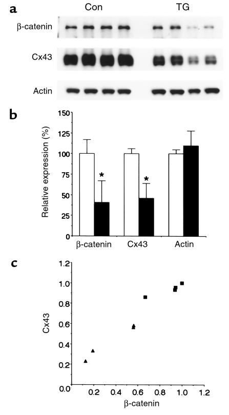
The potent inductive effect of Wnt signaling on Cx43 expression suggested that alterations in β-catenin abundance might contribute to the process of gap junctional remodeling observed in various cardiomyopathies. We therefore examined expression of β-catenin and Cx43 in a recently described transgenic mouse model in which the diphtheria toxin A protein is conditionally expressed in cardiomyocytes. The resulting cardiomyopathy is associated with a high incidence of spontaneous and inducible ventricular arrhythmias (10). As shown in Figure 6, a and b, and consistent with our previous report (10), Cx43 levels were significantly reduced in these transgenic hearts to 46 ± 18% of control levels (P < 0.05). Interestingly, β-catenin levels were concordantly downregulated in these myopathic samples to 41 ± 26% of control levels (P < 0.05). The effect was specific, in that actin levels were not significantly different in transgenics compared with age-matched controls (P = NS). Moreover, there was a significant correlation (r = 0.96) between β-catenin levels and Cx43 levels in individual hearts, both in the control and transgenic samples (Figure 6c).
Figure 6.
Expression of β-catenin and Cx43 in myopathic hearts. (a) Representative Western blot of Cx43, β-catenin and actin expression in lysates from control (Con) and cardiomyopathic transgenic (TG) hearts. (b) Quantification of expression in control (open bars) and TG hearts (filled bars). Values shown are mean ± SD (n = 4). (c) Correlation of β-catenin and Cx43 expression in individual control (filled squares) and transgenic (filled triangles) cardiac lysates. Data are presented relative to the sample with maximal expression.
Cx43 associates with β-catenin in the cell membrane.
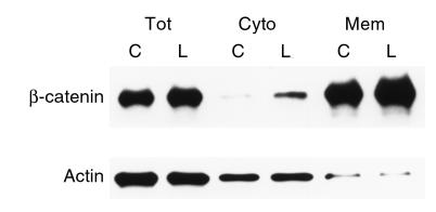
β-Catenin is a multifunctional protein, whose activities depend on its subcellular localization. Cytosolic accumulation frequently correlates with entry of β-catenin into the nucleus and transcriptional activity (40), whereas plasma membrane–associated β-catenin acts as a component of cell-adhesive junctions (41). We therefore examined the subcellular localization of β-catenin in cardiac myocytes after exposure to lithium. As shown in Figure 7, in response to lithium treatment, β-catenin accumulated both in the cytosolic and membrane pools, consistent with the observed transcriptional effect and suggesting possible additional effects from the increase in membrane-localized component.
Figure 7.
Subcellular fractionation of β-catenin. Myocytes were either untreated (C) or treated with 20 mM LiCl (L) for 48 hours. The cells were harvested, disrupted by Dounce homogenization, and the total cellular lysates (Tot) were fractionated into cytosolic (Cyto) and membrane (Mem) fractions. Each fraction was probed for expression of β-catenin (upper). Equal amounts of protein were loaded in each lane. Equivalency of loading in total lysates and among each fraction was verified with an antibody against actin (lower).
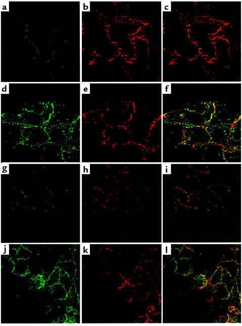
To investigate further the fate of Cx43 and β-catenin that accumulated in response to Wnt-1 signaling, we used confocal microscopy to examine the subcellular localization of the 2 proteins. Control myocytes showed the expected pattern of Cx43 junctional staining, as shown in Figure 8a. β-Catenin was also largely found in a similar membrane location (Figure 8b), although merging of the images suggested only a modest degree of colocalization of the 2 proteins (Figure 8c). Treatment with Li+ resulted in a marked induction of Cx43 protein (Figure 8d), consistent with the Western blot analysis (see Figure 1). Moreover, Cx43 and β-catenin now appeared to colocalize extensively within the junctional membrane (Figure 8f). Similar experiments were carried out with myocytes cocultured with either parental N2A cells or those secreting Wnt-1. In myocytes adjacent to parental N2A cells, moderate levels of Cx43 and β-catenin were both present in cardiac myocytes (Figure 8, g and h), but there was only a faint indication of colocalization (Figure 8i). As with the Li+ studies, coculture with Wnt-1–expressing cells led to a marked induction of Cx43 expression in cardiac myocytes and a substantial degree of colocalization with β-catenin (Figure 8, j–l).
Figure 8.
Wnt signaling leads to colocalization of Cx43 and β-catenin. Myocytes were grown on coverslips in the absence (a–c) or presence (d–f) of 20 mM LiCl or cocultured with N2A cells transduced with the parental retroviral vector (g–i) or with the Wnt-1–expressing retrovirus (j–l). Coverslips were examined for expression of Cx43 and β-catenin antibody by confocal immunomicroscopy. The green channel reflects Cx43 expression (a, d, g, j), and the red channel reflects β-catenin expression (b, e, h, k). The green and red channels were merged to view the extent of colocalization of the 2 proteins (c, f, i, l).
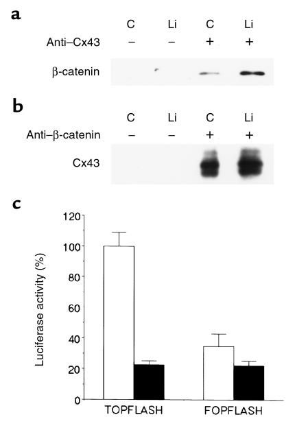
To confirm the association of Cx43 with β-catenin using biochemical techniques, we carried out coimmunoprecipitation-Western analysis of 1% Triton X-100–soluble fractions of cardiac myocyte lysates. Fractions were first immunoprecipitated with either anti-Cx43 antisera or irrelevant rabbit IgG, and the immunoprecipitates were examined for the presence of β-catenin. As shown in Figure 9a, anti-Cx43 antisera specifically coprecipitated β-catenin from myocytes. In contrast, no β-catenin was recovered when lysates were immunoprecipitated with irrelevant immunoglobulin. Moreover, the recovery of β-catenin by Cx43 antisera appeared greater in myocytes treated with Li+, consistent with the enhanced association between the 2 proteins suggested by the confocal analysis. Similarly, antibodies to β-catenin successfully immunoprecipitated Cx43 (Figure 9b). Once again, the recovery of the coprecipitated protein appeared greater in myocytes treated with Li+, further supporting a physical interaction between the 2 proteins that is strengthened in response to activation of the Wnt signaling pathway.
Figure 9.
Biochemical association between Cx43 and β-catenin and repression of transactivation. Triton X-100 (1%) soluble lysates were prepared from myocytes grown in the absence (C) or presence of 20 mM LiCl (Li) and immunoprecipitated with irrelevant IgG (–) or with specific antisera, and the recovered proteins were immunoblotted with the indicated antibody. (a) Lysates immunoprecipitated with anti-Cx43 antisera and probed for β-catenin. (b) Lysates immunoprecipitated with β-catenin antisera and probed for Cx43. (c) Neonatal myocytes were treated with LiCl and transfected with 2 μg of either pTOPFLASH or pFOPFLASH, 9.8 μg of either a Cx43 expression plasmid (filled squares) or its parental vector (open squares), and 0.2 μg of a β-galactosidase expression plasmid to normalize transfection efficiency. Transactivation of the pTOPFLASH reporter gene was significantly reduced by forced expression of Cx43.
These results suggested that augmented expression of Cx43 might sequester β-catenin in the cell membrane and negatively regulate β-catenin transcriptional activity. To test this hypothesis, we transfected LiCl-treated neonatal cardiomyocytes with either a control vector or a Cx43 expression plasmid and determined the degree of β-catenin–mediated transactivation, using the pTOPFLASH and pFOPFLASH reporter genes. Forced expression of Cx43 in cardiomyocytes reduced pTOPFLASH activity by almost 80% (77 ± 2.4%; P < 0.05), to levels no different from the pFOPFLASH reporter (Figure 9c).
Discussion
Wnt signaling regulates a variety of cell fate decisions and developmental processes in species ranging from flies and worms to humans. Studies in these varied model systems have uncovered much of the circuitry linking this family of secreted ligands to their downstream effectors. Several converging lines of evidence have suggested that the effects of Wnt signaling may result in part via modulation of gap junction channel activity. We found that Wnt-1 is a specific and potent inducer of Cx43 expression in cardiomyocytes and that this effect results in enhanced accumulation of Cx43 protein and formation of functional gap junction channels. The induction of Cx43 gene expression appears substantially greater than that observed with other stimuli previously shown to enhance Cx43 transcript accumulation in myocytes, including cyclic AMP or angiotensin II (39, 42). As anticipated, based on the current understanding of the Wnt signal transduction pathway, the effect is mediated, at least in part, through Wnt-dependent accumulation of β-catenin and transcriptional activation via a β-catenin/Tcf nuclear complex.
Wnt ligands act via the Frizzled class of cell-surface receptors. In addition, a class of secreted Frizzled-like proteins, in which the transmembrane domain is absent, have recently been identified, and these appear to function as soluble Wnt antagonists (43). Expression of several Frizzled-related genes have already been identified in the heart (e.g., refs. 44–46), and our studies here suggest that at least some member(s) of this family must act as functional receptors to account for the observed effect of ectopically expressed Wnt-1 on Cx43 expression. Wnt proteins have been divided into 2 broad classes on the basis of their activity profiles. Members of the Wnt-1 class all lead to stabilization of β-catenin and downstream signaling, whereas the effects of Wnt5A class members are less well understood (15, 47). We do not yet know the identity of all endogenous Wnt ligands present in the developing or adult heart, but expect that at least some members of the Wnt-1 class are likely to be present during most stages of heart formation.
In response to Wnt signaling, not only does β-catenin interact with the Cx43 gene and augment transcription, but it also appears to interact with the Cx43 protein itself, likely as part of a complex within the junctional membrane. Whether this interaction is direct, or instead requires other components of cell junctions such as members of the cadherin family, the PDZ-domain–containing ZO-1 protein (48), or other cellular constituents remains to be determined. Regardless, the sequestration of β-catenin by Cx43 raised the possibility that Cx43 may serve to negatively regulate its own transcription, as well as other β-catenin/Tcf-dependent transcriptional targets. Our results are consistent with this hypothesis and build on previous studies that propose to integrate aspects of cell adhesion and gene regulation. For example, E-cadherin, a constituent of adherens junctions, also interacts β-catenin and may influence Tcf-dependent transcription (49, 50).
Dysregulated β-catenin levels, as seen for example in colon cancers with mutations in the upstream regulator protein APC, appears to be an important event in the development of the neoplastic phenotype (31). Interestingly, connexin gene products have been considered as “tumor suppressors” in that loss of intercellular communication is frequently observed in transformed cells and restoration of coupling by transfection with connexin genes may serve to slow cell growth (51, 52). The ability of Cx43 to modulate β-catenin signaling suggests a novel mechanism through which connexin gene products may regulate cell growth in a manner quite distinct from their channel-forming properties.
Finally, recent data from several groups have documented downregulation and/or abnormal localization of connexins in experimental and human cardiomyopathies. This process, referred to as gap junction remodeling, is thought to predispose to some forms of cardiac arrhythmias, including reentrant ventricular tachycardia (9). The robust response of the Cx43 promoter to β-catenin–dependent transactivation suggested that alterations in Wnt signaling, and perhaps other pathways that converge to influence β-catenin function, might influence gap junction channel gene expression in the heart. Our results showing a concordant decrease in β-catenin and Cx43 abundance in arrhythmogenic myocardium is consistent with this hypothesis. Interestingly, enhanced expression of frizzled transcripts has been reported in several cardiomyopathies, including rats with experimentally induced left ventricular hypertrophy or myocardial infarction, where the increased expression has been localized to the expanded population of myofibroblasts (53, 54). A corresponding increase in Frizzled protein expression by the expanded fibroblast population could potentially compete with myocytes for limiting amounts of Wnt ligands, thereby blunting myocyte-specific Cx43 expression and providing a mechanistic explanation for at least a component of Cx43 down-regulation and gap junctional remodeling.
In summary, our results demonstrate an important reciprocal relationship between Wnt signaling and Cx43 expression and function in cardiomyocytes. Our current studies are directed toward more fully elucidating the potential contribution of dysregulated Wnt signaling to abnormal connexin gene expression and gap junctional remodeling, both during cardiac development and particularly in the setting of various forms of heart disease.
Acknowledgments
Supported in part by grants from the National Institutes of Health (NIH; RO1-HL38499 to G.I. Fishman, and RO1-CA47207 to A.M.C. Brown). The confocal laser scanning microscopy was performed at the Mount Sinai School of Medicine core facility supported by NIH grant 1 S10 RR09145-01 and NSF Major Research Instrumentation grant DBI-9724504. G.I. Fishman is a recipient of the Established Investigator Award of the American Heart Association. We thank J. Zhang for excellent technical assistance, E. Beyer for providing the rat Cx43 probe, E. Hertzberg for Cx43 antisera, R. Werner for the Cx43 reporter gene construct, J. Kitajewski for the pTOPFLASH, pFOPFLASH, and β-catenin expression plasmids, and S. Henderson for assistance with the confocal microscopy.
References
- 1.Loewenstein WR. Junctional intercellular communication: the cell-to-cell membrane channel. Physiol Rev. 1981;61:829–913. doi: 10.1152/physrev.1981.61.4.829. [DOI] [PubMed] [Google Scholar]
- 2.Bennett MV, et al. Gap junctions: new tools, new answers, new questions. Neuron. 1991;6:305–320. doi: 10.1016/0896-6273(91)90241-q. [DOI] [PubMed] [Google Scholar]
- 3.Kumar NM, Gilula NB. The gap junction communication channel. Cell. 1996;84:381–388. doi: 10.1016/s0092-8674(00)81282-9. [DOI] [PubMed] [Google Scholar]
- 4.Spray, D.C., Suadicani, S., Srinivas, M., and Fishman, G.I. 1999. Gap junctions in the cardiovascular system. In Handbook of physiology: The heart. H. Fozzard, E. Page, and J. Solaro, editors. Oxford University Press. San Francisco, CA. In press.
- 5.Saffitz JE, et al. The molecular basis of anisotropy: role of gap junctions. J Cardiovasc Electrophysiol. 1995;6:498–510. doi: 10.1111/j.1540-8167.1995.tb00423.x. [DOI] [PubMed] [Google Scholar]
- 6.Coppen SR, Severs NJ, Gourdie RG. Connexin45 (alpha 6) expression delineates an extended conduction system in the embryonic and mature rodent heart. Dev Genet. 1999;24:82–90. doi: 10.1002/(SICI)1520-6408(1999)24:1/2<82::AID-DVG9>3.0.CO;2-1. [DOI] [PubMed] [Google Scholar]
- 7.Smith JH, Green CR, Peters NS, Rothery S, Severs NJ. Altered patterns of gap junction distribution in ischemic heart disease. An immunohistochemical study of human myocardium using laser scanning confocal microscopy. Am J Pathol. 1991;139:801–821. [PMC free article] [PubMed] [Google Scholar]
- 8.Peters NS, Green CR, Poole-Wilson PA, Severs NJ. Reduced content of connexin43 gap junctions in ventricular myocardium from hypertrophied and ischemic human hearts. Circulation. 1993;88:864–875. doi: 10.1161/01.cir.88.3.864. [DOI] [PubMed] [Google Scholar]
- 9.Peters NS, Coromilas J, Severs NJ, Wit AL. Disturbed connexin43 gap junction distribution correlates with the location of reentrant circuits in the epicardial border zone of healing canine infarcts that cause ventricular tachycardia. Circulation. 1997;95:988–996. doi: 10.1161/01.cir.95.4.988. [DOI] [PubMed] [Google Scholar]
- 10.Lee P, et al. Conditional lineage ablation to model human diseases. Proc Natl Acad Sci USA. 1998;95:11371–11376. doi: 10.1073/pnas.95.19.11371. [DOI] [PMC free article] [PubMed] [Google Scholar]
- 11.Hagendorff A, Schumacher B, Kirchhoff S, Luderitz B, Willecke K. Conduction disturbances and increased atrial vulnerability in Connexin40-deficient mice analyzed by transesophageal stimulation. Circulation. 1999;99:1508–1515. doi: 10.1161/01.cir.99.11.1508. [DOI] [PubMed] [Google Scholar]
- 12.Guerrero PA, et al. Slow ventricular conduction in mice heterozygous for a connexin43 null mutation. J Clin Invest. 1997;99:1991–1998. doi: 10.1172/JCI119367. [DOI] [PMC free article] [PubMed] [Google Scholar]
- 13.Reaume AG, et al. Cardiac malformation in neonatal mice lacking connexin43. Science. 1995;267:1831–1834. doi: 10.1126/science.7892609. [DOI] [PubMed] [Google Scholar]
- 14.Waldo KL, Lo CW, Kirby ML. Connexin 43 expression reflects neural crest patterns during cardiovascular development. Dev Biol. 1999;208:307–323. doi: 10.1006/dbio.1999.9219. [DOI] [PubMed] [Google Scholar]
- 15.Cadigan KM, Nusse R. Wnt signaling: a common theme in animal development. Genes Dev. 1997;11:3286–3305. doi: 10.1101/gad.11.24.3286. [DOI] [PubMed] [Google Scholar]
- 16.Nusse R. WNT targets. Repression and activation. Trends Genet. 1999;15:1–3. doi: 10.1016/s0168-9525(98)01634-5. [DOI] [PubMed] [Google Scholar]
- 17.Olson DJ, Christian JL, Moon RT. Effect of wnt-1 and related proteins on gap junctional communication in Xenopus embryos. Science. 1991;252:1173–1176. doi: 10.1126/science.252.5009.1173. [DOI] [PubMed] [Google Scholar]
- 18.Olson DJ, Moon RT. Distinct effects of ectopic expression of Wnt-1, activin B, and bFGF on gap junctional permeability in 32-cell Xenopus embryos. Dev Biol. 1992;151:204–212. doi: 10.1016/0012-1606(92)90227-8. [DOI] [PubMed] [Google Scholar]
- 19.Meyer RA, et al. Developmental regulation and asymmetric expression of the gene encoding Cx43 gap junctions in the mouse limb bud. Dev Genet. 1997;21:290–300. doi: 10.1002/(SICI)1520-6408(1997)21:4<290::AID-DVG6>3.0.CO;2-2. [DOI] [PubMed] [Google Scholar]
- 20.van der Heyden MA, et al. Identification of connexin43 as a functional target for Wnt signalling. J Cell Sci. 1998;111:1741–1749. doi: 10.1242/jcs.111.12.1741. [DOI] [PubMed] [Google Scholar]
- 21.Park M, Wu X, Golden K, Axelrod JD, Bodmer R. The wingless signaling pathway is directly involved in Drosophila heart development. Dev Biol. 1996;177:104–116. doi: 10.1006/dbio.1996.0149. [DOI] [PubMed] [Google Scholar]
- 22.Park M, Venkatesh TV, Bodmer R. Dual role for the zeste-white3/shaggy-encoded kinase in mesoderm and heart development of Drosophila. Dev Genet. 1998;22:201–211. doi: 10.1002/(SICI)1520-6408(1998)22:3<201::AID-DVG3>3.0.CO;2-A. [DOI] [PubMed] [Google Scholar]
- 23.Clark CC, et al. Molecular cloning of the human proto-oncogene Wnt-5A and mapping of the gene (WNT5A) to chromosome 3p14-p21. Genomics. 1993;18:249–260. doi: 10.1006/geno.1993.1463. [DOI] [PubMed] [Google Scholar]
- 24.Zakin LD, et al. Structure and expression of Wnt13, a novel mouse Wnt2 related gene. Mech Dev. 1998;73:107–116. doi: 10.1016/s0925-4773(98)00040-9. [DOI] [PubMed] [Google Scholar]
- 25.Duplaa C, Jaspard B, Moreau C, D’Amore PA. Identification and cloning of a secreted protein related to the cysteine-rich domain of frizzled. Evidence for a role in endothelial cell growth control. Circ Res. 1999;84:1433–1445. doi: 10.1161/01.res.84.12.1433. [DOI] [PubMed] [Google Scholar]
- 26.Augustine K, Liu ET, Sadler TW. Antisense attenuation of Wnt-1 and Wnt-3a expression in whole embryo culture reveals roles for these genes in craniofacial, spinal cord, and cardiac morphogenesis. Dev Genet. 1993;14:500–520. doi: 10.1002/dvg.1020140611. [DOI] [PubMed] [Google Scholar]
- 27.Wang YK, Sporle R, Paperna T, Schughart K, Francke U. Characterization and expression pattern of the frizzled gene Fzd9, the mouse homolog of FZD9 which is deleted in Williams-Beuren syndrome. Genomics. 1999;57:235–248. doi: 10.1006/geno.1999.5773. [DOI] [PubMed] [Google Scholar]
- 28.De Leon JR, Buttrick PM, Fishman GI. Functional analysis of the connexin43 gene promoter in vivo and in vitro. J Mol Cell Cardiol. 1994;26:379–389. doi: 10.1006/jmcc.1994.1047. [DOI] [PubMed] [Google Scholar]
- 29.Harding P, Carretero OA, LaPointe MC. Effects of interleukin-1 beta and nitric oxide on cardiac myocytes. Hypertension. 1995;25:421–430. doi: 10.1161/01.hyp.25.3.421. [DOI] [PubMed] [Google Scholar]
- 30.Yu W, Dahl G, Werner R. The connexin43 gene is responsive to oestrogen. Proc R Soc Lond B Biol Sci. 1994;255:125–132. doi: 10.1098/rspb.1994.0018. [DOI] [PubMed] [Google Scholar]
- 31.Korinek V, et al. Constitutive transcriptional activation by a beta-catenin-Tcf complex in APC–/– colon carcinoma. Science. 1997;275:1784–1787. doi: 10.1126/science.275.5307.1784. [DOI] [PubMed] [Google Scholar]
- 32.Young CS, Kitamura M, Hardy S, Kitajewski J. Wnt-1 induces growth, cytosolic beta-catenin, and Tcf/Lef transcriptional activation in Rat-1 fibroblasts. Mol Cell Biol. 1998;18:2474–2485. doi: 10.1128/mcb.18.5.2474. [DOI] [PMC free article] [PubMed] [Google Scholar]
- 33.McDonald TV, et al. A minK-HERG complex regulates the cardiac potassium current I(Kr) Nature. 1997;388:289–292. doi: 10.1038/40882. [DOI] [PubMed] [Google Scholar]
- 34.Brown, A.M.C., and Dougherty, J.P. 1995. Retroviral vectors. In DNA cloning IV, mammalian systems: a practical approach. D.M. Glover and D.B. Hames, editors. Oxford University Press. Oxford, UK. 113–142.
- 35.Jue SF, Bradley RS, Rudnicki JA, Varmus HE, Brown AM. The mouse Wnt-1 gene can act via a paracrine mechanism in transformation of mammary epithelial cells. Mol Cell Biol. 1992;12:321–328. doi: 10.1128/mcb.12.1.321. [DOI] [PMC free article] [PubMed] [Google Scholar]
- 36.Giarre M, Semenov MV, Brown AM. Wnt signaling stabilizes the dual-function protein beta-catenin in diverse cell types. Ann NY Acad Sci. 1998;857:43–55. doi: 10.1111/j.1749-6632.1998.tb10106.x. [DOI] [PubMed] [Google Scholar]
- 37.Stambolic V, Ruel L, Woodgett JR. Lithium inhibits glycogen synthase kinase-3 activity and mimics wingless signalling in intact cells [erratum 1997, 7:196] Curr Biol. 1996;6:1664–1668. doi: 10.1016/s0960-9822(02)70790-2. [DOI] [PubMed] [Google Scholar]
- 38.Hedgepeth CM, et al. Activation of the Wnt signaling pathway: a molecular mechanism for lithium action. Dev Biol. 1997;185:82–91. doi: 10.1006/dbio.1997.8552. [DOI] [PubMed] [Google Scholar]
- 39.Darrow BJ, Fast VG, Kleber AG, Beyer EC, Saffitz JE. Functional and structural assessment of intercellular communication. Increased conduction velocity and enhanced connexin expression in dibutyryl cAMP-treated cultured cardiac myocytes. Circ Res. 1996;79:174–183. doi: 10.1161/01.res.79.2.174. [DOI] [PubMed] [Google Scholar]
- 40.Yokoya F, Imamoto N, Tachibana T, Yoneda Y. Beta-catenin can be transported into the nucleus in a Ran-unassisted manner. Mol Biol Cell. 1999;10:1119–1131. doi: 10.1091/mbc.10.4.1119. [DOI] [PMC free article] [PubMed] [Google Scholar]
- 41.McCrea PD, Turck CW, Gumbiner B. A homolog of the armadillo protein in Drosophila (plakoglobin) associated with E-cadherin. Science. 1991;254:1359–1361. doi: 10.1126/science.1962194. [DOI] [PubMed] [Google Scholar]
- 42.Dodge SM, et al. Effects of angiotensin II on expression of the gap junction channel protein connexin43 in neonatal rat ventricular myocytes. J Am Coll Cardiol. 1998;32:800–807. doi: 10.1016/s0735-1097(98)00317-9. [DOI] [PubMed] [Google Scholar]
- 43.Bafico A, et al. Interaction of frizzled related protein (FRP) with wnt ligands and the frizzled receptor suggests alternative mechanisms for FRP inhibition of wnt signaling. J Biol Chem. 1999;274:16180–16187. doi: 10.1074/jbc.274.23.16180. [DOI] [PubMed] [Google Scholar]
- 44.Chan SD, et al. Two homologs of the Drosophila polarity gene frizzled (fz) are widely expressed in mammalian tissues. J Biol Chem. 1992;267:25202–25207. [PubMed] [Google Scholar]
- 45.Hu E, et al. Tissue restricted expression of two human Frzbs in preadipocytes and pancreas [erratum 1998, 248:941–943] Biochem Biophys Res Commun. 1998;247:287–293. doi: 10.1006/bbrc.1998.8784. [DOI] [PubMed] [Google Scholar]
- 46.Sagara N, et al. Molecular cloning, differential expression, and chromosomal localization of human frizzled-1, frizzled-2, and frizzled-7. Biochem Biophys Res Commun. 1998;252:117–122. doi: 10.1006/bbrc.1998.9607. [DOI] [PubMed] [Google Scholar]
- 47.Shimizu H, et al. Transformation by Wnt family proteins correlates with regulation of beta-catenin. Cell Growth Differ. 1997;8:1349–1358. [PubMed] [Google Scholar]
- 48.Toyofuku T, et al. Direct association of the gap junction protein connexin-43 with ZO-1 in cardiac myocytes. J Biol Chem. 1998;273:12725–12731. doi: 10.1074/jbc.273.21.12725. [DOI] [PubMed] [Google Scholar]
- 49.Heasman J, et al. Overexpression of cadherins and underexpression of beta-catenin inhibit dorsal mesoderm induction in early Xenopus embryos. Cell. 1994;79:791–803. doi: 10.1016/0092-8674(94)90069-8. [DOI] [PubMed] [Google Scholar]
- 50.Orsulic S, Huber O, Aberle H, Arnold S, Kemler R. E-cadherin binding prevents beta-catenin nuclear localization and beta-catenin/LEF-1-mediated transactivation. J Cell Sci. 1999;112:1237–1245. doi: 10.1242/jcs.112.8.1237. [DOI] [PubMed] [Google Scholar]
- 51.Hotz-Wagenblatt A, Shalloway D. Gap junctional communication and neoplastic transformation. Crit Rev Oncog. 1993;4:541–558. [PubMed] [Google Scholar]
- 52.Mehta PP, Hotz-Wagenblatt A, Rose B, Shalloway D, Loewenstein WR. Incorporation of the gene for a cell-cell channel protein into transformed cells leads to normalization of growth. J Membr Biol. 1991;124:207–225. doi: 10.1007/BF01994355. [DOI] [PubMed] [Google Scholar]
- 53.Blankesteijn WM, Essers-Janssen YP, Ulrich MM, Smits JF. Increased expression of a homologue of Drosophila tissue polarity gene “frizzled” in left ventricular hypertrophy in the rat, as identified by subtractive hybridization. J Mol Cell Cardiol. 1996;28:1187–1191. doi: 10.1006/jmcc.1996.0109. [DOI] [PubMed] [Google Scholar]
- 54.Blankesteijn WM, Essers-Janssen YP, Verluyten MJ, Daemen MJ, Smits JF. A homologue of Drosophila tissue polarity gene frizzled is expressed in migrating myofibroblasts in the infarcted rat heart. Nat Med. 1997;3:541–544. doi: 10.1038/nm0597-541. [DOI] [PubMed] [Google Scholar]


