Abstract
A soft rot of fruits caused by Mucor racemosus occurred on cherry tomato collected in Agricultural Products Wholesale Market in Jinju, Korea. The disease infection usually occurred wounded areas after cracking of fruits. At first, the lesions started with water soaked and rapidly softened and diseased lesion gradually expanded. Colonies were white to brownish to gray in color. Sporangia were 32~54 µm in size and globose in shape. Sporangiophores were 8~14 µm in width. Sporangiospores were 5~12 × 4~8 µm in size, ellipsoidal to subglobose in shape. Columella was 27~42 µm in size, obovoid, ellipsoidal, cylindrical-ellipsoidal, slightly pyriform in shape. Chlamydospores were numerous in sporangiophores and barrelshaped when young, subglobose in old cultures. Optimum growth temperature was about 25℃. The fungus was identified as M. racemosus Fres. This is the first report of soft rot on cherry tomato caused by M. racemosus in Korea.
Keywords: Cherry tomato, Mucor racemosus, Soft rot
Mucor mainly occurs during sale after harvest, transports, market shelf and storage when moisture conditions are favorable. The soft rot on the succulent, fleshy fruits and vegetables, cut flowers, bulbs and corms are often affected by postharvest diseases (Agrios, 1997). Mucor is omnipresent as a saprophyte and sometimes as a weak parasite on stored organs of plants. When the epidermal cells are collapsed, the fungus emerges through the wounds, fruit drop, step on fruits and cracks of matured fruits of cherry tomato, and produces aerial sporangiophores and sporangia.
Disease occurrence
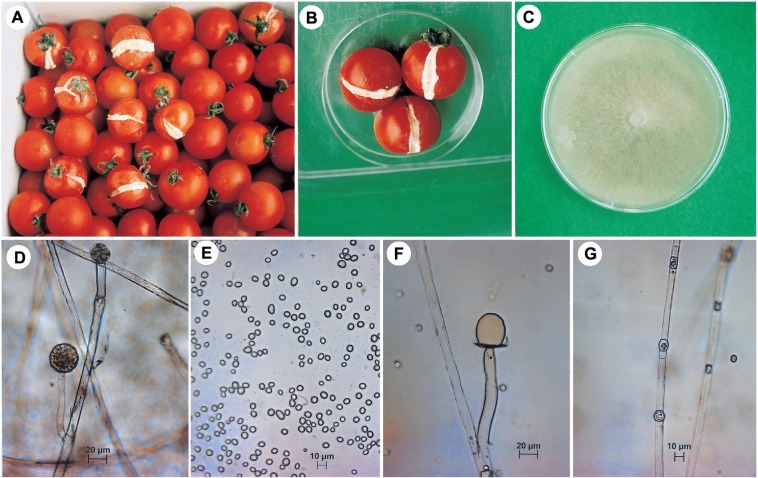
Diseased fruits were collected from cherry tomato (cv. Pepe) in the containers in the spring of 2002. A disease suspected as Mucor soft rot occurred on cherry tomato (Lycopersicon esculentum) in Agricultural Products Wholesale Market in Jinju. Recently, the fungus was occurred from cherry tomato(cv. Suncherry) in the box after postharvest in the April of 2005. The fungus attacked only dropping fruits in the field or cracks of matured fruits of cherry tomato (Fig. 1A), but not occurred young and immatured ones. The infected parts of cracks on matured fruits appeared water soaked at first, then became soften. White hyphae grew from the site where the fungus invaded primarily and covered the affected portions by producing tuft brownish to gray sporangiophores and sporangia. The infected tissues finally were broken down and disintegrated in watery rot. The bush growth of the fungus often extends to the surface of the healthy portions of affected fruits and even to the surface of the containers. The fungus emerged through the wounds and produces aerial sporangiophores sporangia. The causal organism was isolated from mycelial tips on the diseased fruits.
Fig. 1.
Symptoms of soft rot on cherry tomato and mycological characteristics of the pathogenic fungus Mucor racemosus. A: Symptoms started from the cracks of fruits in the box postharvest, B: Artificially inoculated fruits after 3 days inoculation, C: Mycelial growth on PDA after 7 days, D: Sporangium and sporangiophore, E: Sporangiospores, F: Columella, G: Chlamydospores.
Mycological characteristics
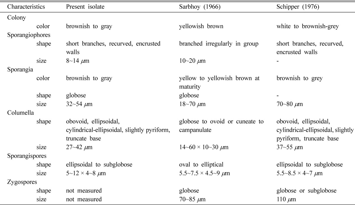
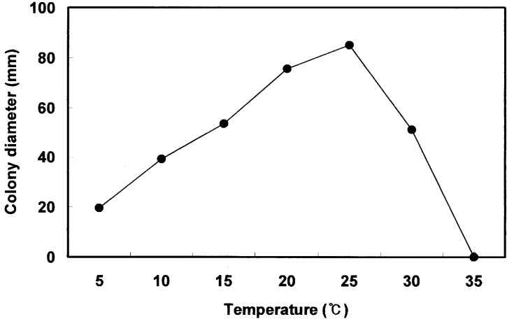
The fungal colonies grown on potato dextrose agar at 25℃ were white at first to brownish to gray with age, and became heavily speckled with the appearance of sporangia (Fig. 1C). Sporangia were 32~54 µm in size and became brownish to gray at maturity. The sporangia contained thousands of ellipsoidal to subglobose sporangiospores. Sporangiophores were 8~14 µm in size and short branches sometimes recurved, with encrusted walls, globose in shape (Fig. 1D). Sporangiospores were 5~12 × 4~8 µm in size and broadly ellipsoidal to subglobose, smooth walled, grayish in color (Fig. 1E). Most of the sporangiospores were appeared to be readily dispersed in the air. Numerous sporangiospores were produced on the diseased fruits. Columella was 27 × 42 µm variable in size and obovoid, ellipsoidal, cylindrical-ellipsoidal, slightly pyriform, usually with truncate base and light brown in color (Fig. 1F, Table 1). Chlamydospores were numerous in sporangiophores and barrel-shaped when young, subglobose in old cultures, yellowish in color and 8~18 µm in size (Fig. 1G). Zygospores were not measured on potato dextrose agar media culture. The maximum temperature for mycelial growth of the fungus was 30℃, minimum temperature was 5℃, optimum growth temperature was 25℃ (Fig. 2).
Table 1.
Comparison of morphological characteristics of the pathogenic fungus isolated from soft rot of cherry tomato with Mucor racemosus described previously

Fig. 2.
Effect of temperature on the mycelial growth of Mucor racemosus, the causal organism of soft rot of cherry tomato. Linear mycelial growth was measured 50 hours after incubation on PDA. Data are means of three replications.
Most characteristics of the fungus examined in this study were almost identical to M. racemosus described by the previous workers (Gobayashi, 1992; Lunn, 1977; Samson et al., 2000; Sarbhoy, 1966; Schipper, 1976). Accordingly, we identified the causal fungus of soft rot of cherry tomato as M. racemosus Fresenius.
Pathogenicity test
The conidial suspension of 100 ml was sprayed to cracked fruits in matured. Inoculated fruits were placed in a humid chamber with 100% relative humidity at 25℃ for 24 hours. The typical symptoms on cherry tomato (cv. Suncherry) were appeared at 3 days after inoculation in plastic fruit container (29 × 22 × 14 cm) (Fig. 1B). The disease infection usually started from cracked parts of matured fruits. The symptoms were identical to those of naturally infected tomatoes. Morphological characteristics of conidia and mycelia of the fungi that were reisolated from inoculated fruits were same as those of naturally infected fruits confirming Koch's postulation. Farr et al. (1995) have reported M. circinelloides, M. hiemalis, and Mucor sp. on tomato.
The soft rot disease caused by Mucor sp. has been reported in Panax ginseng in Korea (The Korean Society of Plant Pathology. 2004), but no records in tomato. Generally, cherry tomato is cultivated by forcing culture or semi-forcing culture in greenhouses near to urban area where normal temperature and humid condition were kept. Such environments are favorable for occurring Mucor soft rot of cherry tomato. Many of the postharvest diseases of fruits, vegetables, grains, and legumes are the results of infection by the pathogens in the field (Agrios, 1997). The world-wide distribution of Mucor was ranged in soil, dung, food, seeds of wheat, barley, rice, tomatoes, tea, groundnuts and animals (Schipper, 1976).
The type culture of the fungus is stored at the Korean Agricultural Culture Collection (KACC No. 41012), National Institute of Agricultural Biotechnology, Rural Development Administration, Suwon, Korea.
Acknowledgement
The authors wish to acknowledge the financial support from Korea Science and Engineering Foundation (KOSEF) to Entomopathogenic Fungal Culture Collection (EFCC), Kangwon National University, Korea. Institute for Cordyceps Research of Kangwon National University is also thanked for providing research facilities to carry out this study. We also wishes acknowledge the permission for exploration of Cordyceps species in their respective areas from Department of Forests and Department of National Parks and Wildlife Conservation of the Government of Nepal.
References
- 1.Agrios GN. Plant pathology. 4th ed. Academic Press; 1997. p. 635. [Google Scholar]
- 2.Farr DF, Bills GF, Chamuris GP, Rossman AY. Fungi on plants and plant products in the United States. APS Press; 1995. [Google Scholar]
- 3.Gobayashi T, Katumoto K, Abiko K, Abe Y, Kakishima M. Illustrated genera of plant pathogenic fungi in Japan. The Whole Farming Educational Association; 1992. (in Japanese) [Google Scholar]
- 4.Lunn JA. CMI Descriptions of pathogenic fungi and bacteria. No. 529. 1977. [Google Scholar]
- 5.The Korean Society of Plant Pathology. List of plant diseases in Korea. 4th ed. 2004. (in Korean) [Google Scholar]
- 6.Schipper MAA. On Mucor circinelloides, Mucor racemosus and related species. Studies in Mycol. 1976;12:1–40. [Google Scholar]
- 7.Samson RA, Hoekstra ES, Frisvad JC, Filtenborg O. Introduction to food-and airborne fungi. Utrecht: Centraalbureau voor schimmelcultures; 2000. [Google Scholar]
- 8.Sarbhoy AK. CMI Descriptions of pathogenic fungi and bacteria. No. 107. 1966. [Google Scholar]


