Abstract
Objective. To investigate the association of FXYD-3 expression with clinicopathological variables and PINCH in patients with ESCC. Patients and Methods. Expression of FXYD-3 protein was immunohistochemically examined in normal esophageal mucous (n = 20) and ESCC (n = 64). Results. Expression of FXYD-3 in the cytoplasm markedly increased from normal esophageal epithelial cells to primary ESCC (P = 0.001). The expression of FXYD-3 was correlated with TNM stages and depth of tumor invasion. Furthermore, the cases with lymph node metastasis tended to show a higher frequency of positive expression than those without metastasis (P = 0.086), and FXYD-3 expression tended to be positively related to the expression of PINCH (P = 0.063). Moreover, the cases positive for both proteins had the highest frequency of lymph node metastasis (P = 0.001). However, FXYD-3 expression was not correlated with patient's gender (P = 0.847), age (P = 0.876), tumor location (P = 0.279), size (P = 0.771), grade of differentiation (P = 0.279), and survival (P = 0.113). Conclusion. Overexpression of FXYD-3 in the cytoplasm may play an important role in the tumorigenesis and development in the human ESCC, particularly in combination with PINCH expression.
1. Introduction
Esophageal squamous cell carcinoma (ESCC) ranks among the 10 most common cancers in the world and also is one of the most poorly controlled malignancies in the People Republic of China, especially in the area of the Taihang Mountains. To date, surgical resection remains the first treatment. However, nearly 95% of surgically resected patients with advanced esophageal cancer succumb to recurrent or metastatic disease within 5 years [1]. Accordingly, it is necessary to investigate the mechanism of tumorigenesis and metastasis of ESCC.
The FXYD proteins constitute a family of conserved auxiliary subunits of the Na, K-ATPase and have been focused in biomedicine field recently due to their ability to finely regulate the activity of the enzyme complex in various physiological and pathological settings [2]. In mammals there are a total of seven FXYD proteins including phospholemman (FXYD-1), of which all but FXYD-6 have been shown to be tissue-specific modulators of Na, K-ATPase [3]. FXYD3, also known as Mat-8 (mammary tumor, 8 kDa), is a member of FXYD family and was originally cloned from murine mammary tumors induced by the neu and ras oncogenes [4]. Although it is a member of the FXYD family, it differs from the most of the other members. It has a signal peptide that is uncleaved and completely different by being the only one with two transmembrane domains. The other members have only one transmembrane domain [2]. It is reported that FXYD-3 is expressed not only in normal tissues but also in tumors. In normal tissues it is mainly expressed in the urinary bladder, uterus, lung, stomach, colon, and skin [4, 5]. And in the tumors, it has been found in breast cancer, colon cancer, bladder cancer, benign and malignant prostate tumors, as well as pancreatic ductal adenocarcinoma [5–9]. Interestingly, in some types of cancers such as breast, pancreatic, and androgen-dependent prostate cancer, it is overexpressed, whereas it seems to be downregulated in androgen-independent prostate cancer and kidney cancer [5–7]. Moreover, some recent studies showed that FXYD-3 might be a promising biomarker for detecting metastasis in some cancers [8–12]. However, to the best of our knowledge, no study has been done in ESCC.
Particularly interesting is that new cysteine-histidine rich protein (PINCH) is an adapter protein, which consists primarily of five LIM (double zinc finger) domains, and the gene is located on chromosome 2q12.2. Studies showed that PINCH was involved in cell adhesion, migration, and apoptosis [13, 14]. PINCH protein can interact directly with integrin-linked kinase (ILK) and Nck-2 protein and is associated with integrin signaling and growth factor signaling pathway [15–18]. It has been observed that PINCH expression is upregulated in many types of malignancies, including oral and ESCC, colorectal, pancreatic, skin, breast, lung, and prostate cancer and endometrioid endometrial carcinoma, as well as gliomas [19–27]. PINCH localizes to the peritumoral stromal cells, especially at the tumors invasive edges [19]. Furthermore, PINCH is an independently prognostic factor in patients with colorectal cancer [20]. Our previous study on the same series of the cases used in the present study demonstrated that PINCH expression was upregulated in ESCC compared to normal esophageal squamous cells, and further the strong expression of PINCH was related to lymph node metastasis [25]. Recent studies have shown that genesis and metastasis of tumors are the result of the interaction between tumor cells and tumor-associated stromal cells [28]. Therefore, it is of significance to explore whether there is a correlation between FXYD-3 expression in tumor cells and PINCH expression in the stromal cells in human ESCC.
The aim of the present study was to investigate FXYD-3 expression in ESCC compared with normal esophageal mucosa and further to analyze the relationship of FXYD-3 expression in ESCC with PINCH expression or clinicopathological variables, including patients' gender, age, tumor location, size, depth of invasion, lymph node status, the grade of differentiation, TNM stages, and survival.
2. Patients and Methods
2.1. Patients
Formalin-fixed paraffin-embedded tissue samples were obtained from 64 ESCC patients who underwent surgical resection at the First Hospital of Hebei Medical University (Shijiazhuang, Hebei Province, China), between 2000 and 2004. The study included 20 distant normal mucosa specimens (all of which were matched with the primary tumors) taken from the margin of distant resection. The primary tumors were located in the upper, middle, and lower sections of the esophagus in 7, 36, and 21 cases, respectively, and 20 cases with lymph node metastasis. None of the patients had received preoperative radiotherapy or chemotherapy. The patients' gender, age, tumor location, size, depth of invasion, lymph node status, the grade of differentiation, and TNM stage [29] were obtained from surgical and/or pathological records at the hospital. The mean age of the patients was 59.5 years old (range 41–78 years). According to the WHO classification, the tumor differentiation was graded as grade I (high differentiation: 20 cases), grade II (moderate differentiation: 39 cases), and grade III (low differentiation: 5 cases), respectively [1]. The patients were followed up until March 2013, and 26 patients were lost to followup. All pathological slides, including normal specimens and tumors, were confirmed by two pathologists (Z. L. Zhu and Z. M. Wang). The study was approved by the ethical committee of the First Hospital of Hebei Medical University, Shijiazhuang, Hebei, China. Written informed consent was obtained from the patients.
Data of PINCH immunohistological staining in ESCC were obtained from our previous study carried out at our laboratory, the First Hospital of Hebei Medical University. According to the intensity of PINCH staining in the tumor-associated stromal cells, PINCH expression was graded as negative group (none or <20% of positive cells) and positive group (≥20% positive cells) [25].
2.2. Immunohistological Staining and Evaluation
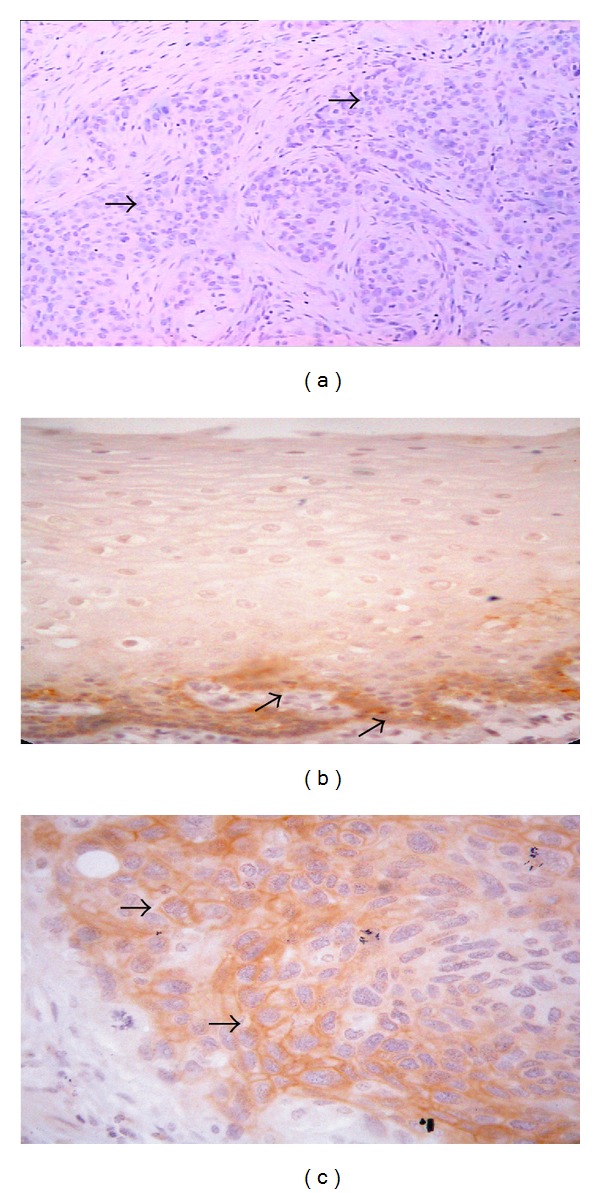
Immunohistochemical staining was performed on 5 um thick formalin-fixed paraffin-embedded sections. The sections were incubated at 60°C for 12 hours, deparaffinized, and then rehydrated. The sections were transferred to 0.01 M Tris-EDTA buffer (pH9.0) and subjected to high pressure cooker (100°C) for 8 minutes and incubated at room temperature for 30 minutes for antigen retrieval. The sections were then washed in phosphate buffered saline (PBS, pH7.4) and incubated with 3% H2O2 in methanol for 20 minutes, to block endogenous peroxidase activity. Nonspecific binding of antibody was prevented by preincubating the sections with 1.5% horse serum (Fuzhou Maxim Biotechnology Co., Ltd., Fuzhou, Fujian, China) in PBS for 10 minutes. After removing the blocking solution, the sections were incubated with a monoclonal anti-FXYD-3 primary antibody (provided kindly by Professor Hanswalter Zentgraf, Applied Tumor Virology, University of Heidelberg, Heidelberg Germany) in 1 : 2 diluted in PBS (pH7.4) over night at 4°C in a moist chamber. Subsequently, the sections were incubated with biotinylated anti-rabbit IgG antibody (Fuzhou Maxim Biotechnology Co., Ltd.) at room temperature for 30 minutes, followed by an incubation of an avidin-biotin-peroxidase complex (Fuzhou Maxim Biotechnology Co., Ltd.) at room temperature for 30 minutes. The sections were washed with PBS between each incubation step. The peroxidase reaction was developed using 3.3 diaminobenzidine (DAB) (Fuzhou Maxim Biotechnology Co., Ltd.) at room temperature for 8 minutes. Then, the sections were rinsed with water and counterstained with Mayer's haematoxylin and then washed, dehydrated in ethanol, and mounted with xylene-based mounting medium. The breast cancer sections known to stain for positive FXYD-3 were included as negative (using PBS instead of the primary antibody) and positive controls in all runs. There was no staining in the negative controls (Figure 1(a)), while the positive controls showed clear staining.
Figure 1.

A negative control (a breast cancer known for positive FXYD-3), where the primary FXYD-3 was replaced by PBS, showed no staining of the FXYD-3 in tumor cells (→) (a). Immunohistochemical assay for FXYD-3 expression in esophageal squamous cell carcinoma (ESCC). FXYD-3 was weak expression in normal mucosa, mainly located in the basal cell layer (→) (b), and strong expression in primary tumor cells (→) (c).
The stained sections were microscopically examined and evaluated independently by two pathologists (Z. L. Zhu and Z. M. Wang) with no knowledge of the clinicopathological information. Cytoplasmic staining was considered as FXYD-3 positive expression. According to the percentage of the positive staining cells, we graded FXYD-3 expression as negative (no positive cells or <5% positive cells), weak (5–25% positive cells), moderate (26–50% positive cells), and strong positive (>50% positive cells), irrespective of the staining intensity [30]. In statistical analysis, we considered negative and weak staining as negative group and moderate and strong staining as positive group. In order to avoid artificial effects, tissues in the areas with poor morphology and necrosis and in the margins of the sections were not considered.
2.3. Statistical Analysis
The statistical analyses were performed by using SPSS version 13.0 software. The Chi-square test was used to examine the correlation between the frequencies of FXYD-3 expression in normal esophageal mucosa and ESCC, the correlation between FXYD-3 expression in cancer and clinicopathological variables or PINCH expression, and the relative risk of FXYD-3 expression related to ESCC. Spearman rank correlation analysis was used to examine the correlation between FXYD-3 expression and PINCH expression in lymph node metastasis. All P values cited were two sided and P < 5% was considered to indicate a statistically significant difference.
3. Results
3.1. FXYD-3 Expression in Normal Mucosa and Primary Tumor
We examined FXYD-3 protein expression in normal esophageal mucosa and ESCC and found that the positive expression of FXYD-3 was in the cytoplasm of normal epithelial cells (Figure 1(b)) and cancer cells (Figure 1(c)), and there was no nuclear staining. In the 20 specimens of normal mucosa, 11 cases were negative (55%) and 9 were positive, including 6 (30%) cases with weak, 2 (10%) moderate, and 1 (5%) strong staining; positive staining was mainly located in the basal cell layer (Figure 1(a)). Among 64 cancers, there were 16 (25%) negative, 10 (16%) weak, 10 (16%) moderate, and 28 (43%) strong expressed cases.
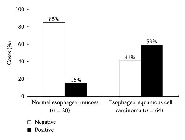
Figure 2 presents the frequency of FXYD-3 expression in normal mucosa and ESCC; the rate of positive FXYD-3 expression in cancer was 59% (38/64), which was significantly higher than that in the normal mucosa (15%, 3/20, X2 = 12.009, P = 0.001). The relative risk of FXYD-3 expression related to ESCC was 8.282; 95% CI was 2.202–31.155.
Figure 2.

Frequency of FXYD-3 immunohistochemical staining in normal esophageal mucosa and esophageal squamous cell carcinoma (ESCC) (P = 0.001).
Furthermore, we also observed the expression of FXYD-3 at the invasive margin and the inner part of the tumor in all 64 ESCCs; there was no obvious difference between the two sites.
3.2. FXYD-3 Protein Expression in Relation to Clinicopathological Variables and PINCH Expression in ESCCs
Table 1 shows the relationship between FXYD-3 expression in tumors and the clinicopathological variables. The expression of FXYD-3 was correlated with TNM stages and depth of tumor invasion. There was a higher frequency of FXYD-3 expression in stage II + III + IV, 68%, and in group muscularis and adventitia, 66%, than in stage I, 29%, and in group mucosa and submucosa, 36%, respectively (P = 0.008 and P = 0.041). The cases with lymph node metastasis tended to show a higher frequency of positive expression than those without metastasis (75 versus 52%, P = 0.086). The FXYD-3 expression was not significantly correlated with patients' gender (P = 0.847), age (P = 0.876), tumor location (P = 0.279), tumor size (P = 0.771), grade of differentiation (P = 0.279), and survival (P = 0.113).
Table 1.
The relationship of FXYD-3 protein expression with clinicopathological and biological variables in the patients with ESCC.
| Variables | N | FXYD-3 expression | χ 2 | P value | |
|---|---|---|---|---|---|
| Negative (%) | Positive (%) | ||||
| Gender | |||||
| Male | 50 | 20 (40) | 30 (60) | 0.037 | 0.847 |
| Female | 14 | 6 (43) | 8 (57) | ||
| Age (years) | |||||
| ≤50 | 19 | 8 (42) | 11 (58) | 0.025 | 0.876 |
| >50 | 45 | 18 (40) | 27 (60) | ||
| Tumour location | |||||
| Upper | 7 | 3 (43) | 4 (57) | 2.556 | 0.279 |
| Middle | 36 | 14 (39) | 22 (61) | ||
| Lower | 21 | 9 (43) | 12 (57) | ||
| Tumour size (cm) | |||||
| ≤3 | 26 | 10 (38) | 16 (62) | 0.085 | 0.771 |
| >3 | 38 | 16 (42) | 22 (58) | ||
| Lymph node status | |||||
| Nonmetastasis | 44 | 21 (48) | 23 (52) | 2.944 | 0.086 |
| Metastasis | 20 | 5 (25) | 15 (75) | ||
| Grade of differentiation | |||||
| I | 20 | 11 (55) | 9 (45) | 2.556 | 0.279 |
| II | 39 | 13 (33) | 26 (67) | ||
| III | 5 | 2 (40) | 3 (60) | ||
| Depth of invasion | |||||
| Mucosa and submucosa | 14 | 9 (64) | 5 (36) | 4.159 | 0.041 |
| Muscularis and adventitia | 50 | 17 (34) | 33 (66) | ||
| TNM stage | |||||
| I | 14 | 10 (71) | 4 (29) | 7.049 | 0.008 |
| II + III + IV | 50 | 16 (32) | 34 (68) | ||
| Survival (years) | |||||
| ≥5 | 11 | 8 (73) | 3 (27) | 2.508 | 0.113 |
| <5 | 27 | 12 (44) | 15 (56) | ||
| PINCH | |||||
| Negative | 28 | 15 (54) | 13 (46) | 3.459 | 0.063 |
| Positive | 36 | 11 (31) | 25 (69) | ||
ESCC: esophageal squamous cell carcinoma.
PINCH: particularly interesting new cysteine-histidine rich protein.
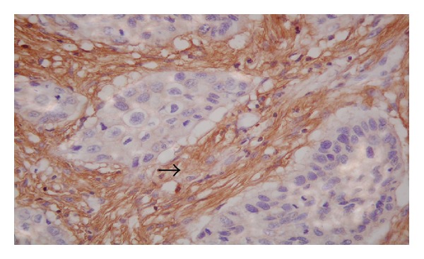
In addition, in our previous study [25], we found that PINCH expression was not correlated with patients' gender, age, tumor location, tumor size, and grade of differentiation (P > 0.05). However, we also found that PINCH expression was upregulated in the stromal cells of ESCC, and the strong expression of PINCH (Figure 3) was related to lymph node metastasis. In the present study, we further analyzed the relationship between FXYD-3 expression and PINCH expression which was studied in our previous paper on the same samples of ESCCs. The results showed that FXYD-3 expression tended to be positively related to the PINCH expression in all 64 ESCCs (Table 1). Of the 36 cases with PINCH positive expression, 25 (69%) cases were FXYD-3 positive and 11 (31%) cases were FXYD-3 negative. However, in 28 cases with PINCH negative expression, there were 13 (46%) cases for FXYD-3 positive and 15 (54%) cases for FXYD-3 negative (X2 = 3.459, P = 0.063). Moreover, we also analyzed the relationship of both of the expressions FXYD-3 and PINCH with the lymph node metastasis and found that the cases positive for both the proteins had the highest frequency of lymph node metastasis (15/20, 75%), cases negative for both the proteins had the lowest frequency of the metastasis (2/20, 10%), and cases positive for either protein had a moderate frequency (3/20, 15%) of lymph node metastasis. Table 2 showed the correlation between FXYD-3 expression and PINCH expression in 20 cases with lymph node metastasis.
Figure 3.

Particularly interesting new cysteine-histidine rich protein (PINCH), as a marker for stromal cells, was upregulated in the stromal cells (→), predicting the ability of invasion and metastasis of ESCC.
Table 2.
Correlation between FXYD-3 expression and PINCH expression in 20 cases with lymph node metastasis.
| Variables | N | PINCH expression | r | P value | |
|---|---|---|---|---|---|
| Negative (%) | Positive (%) | ||||
| FXYD-3 | |||||
| Negative | 5 | 2 (40) | 3 (60) | 0.577 | 0.008 |
| Positive | 15 | 0 (0) | 15 (100) | ||
4. Discussion
The FXYD proteins constitute a family of conserved auxiliary subunits of the Na, K-ATPase and have been the study focus in biomedicine field recently due to their ability to finely regulate the activity of the enzyme complex in various physiological and pathological settings [2]. The FXYD protein family contains seven members that are small, single-span membrane proteins characterized by a signature sequence containing an FXYD motif and three other conserved amino acid residues [2, 31]. Recent evidence suggests that all members including FXYD-1 (phospholemman) [32], FXYD-2 (gamma subunit of Na, K-ATPase) [33], FXYD-3 (phospholemman-like protein, PLML) [4], FXYD-4 (corticosteroid hormone-induced factor, CHIF) [34], FXYD-5 (protein related to ion channel, Ric) [35], FXYD-6 (phosphohippolin) [36], and FXYD-7 [37] associate with Na, K-ATPase in a tissue-specific way and modulate its transport properties.
FXYD-3 has been reported to be located on the both surface and cytoplasm in some cancer cells such as gastric and colon cancer cells [38, 39]. The present study also showed that the positive staining of FXYD-3 was located in the cytoplasm of ESCC. These results suggested that FXYD-3 might be a marker for tumor cells. In order to investigate the specific location of FXYD-3 expression in cells, Arimochi research group [38, 40] detected the expression of FXYD-3 in Chinese hamster ovary-K1 (CHO-k1) cells and human colorectal cancer cells and reported that the FXYD-3 expression was distributed in the intracellular membranes, being not only detected around the nuclear envelop but also partly overlapping with an endoplasmic reticulum marker. Furthermore, the spherical structures observed were not co-localized with markers for lysosomes, endosomes, and Golgi bodies, suggesting that FXYD-3 is distributed in a distinct endoplasmic reticulum region and the nuclear envelope after synthesis on membrane-bound ribosomes.
FXYD-3 has been detected in some normal tissues (such as the brain, lung, stomach, colon, intestine, liver, pancreas, prostate, skin, etc.) [31, 41, 42] but overexpressed in a growing number of tumors and tumor cell lines including cancer in the breast [4], androgen-dependent prostate [7], pancreas [31], stomach [39], kidney, and bladder [8]. Furthermore, siRNA-mediated knockdown of FXYD-3 showed a reduction in the proliferative activity of both PC-3 and LNCaP prostate cancer cells [6] and T3 M4 pancreatic cancer cells [5] in vitro. These findings suggested a potential role of FXYD-3 in the development and progression of these cancers.
In the present study, we found that the positive staining of FXYD-3 in the normal mucosa was mainly located in the basal cell layer where the basal cells have more strong capacity of proliferation and repair and can differentiate into squamous epithelial cells. This result, to a certain extent, indicates that FXYD-3 may involve in the cellular proliferation and differentiation. In addition, we found that the frequency of the positive FXYD-3 expression was significantly higher in the primary tumor compared to the normal mucosa and in the cases with lymph node metastasis compared to those without metastasis. Moreover, we also found that the expression of FXYD-3 was correlated with TNM stages and depth of tumor invasion. These results indicate that there may be a change in FXYD-3 expression during tumor development from normal tissue to primary tumor, and overexpression of FXYD-3 may promote cell growth, acting as an oncogene, and be related to the tumorigenesis and development.
However, interestingly, in contrast to the above results, FXYD-3 expression was reported to be downregulated in androgen-independent prostate cancer [7] and lung cancer [43], suggesting that FXYD-3 might be a potential tumor suppressor gene. Moreover, Okudela et al. [43] also found that the forced expression of oncogene KRAS reduced the expression of FXYD-3 in NHBE-T cells, which was in contrast to the initial observation that FXYD-3 was highly expressed in oncogenic ras-initiated murine breast tumors [41]. So, taken above together, the regulation of FXYD-3 expression and its role in tumorigenesis and development are assumed to differ among the type of cancer, species, or subtype of the RAS oncogene, and the disordered expression itself, whether strong or weak, might affect cellular homeostasis resulting in the transformation.
Recently, in order to study the mechanism of role of FXYD-3 in tumorigenesis, Bibert et al. [44] used human colon adenocarcinoma cell line (Caco-2) to investigate the effect of FXYD-3 silencing on cell proliferation, differentiation, and apoptosis, and Na, K-ATPase activity and expression. Their results showed that FXYD-3 silencing had no effect on cell proliferation but could promote cell apoptosis and prevent cell differentiation of Caco-2 cells. From those results, they supposed that the most possibility was that FXYD-3 silencing prevented a proper regulation of Na, K-ATPase, which led to perturbation of cellular Na+ and K+ homeostasis and changes in the expression of Na, K-ATPase isozymes, whose properties were incompatible with Caco-2 cell differentiation. Overall, the role and its mechanism of FXYD-3 expression in tumorigenesis remain unclear. This issue will promote us to further study in the future.
Gordon et al. [45] studied the expression of FXYD-3 protein in lung adenocarcinomas and found that FXYD-3 expression was positively related to poorer survival of the patients. However, in the present study, our results revealed that there was no correlation between the survival and the expression of FXYD-3 protein, although the FXYD-3 expression was correlated with TNM stages and depth of tumor invasion, which might, at least in part, indicate poor survival. We think these different results might be explained by several reasons such as different case number and clinicopathological features, methods, and criteria for determining positive expression of FXYD-3 as well as statistic methods in these studies.
Additionally, it has been proven that FXYD-3 can adjust cell microenvironment, which may affect cellular adhesion and migration, by interaction with Na, K-ATPase [46]. Recent studies [47–49] have also suggested that FXYD-3 could regulate indirectly the development of tight junctions through its interaction with Na, K-ATPase, to promote cellular polarization and differentiation. Moreover, the change of FXYD-3 expression might be involved in the epithelial-mesenchymal transition, namely, a type of interaction between tumor cells and stromal cells, which is linked with the vigorous migration and invasion of cancer cells [48]. While PINCH protein is an adapter protein and localizes to the cytoplasma and cell-matrix adherens junctions in cell lines, especially in peritumoral stromal cells as a marker for tumor-associated stromal cells. And PINCH, also through involvement in the tumor-stromal interaction, can promote tumor invasiveness and development [17, 19]. From these studies, we found that both of these proteins were involved in cell adhesion and migration and participated in the interaction between tumor cells and stromal cells, although FXYD-3 was mainly expressed in the tumor cells, while PINCH was mainly expressed in the stromal cells. Recent studies have shown that genesis and development of tumors are the result of the interaction between tumor cells and stromal cells [28]. Therefore, it is of significance to explore whether there is a correlation between FXYD-3 expression in tumor cells and PINCH expression in the stromal cells. Perhaps, this may open up a new idea for the study on tumorigenesis and development.
In our previous study, we found that PINCH expression was not correlated with patients, gender, age, tumor location, size, and differentiation, while we also found that PINCH expression was upregulated in the stromal cells of ESCCs and the strong expression of PINCH was related to lymph node metastasis. So, in order to explore the correlation between expression of FXYD-3 and expression of PINCH in ESCCs, in the present study, we further compared the FXYD-3 expression with the PINCH expression in ESCCs, and found that FXYD-3 expression tended to be positively related to the expression of PINCH in the primary tumors, although the correlation coefficient is lower (r = 0.232, P = 0.063). More importantly, we further observed that the cases positive for both the proteins had the highest frequency of the lymph node metastasis (15/20, 75%), the cases negative for both the proteins had the lowest frequency of the metastasis (2/20, 10%), and the cases positive for either protein had a moderate frequency (3/20, 15%, P = 0.008). The results, at least in part, suggested that FXYD-3 might cooperate with PINCH in the metastasis of ESCC, although the samples were smaller. Taking together with our previous study of PINCH expression in ESCC [25], we guess that, during tumor development and metastasis, FXYD-3 in the tumor cells, through involvement in cell adhesion and interaction between tumor cells and stromal cells, may interact with PINCH, although the mechanism remains unclear. FXYD-3 may act as a driving machine, while PINCH acts as a tractor; both of them cooperate to promote tumor invasiveness and development. This hypothesis needs to be proven in the future.
5. Conclusion
The results suggest that overexpression of FXYD-3 in the cytoplasm may play an important role in the tumorigenesis and development in the human ESCC, at least in part, particularly in combination with PINCH expression.
Acknowledgment
This study is supported by the Science and Technology Research and Development Program of Hebei Province, China, 2011, no. 11276103D-40.
References
- 1.Wu ZB, Yang GH. Chinese Surgical Pathology. Beijing, China: People's Health Press; 2002. [Google Scholar]
- 2.Geering K. FXYD3 proteins: new regulators of Na–K-ATPase. American Journal of Physiology. Renal Physiology. 2006;29:241–250. doi: 10.1152/ajprenal.00126.2005. [DOI] [PubMed] [Google Scholar]
- 3.Bibert S, Roy S, Schaer D, Felley-Bosco E, Geering K. Structural and functional properties of two human FXYD3 (Mat-8) isoforms. Journal of Biological Chemistry. 2006;281(51):39142–39151. doi: 10.1074/jbc.M605221200. [DOI] [PubMed] [Google Scholar]
- 4.Morrison BW, Randall Moorman J, Kowdley GC, Kobayashi YM, Jones LR, Leder P. Mat-8, a novel phospholemman-like protein expressed in human breast tumors, induces a chloride conductance in Xenopus oocytes. Journal of Biological Chemistry. 1995;270(5):2176–2182. doi: 10.1074/jbc.270.5.2176. [DOI] [PubMed] [Google Scholar]
- 5.Kayed H, Kleeff J, Kolb A, et al. FXYD3 is overexpressed in pancreatic ductal adenocarcinoma and influences pancreatic cancer cell growth. International Journal of Cancer. 2006;118(1):43–54. doi: 10.1002/ijc.21257. [DOI] [PubMed] [Google Scholar]
- 6.Grzmil M, Voigt S, Thelen P, Hemmerlein B, Helmke K, Burfeind P. Up-regulated expression of the MAT-8 gene in prostate cancer and its siRNA-mediated inhibition of expression induces a decrease in proliferation of human prostate carcinoma cells. International Journal of Oncology. 2004;24(1):97–105. [PubMed] [Google Scholar]
- 7.Vaarala MH, Porvari K, Kyllönen A, Vihko P. Differentially expressed genes in two LNCaP prostate cancer cell lines reflecting changes during prostate cancer progression. Laboratory Investigation. 2000;80(8):1259–1268. doi: 10.1038/labinvest.3780134. [DOI] [PubMed] [Google Scholar]
- 8.Zhang Z, Pang S-T, Kasper KA, et al. FXYD3: a promising biomarker for urothelial carcinoma. Biomarker Insights. 2011;6:17–26. doi: 10.4137/BMI.S6487. [DOI] [PMC free article] [PubMed] [Google Scholar]
- 9.Gazquez C, Ribal MJ, Marin-Aguilern M, et al. Biomarkers vs conventional histological analysis to detect lymph node micremetastases in bladder cancer: a real improvement? British Journal of Urology International. 2012;110:1310–1316. doi: 10.1111/j.1464-410X.2012.11025.x. [DOI] [PubMed] [Google Scholar]
- 10.Meding S, Balluff B, Elsner M, et al. Tissue-based protemics reveals FXYD3, S100A11 and GSTM3 as novel markers for regional lymph node metastasis in colon cancer. The Journal of Pathology. 2012 doi: 10.1002/path.4021. [DOI] [PubMed] [Google Scholar]
- 11.Kiyamova R, Garifukin O, Gryshkova V, et al. Preliminary study of thyroid and colon cancers-associated antigens and their cognate autoantibodies as potential cancer biomarkers. Biomarkers. 2012;17:362–371. doi: 10.3109/1354750X.2012.677476. [DOI] [PubMed] [Google Scholar]
- 12.Marín-Aguilera M, Mengual L, Ribal MJ, et al. Utility of urothelial mRNA markers in blood for staging and monitoring bladder cancer. Urology. 2012;79(1):240.e9–240.e15. doi: 10.1016/j.urology.2011.09.006. [DOI] [PubMed] [Google Scholar]
- 13.Qin J, Wu C. ILK: a pseudokinase in the cancer stage of cell-matrix adhesion and signaling. Current Opinion in Cell Biology. 2012;24:607–613. doi: 10.1016/j.ceb.2012.06.003. [DOI] [PMC free article] [PubMed] [Google Scholar]
- 14.Montanez E, Karaköse DE, Tischner D, Villunger A, Fässler R. PINCH-1 promotes Bcl-2-dependent survival signalling and inhibits JNK-mediated apoptosis in the primitive endoderm. Journal of Cell Science. 2012;125:5233–5240. doi: 10.1242/jcs.112029. [DOI] [PubMed] [Google Scholar]
- 15.Rearden A. A new LIM protein containing an autoepitope homologous to ‘senescent cell antigen’. Biochemical and Biophysical Research Communications. 1994;201(3):1124–1131. doi: 10.1006/bbrc.1994.1822. [DOI] [PubMed] [Google Scholar]
- 16.Wu C. PINCH, N(i)ck and the ILK: network wiring at cell-matrix adhesions. Trends in Cell Biology. 2005;15(9):460–466. doi: 10.1016/j.tcb.2005.07.002. [DOI] [PubMed] [Google Scholar]
- 17.Tu Y, Li F, Goicoechea S, Wu C. The LIM-only protein PINCH directly interacts with integrin-linked kinase and is recruited to integrin-rich sites in spreading cells. Molecular and Cellular Biology. 1999;19(3):2425–2434. doi: 10.1128/mcb.19.3.2425. [DOI] [PMC free article] [PubMed] [Google Scholar]
- 18.Malan D, Elischer A, Hesse M, Wickström SA, Fleischmann BK, Bloch W. Deletion of integrin linked kinase in endothelial cells results in defective RTK signaling caused by caveolin 1 mislocalization. Development. 2013;140:987–995. doi: 10.1242/dev.091298. [DOI] [PubMed] [Google Scholar]
- 19.Wang-Rodriguez J, Dreilinger AD, Alsharabi GM, Rearden A. The signaling adapter protein PINCH is up-regulated in the stroma of common cancers, notably at invasive edges. Cancer. 2002;95(6):1387–1395. doi: 10.1002/cncr.10878. [DOI] [PubMed] [Google Scholar]
- 20.Gao J, Arbman G, Rearden A, Sun X-F. Stromal staining for PINCH is an independent prognostic indicator in colorectal cancer. Neoplasia. 2004;6(6):796–801. doi: 10.1593/neo.04304. [DOI] [PMC free article] [PubMed] [Google Scholar]
- 21.Zhao Z-R, Zhang Z-Y, Cui D-S, et al. Particularly interesting new cysteine-histidine rich protein expression in colorectal adenocarcinomas. World Journal of Gastroenterology. 2006;12(2):298–301. doi: 10.3748/wjg.v12.i2.298. [DOI] [PMC free article] [PubMed] [Google Scholar]
- 22.Wang M-W, Gu P, Zhang Z-Y, et al. Expression of PINCH protein in gliomas and its clinicopathological significance. Oncology. 2008;72(5-6):343–346. doi: 10.1159/000113064. [DOI] [PubMed] [Google Scholar]
- 23.Zhang J-T, Li Q-X, Wang D, et al. Up-regulation of PINCH in the stroma of oral squamous cell carcinoma predicts nodal metastasis. Oncology Reports. 2005;14(6):1519–1522. [PubMed] [Google Scholar]
- 24.Yan B-Y, Wang D-W, Zhu Z-L, et al. Overexpression of MAC30 in the cytoplasm of oral squamous cell carcinoma predicts nodal metastasis and poor differentiation. Chemotherapy. 2010;56(6):424–428. doi: 10.1159/000317582. [DOI] [PubMed] [Google Scholar]
- 25.Zhu Z, Yang Y, Zhang Y, et al. PINCH expression and its significance in esophageal squamous cell carcinoma. Disease Markers. 2008;25(2):75–80. doi: 10.1155/2008/473860. [DOI] [PMC free article] [PubMed] [Google Scholar]
- 26.Zhang H-Z, Li X-H, Zhang X, et al. PINCH protein expression in normal endometrium, atypical endometrial hyperplasia and endometrioid endometrial carcinoma. Chemotherapy. 2010;56(4):291–297. doi: 10.1159/000319953. [DOI] [PubMed] [Google Scholar]
- 27.Scaife CL, Shea J, Emerson L, et al. Prognostic significance of PINCH signalling in human pancreatic ductal adenocarcinoma. HPB. 2010;12(5):352–358. doi: 10.1111/j.1477-2574.2010.00177.x. [DOI] [PMC free article] [PubMed] [Google Scholar]
- 28.Hwang RF, Moore T, Arumugam T, et al. Cancer-associated stromal fibroblasts promote pancreatic tumor progression. Cancer Research. 2008;68(3):918–926. doi: 10.1158/0008-5472.CAN-07-5714. [DOI] [PMC free article] [PubMed] [Google Scholar]
- 29.Sobin LH, Wittekind C. TNM Classification of the Esophagus. 6th edition. New York, NY, USA: John Wiley & Sons; 2002. [Google Scholar]
- 30.Wang M-W, Gu P, Zhang Z-Y, et al. FXYD3 expression in gliomas and its clinicopathological significance. Oncology Research. 2009;18(4):133–139. doi: 10.3727/096504009790217380. [DOI] [PubMed] [Google Scholar]
- 31.Sweadner KJ, Rael E. The FXYD gene family of small ion transport regulators or channels: cDNA sequence, protein signature sequence, and expression. Genomics. 2000;68(1):41–56. doi: 10.1006/geno.2000.6274. [DOI] [PubMed] [Google Scholar]
- 32.Palmer CJ, Scott BT, Jones LR. Purification and complete sequence determination of the major plasma membrane substrate for cAMP-dependent protein kinase and protein kinase C in myocardium. Journal of Biological Chemistry. 1991;266(17):11126–11130. [PubMed] [Google Scholar]
- 33.Mercer RW, Biemesderfer D, Bliss DP, Jr., Collins JH, Forbush B., III Molecular cloning and immunological characterization of the γ polypeptide, a small protein associated with the Na,K-ATPase. Journal of Cell Biology. 1993;121(3):579–586. doi: 10.1083/jcb.121.3.579. [DOI] [PMC free article] [PubMed] [Google Scholar]
- 34.Attali B, Latter H, Rachamim N, Garty H. A corticosteroid-induced gene expressing an “IsK-like” K+ channel activity in Xenopus oocytes. Proceedings of the National Academy of Sciences of the United States of America. 1995;92(13):6092–6096. doi: 10.1073/pnas.92.13.6092. [DOI] [PMC free article] [PubMed] [Google Scholar]
- 35.Fu X, Kamps MP. E2a-Pbx1 induces aberrant expression of tissue-specific and developmentally regulated genes when expressed in NIH 3T3 fibroblasts. Molecular and Cellular Biology. 1997;17(3):1503–1512. doi: 10.1128/mcb.17.3.1503. [DOI] [PMC free article] [PubMed] [Google Scholar]
- 36.Yamaguchi F, Yamaguchi K, Tai Y, Sugimoto K, Tokuda M. Molecular cloning and characterization of a novel phospholemman-like protein from rat hippocampus. Molecular Brain Research. 2001;86(1-2):189–192. doi: 10.1016/s0169-328x(00)00213-8. [DOI] [PubMed] [Google Scholar]
- 37.Béguin P, Crambert G, Monnet-Tschudi F, et al. FXYD7 is a brain-specific regulator of Na,K-ATPase α1-β isozymes. EMBO Journal. 2002;21(13):3264–3273. doi: 10.1093/emboj/cdf330. [DOI] [PMC free article] [PubMed] [Google Scholar]
- 38.Arimochi J, Ohashi-Kobayashi A, Maeda M. Interaction of mat-8 (FXYD-3) with Na+/K+-ATPase in colorectal cancer cells. Biological and Pharmaceutical Bulletin. 2007;30(4):648–654. doi: 10.1248/bpb.30.648. [DOI] [PubMed] [Google Scholar]
- 39.Zhu Z-L, Zhao Z-R, Zhang Y, et al. Expression and significance of FXYD-3 protein in gastric adenocarcinoma. Disease Markers. 2010;28(2):63–69. doi: 10.3233/DMA-2010-0669. [DOI] [PMC free article] [PubMed] [Google Scholar]
- 40.Arimochi J, Kobayashi A, Maeda M. Stable expression and visualization of Mat-8 (FXYD-3) tagged with a fluorescent protein in Chinese Hamster Ovary (CHO)-K1 cells. Biotechnology Letters. 2005;27(14):1017–1024. doi: 10.1007/s10529-005-7870-4. [DOI] [PubMed] [Google Scholar]
- 41.Morrison BW, Leder P. neu and ras initiate murine mammary tumors that share genetic markers generally absent in c-myc and int-2-initiated tumors. Oncogene. 1994;9(12):3417–3426. [PubMed] [Google Scholar]
- 42.Yamamoto H, Okumura K, Toshima S, et al. FXYD3 protein involved in tumor cell proliferation is overproduced in human breast cancer tissues. Biological and Pharmaceutical Bulletin. 2009;32(7):1148–1154. doi: 10.1248/bpb.32.1148. [DOI] [PubMed] [Google Scholar]
- 43.Okudela K, Yazawa T, Ishii J, et al. Down-regulation of FXYD3 expression in human lung cancers: its mechanism and potential role in carcinogenesis. American Journal of Pathology. 2009;175(6):2646–2656. doi: 10.2353/ajpath.2009.080571. [DOI] [PMC free article] [PubMed] [Google Scholar]
- 44.Bibert S, Aebischer D, Desgranges F, et al. A link between FXYD3 (Mat-8)-mediated Na,K-ATPase regulation and differentiation of caco-2 intestinal epithelial cells. Molecular Biology of the Cell. 2009;20(4):1132–1140. doi: 10.1091/mbc.E08-10-0999. [DOI] [PMC free article] [PubMed] [Google Scholar]
- 45.Gordon GJ, Richards WG, Sugarbaker DJ, Jaklitsch MT, Bueno R. A prognostic test for adenocarcinoma of the lung from gene expression profiling data. Cancer Epidemiology Biomarkers and Prevention. 2003;12(9):905–910. [PubMed] [Google Scholar]
- 46.Widegren E, Önnesjö S, Arbman G, et al. Expression of FXYD3 protein in relation to biological and clinicopathological variables in colorectal cancers. Chemotherapy. 2009;55(6):407–413. doi: 10.1159/000263227. [DOI] [PubMed] [Google Scholar]
- 47.Anderle P, Rakhmanova V, Woodford K, Zerangue N, Sadée W. Messenger RNA expression of transporter and ion channel genes in undifferentiated and differentiated Caco-2 cells compared to human intestines. Pharmaceutical Research. 2003;20(1):3–15. doi: 10.1023/a:1022282221530. [DOI] [PubMed] [Google Scholar]
- 48.Rajasekaran AK, Rajasekaran SA. Role of Na-K-ATPase in the assembly of tight junctions. American Journal of Physiology. Renal Physiology. 2003;285(3):F388–F396. doi: 10.1152/ajprenal.00439.2002. [DOI] [PubMed] [Google Scholar]
- 49.Contreras RG, Shoshani L, Flores-Maldonado C, Lázaro A, Cereijido M. Relationship between Na+,K+-ATPase and cell attachment. Journal of Cell Science. 1999;112(23):4223–4232. doi: 10.1242/jcs.112.23.4223. [DOI] [PubMed] [Google Scholar]


