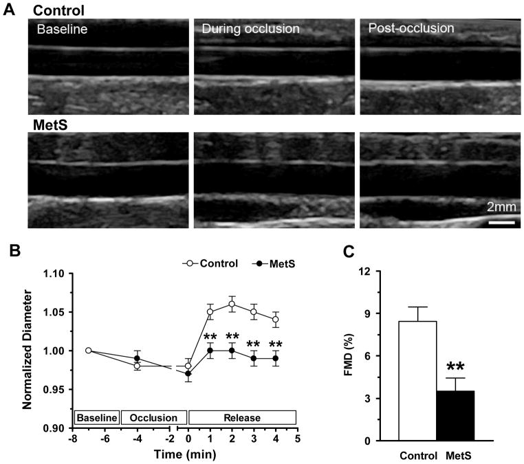
Figure 4. Flow mediated dilation in MetS monkeys.
(A) Ultrasonic images of the brachial artery during FMD measurement. (B) Average time course of arterial diameter during the FMD measurement. The duration of FMD response was abbreviated in the MetS monkeys. (C) The amplitude of FMD was significantly reduced in MetS monkeys. Data are expressed as mean ± SEM. n = 11 in control and n = 15 in MetS group. *p <0.05, ** p <0.01.

