Fig 1.
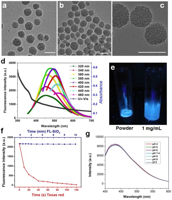
TEM images of mesoporous silica nanoparticles before (a) and after (b&c) calcination. No obvious morphology change was observed. Scale bars: 100 nm. d) Absorption and emission spectra of FL-SiO2 nanoparticles. The fluorescence intensity was at the maximum when the particles were excited at ~380 nm. e) FL-SiO2 nanoparticles in powder and solution under excitation at 365 nm. f) Photostability of Texas Red (red dots) and FL-SiO2 nanoparticles (blue dots). When exposed to excitation light, Texas Red was quickly bleached. FL-SiO2 nanoparticles, on the other hand, showed robust photostability. g) The fluorescence intensities of FL-SiO2 nanoparticles remained constant at different pH (from 4 to 9), and in a highly reducing environment. The incubation was carried out at room temperature for 24 h.

