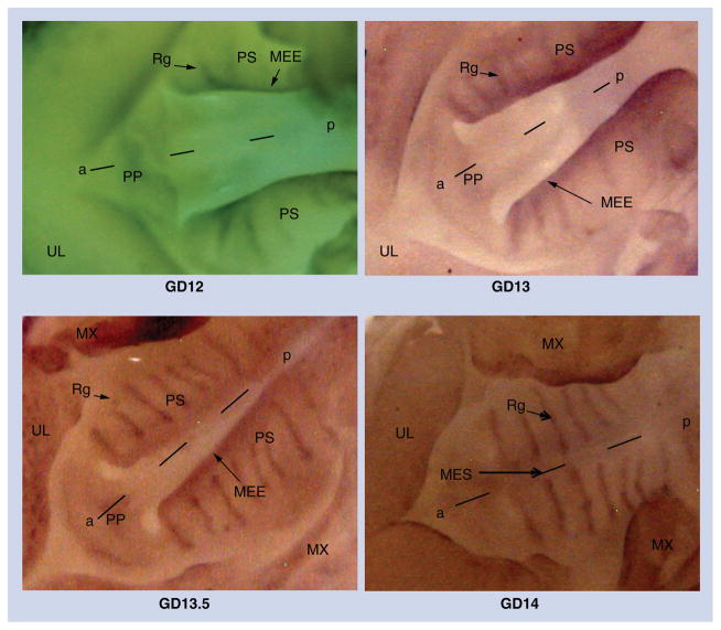
Figure 1. Whole-mount in situ hybridization with Sox4 riboprobes on GD12–GD14 palatal processes.
Sox4 expression in the developing murine secondary palate was determined from GD12 to GD14 using antisense riboprobes. On GD12 and GD13, Sox4 expression is visible in the MEE and the developing rugae. At GD13.5, Sox4 expression is more distinct in the rugae and expression persists at the medial edge epithelium. After palatal fusion on GD14, expression is confined only to the rugae with no evidence of expression at the MES where the palatal shelves have fused. Representative photomicrographs depict ventral views of the oral region of GD12–GD14 mouse embryos subsequent to removal of the mandible and tongue.
a–p: Anteroposterior axis; MEE: Medial edge epithelium; MES: Medial edge seam; MX: Maxilla; PP: Primary palate; PS: Palatal shelf; Rg: Ruga; UL: Upper lip.

