Abstract
Circulating tumor cells (CTCs) are becoming a scientifically recognized indicator of primary tumors and/or metastasis. These cells can now be accurately detected and characterized as the result of technological advances. We analyzed the presence of CTCs in the peripheral blood of patients with metastatic breast cancer by real-time reverse-transcription PCR (RT-qPCR) using a panel of selected genes. The analysis of a single marker, without an EpCAM based enrichment approach, allowed the positive identification of 35% of the metastatic breast cancer patients. The analysis of five genes (SCGB2, TFF1, TFF3, Muc1, KRT20) performed in all the samples increased the detection to 61%. We describe a sensitive, reproducible and easy to implement approach to characterize CTC in patients with metastasic breast cancer.
Introduction
Breast cancer is one of the most common malignant tumours and is the major cause of death in women in the developed world. Mortality for this disease has decreased in the last years due to screening mammography programs, more precise surgery, and the efficiency of new treatments [1], [2]. Nevertheless, about 20–30% of patients with node negative breast cancer relapse and die in the following years due to the development of metastases during the course of the disease.
The appearance of metastasis depends on the migratory capacity of cells (circulating tumour cells) from the primary tumor across the lymphatic or blood vessels to distant organs [3]. The presence of circulating tumor cells (CTCs) has been associated with survival and disease progression in patients with metastatic breast cancer [4], [5]. CTCs have also been observed in neoadjuvant-treated breast cancer patients and in women with suspected breast cancer [6], [7].
Recent technological advances have allowed the development of a variety of methods to accurately detect and characterize CTCs [8], [9], [10]. These methods are based on an initial enrichment step of the sample to increase sensitivity, followed by later detection of tumour cells. Most CTC enrichment methods include either density-gradient centrifugation to extract mononuclear cells, physical filtration with commercial filter pores (isolation by size of epithelial tumor cells (ISET)) [11] or immunomagnetic separation against surface molecules commonly expressed on malignant epithelial cells. The application of microfluidics-based technologies for CTC separation is an attractive alternative which facilitates automation for high throughput sample processing. Detection of CTCs may involve either the use of monoclonal antibodies specific for epithelial cells combined with image, flow or laser scanning cytometry [12], or real time reverse transcription PCR (RT-qPCR) markers for epithelial specific transcripts [13]. The CellSearch System® (Veridex LLC, Raritan, NJ) is the only FDA-approved technique which allows the detection and quantification of CTCs [14].
The number of articles describing single or multiple markers to characterize CTCs using RT-qPCR in the blood of breast cancer patients has increased greatly in recent years [15]–[19]. The markers most frequently studied are cytokeratins, mammaglobin (SCGB2) and HER2. Cytokeratin 19 (KRT19) is a cytoskeletal component present in normal and cancerous epithelial cells [20]. Mammaglobin is a member of a family of epithelial secretory proteins and is considered to be a specific breast marker whose expression is confined to the mammary gland [21]. HER2 is a member of the epidermal growth factor receptor (EGFR/ErbB) family. Its amplification plays an important role in the pathogenesis and progression of certain aggressive types of breast cancer [22]. Additional markers, often associated with colon, breast, ovarian, lung and pancreatic cancers, have also been studied. These include EpCAM (Epithelial cell adhesion molecule), CEACAM (Carcinoembryonic antigen-related cell adhesion molecule), Muc1 (Mucine 1), KRT20 (cytokeratin 20), Maspin, EGFR (epidermal growth factor receptor), hTERT (human telomerase), and EPHB4 (ephrin receptor) [15], [19], [23].
Two further markers merit inclusion in CTC studies: TFF1 (trefoil factor 1 or pS2) and TFF3 (trefoil factor 3 or human secretory protein p1.B). These markers belong to the family of “trefoil” proteins whose functions are not defined, but they may protect the mucosa from insult, stabilize the mucus layer, and affect healing of the epithelium. The abnormal expression of these genes in several malignancies suggests a promotional role in tumorigeneis [24].
Although much research focuses on the prognostic role of CTCs detected by RT-qPCR in breast cancer, no consensus has been established regarding the biological markers to be used to identify these cells. The heterogeneity of experiments in studies to date does not allow conclusions to be drawn on the superiority of a specific group of markers. This may be due to differences in the detection methods, in the selection of patients, in types and number of target genes, and in the studied blood fraction.
In this study we aimed to develop a specific multimarker panel for the RT-qPCR based characterization of CTC in the blood of metastatic breast cancer patients. We analyzed the role of eight genes (SCGB2, TFF1, TFF3, Muc1, KRT20, KRT19, EpCAM, CEACAM) as CTC markers in these patients.
Materials and Methods
Ethical Standards
The experiments described in the paper comply with the current laws of our country.
Cell Line and Assay Validation
The BTB-474 human breast cancer cell line (ATCC) was used to ensure that all the experimental parameters resulted in a highly efficient, sensitive and reproducible experiment.
RNA was purified from BTB-474 culture cells and diluted to different concentrations: 1000, 100, 10, 1, 0.1 and 0.01 ng/µl equivalent to 105, 104, 103, 102, 10 and 1 circulating tumor cell or CTC equivalent (CTC-EQ) respectively. One µg of each RNA dilution was retrotranscribed and amplified as described in the “Reverse transcription, pre-amplification and quantitative PCR analysis section”. Quantitative PCR was performed on each dilution using at least three replicates. A standard curve was generated by plotting the dilution series of the template against the Cq (quantification cycle) for each dilution. The slope of the curve was used to determine the reaction efficiency.
The sensitivity of each target gene was assessed by measuring whether a given low amount of template (1CTC-EQ) fitted the standard curve while maintaining the desirable efficiency. The standard curve also included a R2 (correlation coefficient) value, a measure of replicate reproducibility.
Blood Collection and Patients
Peripheral blood was collected from 41 metastatic breast cancer patients. All patients had advanced disease confirmed by histological or imagining techniques (all of them had thoracic and abdominal CT and bone scintigraphy) and were treated in the Medical Oncology Department at Hospital de la Santa Creu i Sant Pau. Thirty-five of the 41 patients had positive estrogen receptor status, 10 had HER-2 amplification, of whom 3 had also estrogen receptor positivity. The metastases were located in soft tissues in one case, in bone in 7 cases and in various locations (bone and lung or liver) in 33 cases. All patients were receiving second or third line systemic treatment, together with endocrine therapy in 13, anti-HER-2 targeted therapy combined with chemotherapy in 10 and chemotherapy alone in 18 (10 with taxanes and 8 with capecitabine). Peripheral blood from 34 healthy female volunteers and bone marrow samples from 10 hematological patients were used as negative controls. All samples were obtained after written informed consent was given. All participants provided their written informed consent to participate in this study. The study was performed in accordance with the ethical standards laid down in the declaration of Helsinki and was approved by the Hospital Santa Cruz y Sant Pau (HSCISP) Ethical Committee. Last certification obtained April 19th, 2012.
To avoid contamination with epithelial cells during the blood extraction procedure, the first 5 ml of blood were discarded and 7,5 ml were collected into EDTA-tubes.
Peripheral blood mononuclear cells (PBMC) were isolated from each blood sample by centrifugation through a Ficoll density gradient (Lymphoprep®, Nycomed, Oslo, Norway). Total RNA was extracted from PBMC and quantified by spectrometry.
Reverse Transcription, Pre-amplification and Quantitative PCR Analysis
One µg of RNA was retrotranscribed in a total reaction volume of 20 µl containing MgCl2 5 mM, 10X buffer, DTT 10 mM, dNTP’s 10 mM each, random hexamers 15 µM, RNAsin 20 U and 200 units of MuLV enzyme. Samples were incubated for 10 min at 20°C, 45 min at 42°C, and 3 min at 99°C, followed by 10 min at 4°C.
The resulting cDNA was diluted 5-fold with distilled water, and a volume of 5 µl was used in each amplification reaction. The primers and probes for the study of all the genes were purchased already made (Assay on Demand®) from Applied Biosystems.
To improve the sensitivity of the PCR, a pre-amplification reaction of 10 cycles was performed using a pooled mixture of all the PCR assays. This pre-amplification resulted in a mean improvement of 6.54±0.33 Cq values and revealed no differences in the pre-amplification uniformity values of all the tested assays.
PCR reactions were set up in MicroAmp® optical 96 well reaction plates in a 20 µl reaction volume using TaqMan Universal PCR Master Mix and the Assays on Demand® according to the manufacturer’s instructions. After 2 min at 50°C, the amplification was carried out by 40 cycles at 95°C for 15 s and 65°C for 60 s in the ABI PRISM 7500 Sequence Detector System (Applied Biosystems, Foster City, CA, USA.
The following assays targeting specific mRNAs were included in the study: Hs00761767 for KRT19 (NM_002276), Hs Hs00300643 for KRT20 (NM_019010.2), Hs00267190 for SCGB2 (NM_002411.2), Hs00173625 for TFF3 (NM_003226.2), Hs00907239 for TFF1 (NM_003225.2), Hs00158980 for EpCAM (NM_002354.2), Hs00989786 for CEACAM (NM_001184813.1) and Hs00536495 for Muc1 (NM_058173.2).
Each sample was analyzed in triplicate. Negative controls included samples without reverse transcriptase enzyme, samples where total RNA and cDNA was replaced with genomic DNA and samples where water is used instead of template.
Quantitative values were obtained from the quantification cycle (Cq) at which the fluorescent signal reached the threshold value between 0.1 and 0.5. ΔCT values were calculated by normalizing the average expression of the target gene to the average expression of the reference gene (GAPDH, B2 and HPRT1).
The ΔΔCT method was used for relative gene expression analysis, and the average ΔCT of the healthy controls for each target gene was used as the calibrator sample. The amount of target, normalized to an endogenous reference and relative to a calibrator, is given by 2–ΔΔCT. This method assigns a value of 1 to the calibrator sample, and all other quantities are expressed as an n-fold difference relative to the calibrator.
A sample was considered positive for a target gene if its relative gene expression was between 10 and 15-fold the highest value of the control samples.
Results
Efficiency, Sensitivity and Reproducibility of the Assays
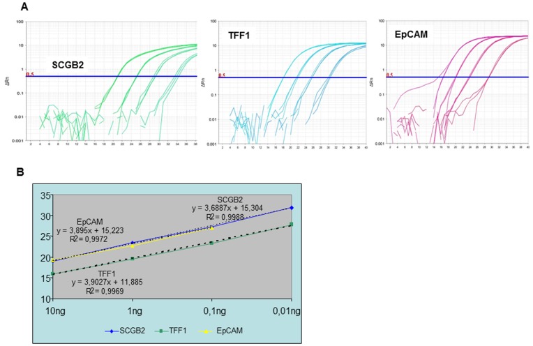
The slopes of the log phase of the amplification reaction for all the tested genes were close to -3.32. This value corresponded to efficiency close to 100%. As an example, Figure 1 shows the amplification plots and the corresponding standard curves for three of the target genes, SCGB2, TFF1 and Muc1. The parallel nature of the blue, yellow and green curves indicated that all the amplification reactions had similar efficiencies and could therefore be accurately compared at any dilution, a requirement for the ΔΔCT calculation. A similar pattern was obtained in the case of the reference genes (GAPDH, B2 and HPRT1).
Figure 1. Efficiency, sensitivity and reproducibility of the assays.
A) Amplification curves for a 10-fold dilution series for SCGB2, TFF1 and Muc1 gene. As the template amount decreases, the cycle number at which significant amplification is seen increase. B) Example of a standard curve and illustration of amplification efficiencies between targets. A standard curve shows the quantification cycle (Cq) on the y-axis and the starting quantify of RNA on the x-axis. Slope, y-intercept, and correlation coefficient values provide information about the performance of the reaction.
The sensitivity of the reaction of each target gene was determined by the most diluted sample that fitted the standard curve. For the 1CTC-EQ point dilution, no amplification was detected for any of the assays. The variation in Cq values in the 10 CTC-EQ samples between triplicates was higher than 2.3, thus not fitting the standard curve. Sensitivity was higher when a pre-amplification step of 10 cycles was added to the protocol. A good amplification curve was obtained for all tested genes in 1 CTC-EQ and the efficiency of the standard curve was close to 100%.
The correlation coefficients, also shown in Figure 1, were between 0.996 and 0.999, reflecting the linearity of the standard curve.
RT-qPCR Analysis
Having established the feasibility of our approach, samples of peripheral blood from 41 metastatic breast cancer patients, 10 haematological tumors, and 34 healthy female volunteers were tested for the expression of the eight selected genes. For the analysis of SCGB2, we additionally studied samples from 9 further female volunteers.
We used the quantitative values of three reference genes (GAPDH, HPRT1 and B2M) to normalize the expression value of our panel of candidate genes. Fourteen out of 41 (34%) blood samples from metastatic breast cancer patients showed positive SCGB2 expression, while no amplification occurred in samples from peripheral blood of the 34 healthy female volunteers or from the bone marrow of 10 hematological patients. Due to the high specificity of these results we classified the SCGB2 gene as an “excellent” CTC biomarker.
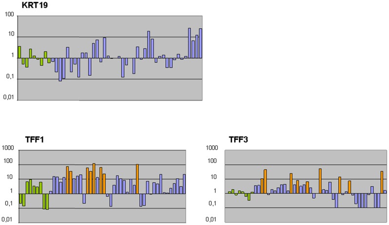
Three genes (KRT19, EpCAM and CEACAM) showed no specificity for breast cancer circulating tumor cells. The observed level of expression for these three biomarkers was similar in metastatic breast cancer patients and healthy volunteers (Fig. 2 and Figure S1). We considered these genes as “poor” CTC markers.
Figure 2. Relative expression of KRT19, TFF1 and TFF3.
Data were calculated using the ΔΔCT method, whereas the average of healthy controls was used as the calibrator sample (value = 1). Green bars indicate the values from nine healthy control samples, purple bars indicate breast cancer samples with no up regulated value and orange bars indicate breast cancer samples over expressing the target gene.
The two genes from the trefoil family (TFF1 and TFF3) showed a positive expression in 7/41 (17%) and 8/41 (20%) respectively, of metastatic breast cancer patients. The expression values observed in the control group were used, as detailed at the end of the Material and Methods section, to define a positive expression value (Fig. 2). We defined these two genes as “good” CTC markers.
Finally, we considered the Muc1 and KRT20 genes as “fair” CTC markers considering their pattern of positive expression in 4/41 (10%) and 3/41 (7%) of metastatic breast cancer patients, respectively. There was a high heterogeneity in CTC expression between the patients. In 25 patients (61%) one or more gene expression was detected. In 16 patients were positive for 1 gene (39%), 7 patients were positive for 2 genes (17%), 1 patient was positive for 3 genes (2.4%), and 1 patient was positive for 4 genes (2.4%) (Table 1).
Table 1. Associations between positively expressed gene markers.
| N° positivegenes | Patient ID(n = 25) | SCGB2+(n = 14) | TFF3+(n = 8) | TFF1+(n = 7) | KRT19+(n = 1) | KRT20+(n = 3) | Muc1+(n = 4) |
| 1 | CM-1 | + | |||||
| 1 | CM-5 | + | |||||
| 1 | CM-8 | + | |||||
| 1 | CM-9 | + | |||||
| 1 | CM-18 | + | |||||
| 1 | CM-20 | + | |||||
| 1 | CM-24 | + | |||||
| 1 | CM-26 | + | |||||
| 1 | CM-32 | + | |||||
| 1 | CM-3 | + | |||||
| 1 | CM-21 | + | |||||
| 1 | CM-27 | + | |||||
| 1 | CM-13 | + | |||||
| 1 | CM-16 | + | |||||
| 1 | CM-2 | + | |||||
| 1 | CM-25 | + | |||||
| 2 | CM-4 | + | + | ||||
| 2 | CM-30 | + | + | ||||
| 2 | CM-6 | + | + | ||||
| 2 | CM-7 | + | + | ||||
| 2 | CM-40 | + | + | ||||
| 2 | CM-41 | + | + | ||||
| 2 | CM-14 | + | + | ||||
| 3 | CM-17 | + | + | + | |||
| 4 | CM-12 | + | + | + | + |
Number of samples with positively and negatively expressed SCGB2 and its association with the other markers. Sixteen patients were positive for 1 of the genes: 9 for SCGB2, 3 for TFF3, 2 for TFF1, 1 for KRT20 and 1 for Muc1. Seven patients were positive for 2 genes: 2 for SCGB2+TF3, 2 for SCGB2+TFF1, 2 for KRT20+Muc1 and 1 for TFF1+TFF3. One patient was positive for 3 genes: TFF1+ TFF3+Muc1 and one patient was positive for four genes: SCGB2+TFF1+TFF3+KRT19.
Discussion
Circulating tumor cells are considered as a promising diagnostic tool in oncology, and much effort is being put into finding sensitive and specific analytical methods for their detection and molecular characterization. Several techniques have been explored in breast cancer to identify CTCs, but in this paper we discuss only the results obtained using molecular biomarkers.
We analyzed the role of eight genes SCGB2, TFF1, TFF3, Muc1, KRT20, KRT19, EpCAM, and CEACAM as CTC markers in a series of metastatic breast cancer patients. We developed a new panel of five genes SCGB2, TFF1, TFF3, Muc1 and KRT20 that identified CTCs in the peripheral blood in 61% of patients, without an EpCAM based enrichment approach. We used a robust quantitative PCR method applying cut-off threshold values to compensate for the low-level illegitimate mRNA expression in hematopoietic cells [25].
Controversy concerning which molecular markers should be tested to identify CTCs in breast cancer patients has been a topic of discussion for many years. In an early series of 133 patients with invasive breast cancer, Grünewald et al [26], found that mammaglobin (SCGB2) transcripts were a specific marker for hematogenous spread of breast cancer cells, but that KRT19 mRNA expression was not specific because it was expressed in 39% of healthy volunteers. In another series, however, Kahn et al [27], found KRT19 expression to be specific for patients with invasive breast cancer. Since then, other studies have reported a significant association between KRT19 and mammaglobin transcript level in CTCs from metastatic breast cancer patients [6], [16]. In contrast, we did not observe any correlation between the expressions of these two genes. Using an experimental design that did not include an enrichment step, we found that the level of KRT19 expression in patients was similar to that in the control group. This lack of specificity may be explained by the fact that KRT19 was expressed in the lymphocyte population in normal PBMC. Immunomagnetic selection for epithelial cells reduced the background KRT19 signal to a frequency of <5% in normal donors [28]. However, it has been demonstrated that during the enrichment procedure normal-like breast cancer cells characterized by an aggressive behavior are lost. Taking this limitation into account we analyzed the non-enriched peripheral blood mononuclear cells.
Since 1999, when Zach reported that mammaglobin was a specific tool to detect CTCs in peripheral blood [29], many papers have confirmed the diagnostic and prognostic value of this gene as a specific molecular marker in breast cancer [15], [16]. We detected mammaglobin expression in 34% of the metastatic breast cancer patients while was absent in control samples (healthy volunteers and patients with hematological disorders). Due to the high specificity of our results using SCGB2 we classified this gene as an “excellent” CTC biomarker.
The TFF1 and TFF3 genes have been poorly studied regarding detection of circulating tumor cells in cancer patients. Molloy et al [15], designed a multimarker platform for CTC detection in patients with early-stage breast cancer. It includes the human secretory protein TFF3. They found a positive QDA score in 20% of their patients, 31% of whom showed EGP and TFF3 expression. In our study, TFF1 and TFF3 were positive in 20% and 17% of patients, respectively. When assessed in addition to SCGB2, CTC detection improved from 34% to 51%. We thus classified TFF1 and TFF3 as “good” CTC markers.
The Muc1 gene is one of the markers included in a commercial test (AdnaTest BreastCancer®) which combines immunomagnetic tumor cell selection targeting EpCAM and MUC1 followed by multiplex RT-qPCR for the transcripts EpCAM, MUC1 and HER2. The overall detection rate for CTC using this test ranges from 22 to 69% [30], [31]. In our study, the analysis of Muc1 expression was positive in 10% of patients and was considered a “fair” marker. KRT20 marker was also included in this category as it was positively expressed in 7% of the patients.
We describe a sensitive, reproducible, low cost, and easy to implement RT-qPCR assay of five markers (SCGB2, TFF1, TFF3, Muc1, KRT20) that allows the characterization of CTCs in 61% of patients with metastatic breast cancer.
Numerous studies have focused on the prognostic role of CTCs detected by RT-qPCR but no consensus has been established regarding the biological markers to be used to identify those cells. The heterogeneity in the design of the “experiments” (selection of patients, detection methods, number of target genes, and studied blood fraction) can explain why those studies describe panels of markers which only rarely have genes in common. To solve this problem a multicenter study that standardizes protocols for the isolation and molecular identification of CTCs is needed.
Although the role of CTCs in clinical practice is not yet clear, their enumeration/profiling could serve as a real time biopsy with strong implications in the clinical management of breast cancer patients. The molecular profile of these cells may provide important information to identify therapeutic and resistance mechanisms in these cells, and add new insight into the biology of metastasis.
Supporting Information
Relative expression of EpCAM and CEACAM . Data were calculated using the ΔΔCT method, whereas the average of healthy controls was used as the calibrator sample (value = 1). Green bars indicate the values from nine healthy control samples, purple bars indicate breast cancer samples with no up regulated value.
(TIF)
Acknowledgments
The final manuscript was reviewed by Carolyn Neway.
Funding Statement
The authors have no support or funding to report.
References
- 1. Pollan M, Pastor-Barriuso R, Ardanaz E, Argüelles M, Martos C, et al. (2009) Recent changes in breast cancer incidence in Spain 1098–2004. J Natl Cancer Inst 101: 1584–1591. [DOI] [PMC free article] [PubMed] [Google Scholar]
- 2. Early Breast Cancer Trialist’s Collaborative Group (2005) Effects of chemotherapy and hormonal therapy for early breast cancer on recurrence and 15-year survival: an overview of the randomized trials. Lancet 365: 1687–1717. [DOI] [PubMed] [Google Scholar]
- 3. Chiang AC, Massagué J (2008) Molecular basis of metastasis. N Engl J Med 359: 2814–23. [DOI] [PMC free article] [PubMed] [Google Scholar]
- 4. Cristofanilli M, Budd GT, Ellis MJ, Stopeck A, Matera J, et al. (2004) Circulating Tumor Cells, Disease Progression, and Survival in Metastatic Breast Cancer. N Engl J Med 351: 781–791. [DOI] [PubMed] [Google Scholar]
- 5. Weigelt B, Bosma AJ, Hart AA, Rodenhuis S, van’t Veer LJ (2003) Marker genes for circulating tumour cells predict survival in metastasized breast cancer patients. Br J Cancer 88: 1091–4. [DOI] [PMC free article] [PubMed] [Google Scholar]
- 6. Reinholz MM, Nibbe A, Jonart LM, Kitzmann K, Suman VJ, et al. (2005) Evaluation of a panel of tumor markers for molecular detection of circulating cancer cells in women with suspected breast cancer. Clin Cancer Res 11: 3722–32. [DOI] [PubMed] [Google Scholar]
- 7. Lucci A, Hall CS, Lodhi AK, Bhattacharyya A, Anderson AE, et al. (2012) Circulating tumour cells in non-metastatic breast cancer: a prospective study. Lancet Oncol 13: 688–95. [DOI] [PubMed] [Google Scholar]
- 8. Mostert B, Sleijfer S, Foekens JA, Gratama JW (2009) Circulating tumor cells (CTCs): detection methods and their clinical relevance in breast cancer. Cancer Treat Rev 35: 463–74. [DOI] [PubMed] [Google Scholar]
- 9. Alix-Panabières C, Pantel K (2013) Circulating tumor cells: liquid biopsy of cancer. Clin Chem 59 (1): 110–118. [DOI] [PubMed] [Google Scholar]
- 10. Arya SK, Lim B, Rahman AR (2013) Rahmana (2013) Enrichment, detection and clinical significance of circulating tumor cells. Lab Chip 13(11): 1995–2027. [DOI] [PubMed] [Google Scholar]
- 11. Vona G, Sabile A, Louha M, Sitruk V, Romana S, et al. (2000) Isolation by size of epithelial tumor cells - A new method for the immunomorphological and molecular characterization of circulating tumor cells. Am. J. Pathol. 156: 57–63. [DOI] [PMC free article] [PubMed] [Google Scholar]
- 12. Riethdorf S, Fritsche H, Müller V, Rau T, Schindlbeck C, et al. (2007) Detection of circulating tumor cells in peripheral blood of patients with metastatic breast cancer: a validation study of the CellSearch system. Clin Cancer Res 13: 920–8. [DOI] [PubMed] [Google Scholar]
- 13. Zhao S, Liu Y, Zhang Q, Li H, Zhang M, et al. (2011) The prognostic role of circulating tumor cells (CTCs) detected by RT-PCR in breast cancer: a meta-analysis of published literature. Breast Cancer Res Treat 130: 809–16. [DOI] [PubMed] [Google Scholar]
- 14. Riethdorf S, Fritsche H, Müller V, Rau T, Schindlbeck C, et al. (2007) Detection of Circulating Tumor Cells in Peripheral Blood of Patients with Metastatic Breast Cancer: A Validation Study of the CellSearch System. Clin. Cancer Res. 13: 920–928. [DOI] [PubMed] [Google Scholar]
- 15. Molloy TJ, Devriese LA, Helgason HH, Bosma AJ, Hauptmann M, et al. (2011) A multimarker QPCR-based platform for the detection of circulating tumour cells in patients with early-stage breast cancer. Br J Cancer 104: 1913–9. [DOI] [PMC free article] [PubMed] [Google Scholar]
- 16. Ignatiadis M, Kallergi G, Ntoulia M, Perraki M, Apostolaki S, et al. (2008) Prognostic value of the molecular detection of circulating tumor cells using a multimarker reverse transcription-PCR assay for cytokeratin 19, mammaglobin A, and HER2 in early breast cancer. Clin Cancer Res 14: 2593–600. [DOI] [PubMed] [Google Scholar]
- 17. Strati A, Markou A, Parisi C, Politaki E, Mavroudis D, et al. (2011) Gene expresión profile of circulating tumor cells in breast cancer by RT-PCR. BMC Cancer 11: 422. [DOI] [PMC free article] [PubMed] [Google Scholar]
- 18. Molloy TJ, Roepman P, Naume B, van’t Veer LJ (2012) A prognostic gene expression profile that predicts circulating tumor cell presence in breast cancer patients. PLoS One 7(2): e32426. [DOI] [PMC free article] [PubMed] [Google Scholar]
- 19. Obermayr E, Sanchez-Cabo F, Tea MK, Singer CF, Krainer M, et al. (2010) Assessment of a six gene panel for the molecular detection of circulating tumor cells in the blood of female cancer patients. BMC Cancer 10: 666. [DOI] [PMC free article] [PubMed] [Google Scholar]
- 20. Stathopoulou A, Vlachonikolis I, Mavroudis D, Perraki M, Kouroussis Ch, et al. (2002) Molecular detection of cytokeratin-19-positive cells in the peripheral blood of patients with operable breast cancer: evaluation of their prognostic significance. J Clin Oncol 20: 3404–12. [DOI] [PubMed] [Google Scholar]
- 21. Watson MA, Dintzis S, Darrow CM, Voss LE, DiPersio J, et al. (1999) Mammaglobin expression in primary, metastatic, and occult breast cancer. Cancer Res 59: 3028–31. [PubMed] [Google Scholar]
- 22. Müller V, Pantel K (2009) HER2 as a marker for the detection of circulating tumor cells. Breast Cancer Res Treat 117: 535–537. [DOI] [PubMed] [Google Scholar]
- 23. Gervasoni A, Monasterio Muñoz RM, Wengler GS, Rizzi A, Zaniboni A, et al. (2008) Molecular signature detection of circulating tumor cells using a panel of selected genes. Cancer Lett 263: 267–79. [DOI] [PubMed] [Google Scholar]
- 24. May FE (2012) The potential of trefoil proteins as biomarkers in human cancer. Biomark Med 6: 301–4. [DOI] [PubMed] [Google Scholar]
- 25. Sieuwerts AM, Kraan J, Bolt-de Vries J, van der Spoel P, Mostert B, et al. (2009) Molecular characterization of circulating tumor cells in large quantities of contaminating leukocytes by a multiplex real-time PCR. Breast Cancer Res Treat 118: 455–468. [DOI] [PubMed] [Google Scholar]
- 26. Grünewald K, Haun M, Urbanek M, Fiegl M, Müller-Holzner E, et al. (2000) Mammaglobin gene expression: a superior marker of breast cancer cells in peripheral blood in comparison to epidermal-growth-factor receptor and cytokeratin-19. Lab Invest 80: 1071–7. [DOI] [PubMed] [Google Scholar]
- 27. Kahn HJ, Yang LY, Blondal J, Lickley L, Holloway C, et al. (2000) RT-PCR amplification of CK19 mRNA in the blood of breast cancer patients: correlation with established prognostic parameters. Breast Cancer Res Treat 60: 143–51. [DOI] [PubMed] [Google Scholar]
- 28. You F, Roberts LA, Kang SP, Nunes RA, Dias C, et al. (2008) Low-level expression of HER2 and CK19 in normal peripheral blood mononuclear cells: relevance for detection of circulating tumor cells. J Hematol Oncol 1: 2. [DOI] [PMC free article] [PubMed] [Google Scholar]
- 29. Zach O, Kasparu H, Krieger O, Hehenwarter W, Girschikofsky M, et al. (1999) Detection of circulating mammary carcinoma cells in the peripheral blood of breast cancer patients via a nested reverse transcriptase polymerase chain reaction assay for mammaglobin mRNA. J Clin Oncol 17: 2015–9. [DOI] [PubMed] [Google Scholar]
- 30. Van der Auwera I, Peeters D, Benoy IH, Elst HJ, Van Laere SJ, et al. (2010) Circulating tumour cell detection: a direct comparison between the CellSearch System, the AdnaTest and CK-19/mammaglobin RT-PCR in patients with metastatic breast cancer. Br J Cancer 102: 276–84. [DOI] [PMC free article] [PubMed] [Google Scholar]
- 31. Hauch S, Zimmermann S, Lankiewicz S, Zieglschmid V, Böcher O, et al. (2007) The clinical significance of circulating tumour cells in breast cancer and colorectal cancer patients. Anticancer Res 27: 1337–41. [PubMed] [Google Scholar]
Associated Data
This section collects any data citations, data availability statements, or supplementary materials included in this article.
Supplementary Materials
Relative expression of EpCAM and CEACAM . Data were calculated using the ΔΔCT method, whereas the average of healthy controls was used as the calibrator sample (value = 1). Green bars indicate the values from nine healthy control samples, purple bars indicate breast cancer samples with no up regulated value.
(TIF)


