
Fig. 5.
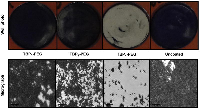
(top) Digital photographs and (bottom) phase-contrast micrographs (Magnification = 630×) of coated and uncoated Ti wells following a 5 h exposure to S. aureus cultures (starting inoculum of ~5 × 10⁷ CFU/mL). Bacteria were stained with 0.1% crystal violet to aid visualization. Scale bars = 20 μm.