Abstract
Alteration of maternal lipid metabolism early in development has been shown to trigger obesity, insulin resistance, type 2 diabetes and cardiovascular diseases later in life in humans and animal models. Here, we set out to determine (i) lipid composition dynamics in single oocytes and preimplantation embryos by high mass resolution desorption electrospray ionization mass spectrometry (DESI-MS), using the bovine species as biological model, (ii) the metabolically most relevant lipid compounds by multivariate data analysis and (iii) lipid upstream metabolism by quantitative real-time PCR (qRT-PCR) analysis of several target genes (ACAT1, CPT 1b, FASN, SREBP1 and SCAP). Bovine oocytes and blastocysts were individually analyzed by DESI-MS in both positive and negative ion modes, without lipid extraction and under ambient conditions, and were profiled for free fatty acids (FFA), phospholipids (PL), cholesterol-related molecules, and triacylglycerols (TAG). Principal component analysis (PCA) and linear discriminant analysis (LDA), performed for the first time on DESI-MS fused data, allowed unequivocal discrimination between oocytes and blastocysts based on specific lipid profiles. This analytical approach resulted in broad and detailed lipid annotation of single oocytes and blastocysts. Results of DESI-MS and transcript regulation analysis demonstrate that blastocysts produced in vitro and their in vivo counterparts differed significantly in the homeostasis of cholesterol and FFA metabolism. These results should assist in the production of viable and healthy embryos by elucidating in vivo embryonic lipid metabolism.
Introduction
The lipid composition of oocytes and preimplantation embryos is crucial for mammalian development and heavily affects the success of embryo freezing in animal production systems. Indeed, in animal reproduction, cryopreservation of oocytes and preimplantation embryos facilitates storage of valuable animal genetic resources and in humans it allows infertile couples to have children [1-4].
Free fatty acids (FFA) are stored as triacylglycerols (TAG); uncharged esters of glycerol arranged as virtually anhydrous cytoplasmic droplets [5] that represent a compact energy reserve [6]. Cholesterol (Chol) and phospholipids (PL) are essential for the formation of cellular membranes and are critically important for cell division after fertilization. However, the lipid abundance in oocytes and embryos seems to be species-specific [7]: porcines show the highest oocyte and embryo lipid content of livestock species, followed by bovines and ovines [6]; mice have the lowest lipid content among animal models [8]. The bovine species has been long accepted as a useful model for humans to study the effects of assisted reproductive techniques (ARTs) [9-11]. Due to ethical constraints, only human oocytes that had failed in fertilization attempts have been analyzed and showed relative low total lipid content [12]. Although the lipid content of bovine oocytes and preimplantation embryos differs from that of the human species, clear similarities in reproductive dynamics exist (i.e. time of blastocyst formation, number of ovulated oocytes per estrous cycle, follicular growing pattern, etc.) that render the bovine species a reliable animal model for studying fertility impairments associated with metabolic disorders in humans [5,13-17].
Various attempts have been made to increase our understanding of lipid metabolism during preimplantation embryonic development and to develop strategies to avoid harmful effects associated to in vitro culture, including supplementation of specific FFA in diets [18-20] or addition of different types and combinations of FFA to in vitro culture media [21-23]. In humans, as in various animal models, metabolic diseases have been associated with assisted reproductive technologies [24] and with particular features of the maternal lipid metabolism [25]. For example, in the case of in vitro culture, TAG droplets and FFA accumulate intracellularly which in turn is associated with significant damage of cellular organelles such as mitochondria and the endoplasmic reticulum, thus impairing survival of oocytes and embryos after freezing [26]. However, the molecular mechanisms underlying altered lipid metabolism during embryo in vitro culture in comparison to in vivo embryo development remain an enigma.
Free fatty acids have been identified in mouse, bovine, pig, sheep and human oocytes and preimplantation embryos by gas-chromatography (GC) and thin-layer chromatography (TLC). These techniques require a large number of individual samples to obtain reliable results [6]. Lipid droplet staining, an alternative technique used for total lipid quantification, requires time-consuming sample preparation and provides no structural information on the detected lipids [4]. Recent progress in mass spectrometry (MS) using matrix-assisted laser desorption/ionization mass spectrometry (MALDI-MS) and desorption electrospray ionization mass spectrometry (DESI-MS) has allowed for the first time resolved, detailed structural analysis of FFA, PL and TAG species present in single oocytes and preimplantation embryos [27-29]. Analysis of large biological datasets like this is facilitated by principal component analysis (PCA), commonly used for exploratory investigation of mass spectral datasets [30]. It allows compression of the data for in-depth characterization of biological samples and has been previously used to analyze DESI-MS data obtained from mouse preimplantation embryos [28].
Gene expression analysis by quantitative real-time PCR (qRT-PCR) has been successfully used to study oocyte and preimplantation embryonic metabolism in relationship to in vitro culture [31-33]. Expression of FA synthase (FASN) was significantly reduced in blastocysts from obese mice, and was associated to changes in Chol synthesis regulation, indicating the physiological importance of enzymes and signaling molecules involved in FA and Chol metabolism in postnatal cellular programming [34]. Moreover, recent findings revealed marked differences in the regulation of Chol biosynthesis, sterol synthesis, and cell differentiation when bovine embryos produced in vivo were compared to their in vitro derived counterparts [35].
Here, we used the bovine model and introduced an analytical approach to gain a better understanding of the complex effects of in vitro culture on embryonic metabolism. A broad lipid structural survey of individual oocytes and blastocysts was obtained by DESI-MS, followed by a robust multivariate analysis, using PCA and LDA, with a data fusion strategy applied for the first time to pure MS data. Important differences between the experimental groups were observed in FFA metabolism and Chol biosynthesis. This information was used to design a qRT-PCR analysis to unravel the relative abundance of genes critically involved in lipid metabolism. Finally, MS and qRT-PCR outcomes were comprehensively evaluated providing novel insights into the metabolic implications based on the molecular data.
Results and Discussion
Representative DESI-mass spectra and abundant lipid species
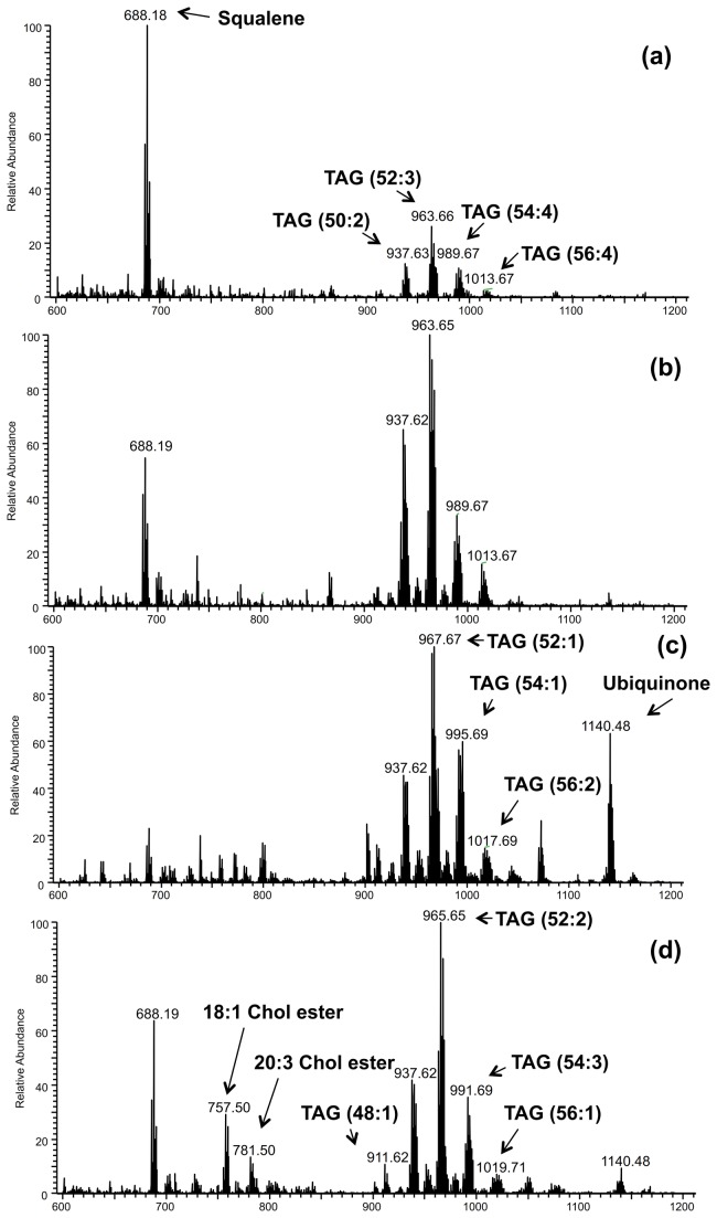
Single bovine oocytes and embryos were subjected to DESI-MS analysis in the positive and negative ion modes. Four different developmental stages were analyzed: immature and in vitro matured oocytes; blastocysts produced in vitro and in vivo. Attribution of the lipid species was made using high mass resolution DESI-MS analysis and collision-induced dissociation (CID) tandem MS experiments. Due to the large amount of chemical information obtained by DESI-MS, many lipid ions of low intensity are present in the mass spectra. Here, we report the identification of the most abundant ions. In the positive ion mode, DESI-MS lipid profiles predominantly displayed cytosolic lipids, such as cholesteryl esters (CE) and TAG, besides squalene (m/z 688.1) and ubiquinone (m/z 1140.4) (see lipid assignments in Table S1 in File S1). The CE species were detected in the m/z range of 700-850 and were observed most abundantly in the DESI-mass spectra obtained from blastocysts (Figure 1). The CE species had 16 to 24 carbons and one to six units of unsaturation (Table S1 in File S1). Oleic (18:1) and linoleic (18:2) CE of m/z 757.5 and m/z 755.4, respectively, gave the most intense CE ions. TAG species containing 48 to 56 carbons and one to five units of unsaturation in the fatty acyl residues, such as TAG (48:1), (48:2), (50:1), (50:2), (52:3), (52:2) predominated in the m/z range of 900-1100 (Table S1 in File S1). Some of these TAG species had been previously detected by MALDI-MS analysis of bovine oocytes, but had not been seen in bovine blastocysts, probably due to the ion suppression caused by the PC species in the MALDI analysis performed in the positive ion mode using 2,5-dihydroxybenzoic acid (DHB) as the organic matrix [29]. Free fatty acids, as well as membrane lipids such as phosphatidylethanolamine (PE), phosphoatidylserine (PS), phosphatidylglycerol (PG) and phosphatidylinositol (PI) were the prevalent species detected in DESI mass spectra in the negative ion mode (see lipid assignments in Table S2in File S1). Most abundant FFA present were palmitic (m/z 255.2), oleic (m/z 281.2), stearic (m/z 283.2) and arachidonic (m/z 303.2) FA. The most abundant phospholipid (PL) species in the DESI-MS spectra were [PG (34:1)] of m/z 747.5, [PS (36:1)] of m/z 788.5, PI with an alkyl ether substituent, [PIo (34:1)] of m/z 821.5, [PI (36:2)] of m/z 861.5, [PI (36:1)] of m/z 863.5 and [PI (38:4)] of m/z 885.5 (Figure S1).
Figure 1. Representative high resolution DESI-MS mass spectra in the positive ion mode.
(a) immature oocyte; (b) in vitro matured oocyte; (c) blastocyst produced in vitro; (d) blastocyst produced in vivo. See text for basis for tentative lipid class assignments of the major peaks.
Differences in lipid signatures clarified by multivariate analysis
The lipid profiles obtained allow for relative quantification of the detected molecular ions based on their intensity changes without use of internal standards. Diverse DESI-MS applications, including cancer diagnosis [36] and grading of human brain cancer tissue [37] have used this analytical strategy and proved it to be highly reproducible, although only relatively quantitative. Nevertheless, relative quantification by DESI-MS has provided good agreement with LC-MS/MS data, which includes the use of internal standards for absolute quantification [38,39].
The multivariate approach used in this work, PCA, provides a global picture of the chemical information embodied in the mass spectrometric data obtained in both the positive and the negative ion modes.
The lipids that best characterize blastocysts and oocytes in the positive ion mode are unsaturated TAG and CE (Figure S2). These compounds are easily recognized by the characteristic isotopic pattern of silver due to the addition of silver nitrate to the DESI spray solution [40,41]. As observed in the representative mass spectra shown in Figure 1, the most abundant silver-adduct ions for oocytes were m/z 963.7/965.7 (TAG 52:3) and m/z 965.7/967.7 (TAG 52:2), whereas m/z 993.7/995.7 (TAG 54:2), m/z 757.5/759.5 (18:1 cholesteryl ester), and m/z 686.2/688.2 (squalene) were more abundant in blastocysts.
In the negative ion mode, the ions that best characterize blastocyst samples were of m/z 281.2 (oleic acid), which was more abundant in blastocysts produced in vivo; m/z 255.2 (palmitic acid) and m/z 283.2 (stearic acid) were more abundant in in vitro derived blastocysts than in their in vivo derived counterparts. Conversely, m/z 747.5 [PG (34:1)], m/z 804.6 [PS (37: 0)], m/z 821.6 [PIo (34:1)] and m/z 883.5 [PI (38:5)] were present in higher relative abundance in oocytes, independent of the maturation state (Figure S1 and Figure S3).
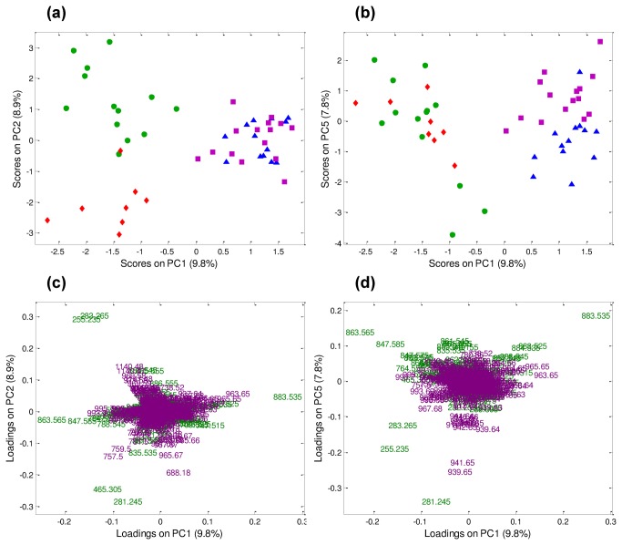
When PCA was performed for the first time by combining both datasets (positive and negative ion modes) using a data fusion strategy (DF-PCA), a clear separation of the four categories was obtained (Figure 2), confirming the synergistic effect of simultaneously considering two independent chemical profiles for sample characterization [42]. Separation between oocytes and blastocysts was visible along PC1 (i.e. highest source of data variability). Moreover, a separation was evident along PC2 for blastocysts produced in vivo and in vitro (Figure 2a). Differences between immature and in vitro matured oocytes were evident along PC5, with only a small contribution to the total data variability (Figure 2b). According to the loading plot, the ions most involved in defining PC1, which separates oocytes and blastocysts, were of m/z 863.6 [PI (36:1)], m/z 883.6 [PI (38:5)], m/z 963.7 [TAG (52:3)], m/z 965.7 [TAG (52:2)] and m/z 993.7 [TAG (54:2)]; whereas, the main contribution for separating blastocysts in vivo and in vitro along PC2 was due to ions of m/z 281.2 (oleic acid), m/z 465.3 (cholesterol sulphate), m/z 283.2 (stearic acid), m/z 255.2 (palmitic acid), and m/z 1140.5 (ubiquinone). Along PC5, m/z 963.7 [TAG (52:3)], m/z 965.7 [TAG (52:2)], m/z 883.5 [PI (38:5)] and m/z 939.7 [TAG (50:1)] were important for separating immature and oocytes matured in vitro (Figure 2c-d).
Figure 2. PCA of the fused datasets from positive and negative ion mode mass spectra.
(a) PC1 vs. PC2 score plot. (b) PC1 vs. PC5 score plot. (c-d) PC1 vs. PC2 (left side) and PC1 vs. PC5 (right side) original loading plots labeled in terms of m/z ratio (green: original negative ion mode dataset; violet: original positive ion mode dataset). Blastocysts produced in vitro (green circles, n= 13), blastocysts produced in vivo (red diamonds, n= 8), immature oocytes (blue triangles, n= 13) and in vitro matured oocytes (violet squares, n= 15).
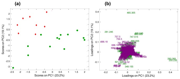
DF-PCA was then performed again, considering only immature and in vitro matured oocytes, in order to better elucidate the differences between these two groups (Figure S4). According to the loading plot, three species of PI (38:5, 34:1 and alkylated 34:1) in the negative ion mode and the polyunsaturated TAG (52:2) in the positive ion mode, were more abundant in in vitro matured than in immature oocytes, in which squalene was present at higher levels. These results are in agreement with differences observed in the representative mass spectra shown in Figure 1 (positive ion mode) and Figure S1 (negative ion mode). Additionally, DF-PCA revealed that in blastocysts produced in vivo, oleic acid, cholesterol sulphate, cholesteryl esters of palmitic and eicosatrienoic acids, TAG (52:2) and squalene were more abundant compared to in vitro produced blastocysts, in which ions of m/z 255.2 (palmitic acid) and m/z 283.2 (stearic acid) were prevalent. The DF-PCA analysis considering exclusively blastocysts produced in vitro or collected in vivo is shown in Figure 3, where PC1 vs. PC2 score and loading plots are shown. Separation between in vitro and in vivo samples goes diagonally through PC1 vs. PC2 score space. These results are in agreement with differences observed in the representative mass spectra shown in Figure 1 (positive ion mode) and Figure S1 (negative ion mode).
Figure 3. DF-PCA considering only blastocysts.
(a) PC1 vs. PC2 score plot. Green circles: blastocysts in vitro (n= 13); red diamonds: blastocysts in vivo (n= 8). (b) PC1 vs. PC2 loading plot labeled in terms of m/z ratio (green: negative ion mode; violet: positive ion mode).
Remarkably, this DF-PCA strategy, which has been employed to combine DESI-MS data obtained in the positive and the negative ion modes, allowed the combination of different datasets collected from the same samples for a common analysis, so as to improve extraction of chemical information useful for sample characterization and/or classification. This strategy provided a novel insight into interactions between cholesteryl esters and TAG with FFA and phospholipids profiles in oocytes and blastocysts, respectively. Moreover, DF-PCA is useful for an unsupervised compression of the fused data, allowing subsequent classification tools to be applied.
Classification of developmental phase by LDA
LDA was performed to classify samples according to their developmental stage based on chemical information obtained by DESI-MS. Prediction performance was validated using a cross-validation (CV) strategy (Table S3 in File S1). LDA was first performed using the positive ion mode data (dataset of 49 samples and 60001 variables) with 10 CV deletion groups and selecting the first 10 PCs to provide a CV global prediction rate of 79.6%. CV prediction rates for the individual categories were: 100% for blastocysts produced in vitro (i.e. all samples assigned to class “in vitro Bla” belong to that class), 100% for blastocysts in vivo, 61.5% for immature oocytes, and 68.8% for oocytes matured in vitro (Table S3 in File S1). When LDA was performed on negative ion mode mass spectra (dataset of 49 samples and 650001 variables), the CV global prediction rate was 89.8% (Table S3 in File S1). In the case of DF-PCA, the LDA strategy leads to 95.9% correct classification (Table S3 in File S1). Although the number of samples is relatively small, the enhancement of the prediction success demonstrates the benefit that data fusion strategy of complementary positive and negative ion mode mass spectra provides to sample characterization and classification. Indeed, the CV confusion matrix for the fused data shows a lower number of falsely assigned samples, consistent with the best results in classification (Table S3 in File S1). The information provided by LDA confirms previous findings of PCA analysis in this work as well as previous reports of MS analysis of individual oocytes and preimplantation embryos [27-29]. These results illustrate the strengths of this analytical approach, which can be applied whenever complementary mass-spectral profiles can be acquired from the same samples to characterize and classify them. Remarkably, ambient ionization MS techniques that allow intact samples to be non-destructively analyzed [43] - without any sample pretreatment steps - represent ideal analytical techniques for data fusion strategies.
Upstream analysis of DESI-MS lipid profile differences evaluated by qRT-PCR
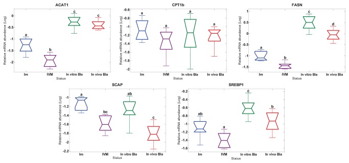
To gain further insight into lipid metabolism, levels of mRNA abundance of genes related to FFA biosynthesis and intracellular Chol homeostasis were examined in individual bovine immature, in vitro matured oocytes and blastocysts collected in vivo and in vitro (Figure 4). The mRNA abundance of Chol acyl transferase (ACAT1), FA synthase (FASN), and SREBP activating protein (SCAP) was significantly (p<0.05) down-regulated in in vitro matured oocytes compared to immature oocytes. Moreover, a trend of decreasing mRNA expression of sterol regulatory element binding protein (SREBP1) was observed in oocytes matured in vitro when compared with the immature group (P=0.06). SCAP serves as a sterol sensor by activating Site-1 protease, which in turn activates SREBP1 and 2, triggering the activation of genes encoding enzymes of Chol and FA biosynthesis in case of low intracellular sterol levels [34,35,44]. Interestingly, up-regulation of transcripts encoding ACAT1, FASN (dramatically over-expressed), SCAP and SREBP1 was observed when blastocysts produced in vitro were compared to their in vivo derived counterparts (P<0.05). Transcript abundance of carnitine palmitoyltransferase-1 (CPT 1b) was not altered among the four analyzed groups. A graphical summary of the gene expression results is shown in Figure 4.
Figure 4. Relative poly (A) + mRNA abundance of target genes.
Box plot of Log converted data (with median, 25th and 75th quartiles and max and mix values) based on single oocytes/embryos preparation: immature oocytes, n=8 (Im); in vitro matured oocytes, n=8 (IVM); in vitro produced blastocysts, n=7 (In vitro Bla); in vivo produced blastocysts, n=8 (In vivo Bla). Different superscripts indicate significant differences (P< 0.05).
Comprehensive interpretation of DESI-MS and qRT-PCR results
Biochemical information present in mass spectra and the use of a multivariate analysis, such as DF-PCA and LDA, in conjunction with gene expression data allowed comprehensive analysis of lipid metabolism.
We observed characteristic differences in lipid profiles between in vitro and in vivo produced blastocysts. Specifically, saturated FFA, palmitic acid and stearic acid, were present in much higher relative abundance within in vitro produced blastocysts than in their in vivo derived counterparts. This observation is in accordance with literature, as palmitic acid had been reported to be the most abundant fatty acid in bovine oocytes, followed by oleic acid, stearic acid and linoleic acid [6]. Linoleic acid is the major fatty acid in bovine follicular fluid, followed by palmitic acid [45,46]. Recent work on the mouse model reported that exposure of cumulus-oocyte complexes (COCs) to palmitic acid at a concentration in the upper range of physiological levels induced endoplasmic reticulum stress, which in turn reduced protein secretion, disrupted mitochondrial activity in oocytes, and impaired oocyte maturation and fertilization [47]. Cell proliferation and the number of inner mass cells in mice blastocysts were reduced concomitantly with an increase in trophoblast cell apoptosis after in vitro culture in medium supplemented with 200 µM of palmitic acid [48]. Moreover, supplementation of the culture medium with stearic and palmitic acid during bovine oocyte in vitro maturation impaired post-fertilization development in a dose-dependent manner [49]. Similarly, supplementation of culture medium during in vitro maturation of bovine oocytes with oleic acid, palmitic acid and stearic acid altered gene expression in blastocysts and reduced embryo quality, based on energy and amino acid metabolism [22]. Presumably, the higher concentrations of FFA within in vitro produced blastocysts are responsible for impaired embryonic development. A recent publication has demonstrated alteration of the REDOX status of bovine oocytes and embryos when high levels of free fatty acids were incorporated to the culture media [50].
In vivo produced blastocysts showed higher concentrations of neutral lipid species, including cholesterol sulphate, cholesteryl esters of palmitic and eicosatrienoic acids, TAG (52:2), squalene and oleic acid. The accumulation of neutral lipid species, which is the normal energy storage of mammalian cells, may potentially contribute to normal developmental rates of mammalian blastocysts produced in vivo. In more detail, squalene, a long-chain poly-unsaturated fatty acid [51], in combination with TAG and sterol esters, has been involved in intracellular lipid droplet formation, clustering in both yeast and mammalian cells [52], probably by regulating membrane properties [53]. To our knowledge, this is the first report of the presence of squalene in mammalian oocytes and preimplantation embryos. Oleic acid is a mono-unsaturated fatty acid proven to be beneficial in bovine post-fertilization development, lipid storage and oocyte maturation [49].
Gene expression analysis was designed based on the DF-PCA analysis of DESI-MS results and genes were selected for this study that encode functional enzymes critically involved in intracellular cholesterol level regulation (SREBP1 and SCAP), long-chain fatty acid biosynthesis (FASN), reversible formation of acetoacyl-CoA from two molecules of acetyl-Coa (ACAT1) and control of the long-chain fatty acid beta-oxidation (CPT 1b). Four of the target genes (SREBP1, FASN, SCAP and ACAT1) were significantly down-regulated after in vitro maturation which is in agreement with previous reports, where the relative abundances of mRNAs of genes crucial for oocyte development (SLC2A8, GDF9, PRDX1 and ZAR1) were down-regulated after in vitro maturation of bovine oocytes [31,32]. Our results indicate that biosynthesis and storage of neutral lipid species, present in higher levels in in vitro matured oocytes than in immature oocytes (PI and TAG of polyunsaturated fatty acids), take place at the acquisition of oocyte developmental competence during folliculogenesis [54]. The presence of high levels of saturated FFA (most likely stearic and palmitic acid) in in vitro produced blastocysts detected by DESI-MS coincides with a significant up-regulation of target genes (SREBP1, FASN, SCAP and ACAT1). Fatty acid synthase, which was over-expressed in in vitro produced blastocysts, is the most relevant enzyme in the biosynthesis of long-chain fatty acids. Palmitic acid is the main product of FASN by the novo synthesis from Acetyl-CoA, Malonyl-CoA and NADPH [55]. The sterol synthesis sensor SREBP/SCAP was clearly activated in in vitro produced blastocysts, which was confirmed by our gene expression results. In agreement with this, a recent publication showed significant up-regulation of 11 genes involved in cholesterol biosynthesis in bovine in vitro compared to the in vivo produced embryos [35]. In contrast to the present and previous results, genes involved in cholesterol metabolism (HSD17B7, CYP11A1), steroid metabolism (HSD3B1, CYP11A1, APOA1), lipid metabolism (MSMO1, ANXA1, ANXA2) and lipid excretion and translocation (ABCC2) were significantly down-regulated in bovine in vitro produced blastocysts in comparison to their in vivo counterparts [56].
Conclusions
This bioanalytical strategy made possible the biological interpretation of DESI and qRT-PCR data for studying the impact of in vitro culture on individual bovine oocytes and blastocysts. Here we provide for the first time a comprehensive and in-depth analysis of the lipid composition and its modulation by in vitro production methods in both the female gamete (oocyte) and the blastocyst. The analysis of two different datasets (i.e. positive and negative ion mode MS) obtained by state-of-the-art ambient MS from single oocytes or blastocysts, followed by application of multivariate statistical analysis and data fusion, makes this work unique and provides new insights into the complex regulation of mammalian oocyte/embryo lipid metabolism, which can be exported to other mammalian species, including humans. Our results demonstrate that current in vitro production systems are associated with profound alterations of the lipid profile and metabolism of preimplantation embryos with possibly deleterious effects on development and health status of the offspring. Using this novel analytical approach to study upstream lipid metabolism, our findings pave the way for monitoring and improvement of in vitro culture systems and reproductive techniques. Further, this work might contribute towards a better understanding of the concept of the embryonic onset of adult disease [57,58] and metabolic impairments associated to lipid metabolism such as obesity and diabetes type II.
Materials and Methods
Cumulus-oocyte complexes (COCs) recovery, in vitro maturation and blastocysts production in vitro
Bovine ovaries were collected from a local abattoir (Westfleisch Lübbecke, Westfalen, Germany) after approval by the local supervisory veterinarian. The ovaries were transported to the host laboratory (Mariensee, Germany) in saline solution (0.9%) at 30°C supplemented with penicillin and streptomycin sulphate. After cumulus cells removal, immature oocytes were stored as described below for DESI-MS analysis. Another group included in vitro matured oocytes; a further subset was used for blastocyst production in vitro [59,60]. Single oocytes (immature, n=8 and in vitro matured, n=8) and blastocysts (produced in vivo, n=8 and produced in vitro, n=7) were frozen in a minimum volume (≤ 5 µL) of PBS supplemented with 1% polyvinyl alcohol (PVA) medium in a 600 µL siliconized cryovials (Biozym Diagnostic GmbH, Hessisch Oldendorf, Germany) and stored at −80 °C until analysis (an extended version of these methods can be found in File S2). A separate ethical approval is not necessary for in vitro experiments with bovine oocytes/embryos in Germany. All experiments were performed in strict accordance with the United States and Germany’ laws and guidelines and approved by institutional committees [61], specifically the guidelines on good scientific practice from DFG (Deutsche Forschungsgemeinschaft).
In vivo embryo production
Dairy cows from the Institute’s experimental herd in Mariensee (Germany) were super-ovulated between 9 and 12 days of the estrous cycle by using intramuscular injections of 2500-3000 IU eCG (Intergonan®; Intervet, Tönisvorst, Germany) and Cloprostenol (Estrumate®; Essex, Munich, Germany) 48 h later. After 48 h, cows were inseminated twice with an interval of 12 h. Eight days later, blastocysts were collected by nonsurgical uterine flushing with 500 mL PBS supplemented with 1% NBCS (newborn calf serum, Invitrogen, Karlsruhe, Germany) [62]. The blastocysts were washed three times in PBS with 0.1% PVA under a stereomicroscope. Single embryos were frozen in individual cups at −80 °C and stored until further analysis.
DESI-MS analysis and attribution of most abundant lipid species of high resolution mass spectrometry
Samples were stored in minimal volume (2-5µL) of PBS supplemented with 0.1% polyvinyl alcohol (PVA) and were shipped on dry ice from the Institute of Farm Animal Genetics (Mariensee, Germany) to Purdue University (West Lafayette, IN, USA; USDA permit 118624 Research). Mouse brain tissue sections and part of the samples have been used for DESI system optimization (Purdue University Animal Care and Use Committee approved protocol is No. 1111000314, as reported in [28]).
The samples (n=82) were submitted to lipid profile analysis in the positive ion mode, in order to detect cholesteryl esters and TAG data by doping the solvent spray with silver ions [40,41,43]. The use of silver nitrate in the DESI-MS solvent spray promotes the formation of lipid silver adducts, which are highly ionizable. Due to the silver isotopic distribution, combinations of ions, such as m/z 963.7/965.7 (TAG 52:3), correspond to the 107Ag and 109Ag adducts of the same lipid molecule. Then the same samples were analyzed in the negative ion mode for FFA and PL profiling [27,28]. A total of 49 samples was analyzed in both positive and negative ion modes.
A Thermo Scientific Exactive (San Jose, CA, USA) mass spectrometer was used for the experiments. DESI-MS profiles were acquired in lab-built stage and the DESI spray was positioned ~2 mm from the surface at an incident angle of 50 °. The DESI spray had 5 kV applied to the stainless steel needle syringe and nitrogen gas pressure was 180 psi (Figure A in File S2). Instrument conditions are described in details in the File S2.
Molecular formula matching and error calculations were performed using the instrument software Xcalibur v.1.0.1.03 (Thermo, Fisher Scientific San Jose, CA, USA) and online search of lipids containing the calculated molecular formulae was carried out in the LIPID MAPS database (Figure B in File S2) [63]. Lipid attributions based on high mass resolution mass measurements data are listed in Table S1 in File S1 (positive ion mode) and Table S2 in File S1 (negative ion mode).
Multivariate data analyses: PCA and LDA
PCA is commonly used for exploratory investigations of the complex information contained in a full mass spectral dataset, to allow simultaneous analysis of all spectral variables and their inter-correlations [30]. Linear discriminant analysis (LDA) can be applied in combination with PCA for sample classification [36]. Moreover, multivariate analysis together with data-fusion allows suitable sample characterization and classification [42]. Considering the 49 samples from which both positive and negative ion mode mass spectra were collected (immature oocytes, n=13; in vitro matured oocytes, n=15; blastocysts produced in vivo, n=8; blastocysts produced in vitro, n=13), PCA was first performed on two separated datasets, as described extensively in File S2. Furthermore, a data fusion strategy (DF-PCA) was applied on the same datasets in order to investigate whether the joined analysis of positive and negative mass spectra improved sample characterization and/or classification [42]. In view of the huge number of variables constituting each mass spectral dataset, a preliminary compression of the individual datasets by means of PCA was performed (as represented in Figure C in File S2) [42]. By applying the same strategy (positive and negative ion mode mass spectra individually considered and thus fused together), LDA was performed as an additional classification method, in order to quantify the ability of DESI-MS lipid profiles in discriminating among oocytes (immature and matured in vitro) and blastocysts (produced in vivo and in vitro). Details are reported in File S2.
Data processing was performed by means of the in-house Matlab (The Math Works, Inc., Natick, USA) routines.
Determination of the relative abundance of mRNA transcripts for ACAT1, CPT 1b, FASN, SREBP1 and SCAP
Poly (A) + RNA from single oocytes (immature, n=8 and matured in vitro, n=8) and blastocysts (produced in vivo, n=8 and produced in vitro, n=7) was isolated using the Dynabeads® mRNA DIRECT™ kit (Invitrogen, Carlsbad, USA) as reported previously [31,64]. Two µL of the RT reaction were used for real-time PCR amplification (for more details see File S2). Gene expression data (Log-transformed) were analyzed for each single gene by one-way ANOVA using JMP (SAS Institute Inc., Cary, NC, USA). The Tukey-Kramer test was applied for a multiple comparison of means. For all tests, a P-value ≤ 0.05 was considered as statistically significant.
Supporting Information
Supplementary tables containing details about (i) the attribution of the lipid species made using high mass resolution DESI-MS analysis and collision-induced dissociation (CID) tandem MS experiments in positive and negative ion modes (Tables S1 and S2); (ii) CV confusion matrix for all LDAs (Table S3). Table S1, Positive ion mode mass spectra. Table S2, Negative ion mode mass spectra. Table S3, CV confusion matrix for all LDAs.
(DOCX)
Extended version of Material and Methods containing details about (i) DESI-MS conditions for lipid profile detection; (ii) PCA and LDA methodology and relative results on both individual and fused datasets; (ii) RNA extraction and quantitative RT-PCR.
(DOCX)
Representative high resolution DESI-MS mass spectra in the negative ion mode. (a) immature oocyte; (b) in vitro matured oocyte; (c) blastocyst produced in vitro; (d) blastocyst produced in vivo. See text and Table S2 in File S1 for tentative lipid class assignments of the major peaks.
(TIF)
PCA of the positive ion mode mass spectral data. Blastocysts in vitro (green circles, n=13), blastocysts in vivo (red diamonds, n=8), immature oocytes (blue triangles, n=13) and in vitro matured oocytes (violet squares, n=15). (a) PC1 vs. PC4 score plot. (b) PC1 vs. PC4 loading plot labeled in terms of m/z ratio.
(TIF)
PCA of the negative ion mode mass spectral data. In vitro blastocysts (green circles, n=13), in vivo blastocysts (red diamonds, n=8), immature oocytes (blue triangles, n=13) and in vitro matured oocytes (violet squares, n=15). (a) PC1 vs. PC4 score plot. (b) PC1 vs. PC4 loading plot labeled in terms of m/z ratio.
(TIF)
DF-PCA considering only oocytes. (a) PC1 vs. PC5 score plot. Blue triangles: immature oocytes (n=13); violet squares: in vitro matured oocytes (n= 15). (b) PC1 vs. PC5 loading plot labeled in terms of m/z ratio (green: negative ion mode; violet: positive ion mode).
(TIF)
Acknowledgments
We thank Klaus-Gerd Hadeler for assistance in embryo production, Doris Herrmann for assisting in gene expression analysis, and Alan K. Jarmusch for assistance in manuscript preparation.
Funding Statement
AFGS was supported from the German Academic Exchange Service (DAAD) and the Fundación Gran Mariscal Ayacucho (FUNDAYACUCHO); VP was supported from the “L’Oreal/UNESCO Italia per le Donne e la Scienza 2013” grant; CF was supported from the Purdue University Center for Cancer Research Small Grants and from the Brazilian National Council for Scientific and Technological Development (CNPq); RGC by NSF IDBR 0852740. The funders had no role in study design, data collection and analysis, decision to publish, or preparation of the manuscript.
References
- 1. Rienzi L, Cobo A, Paffoni A, Scarduelli C, Capalbo A et al. (2012) Consistent and predictable delivery rates after oocyte vitrification: an observational longitudinal cohort multicentric study. Hum Reprod 27: 1606-1612. doi:10.1093/humrep/des088. PubMed: 22442248. [DOI] [PubMed] [Google Scholar]
- 2. Anonymous. (2013) Mature oocyte cryopreservation: a guideline. Fertil Steril 99: 37-43. doi:10.1016/j.fertnstert.2012.09.028. PubMed: 23083924. [DOI] [PubMed] [Google Scholar]
- 3. Pereira R, Marques CC (2008) Animal oocyte and embryo cryopreservation. Cell Tissue Bank 9: 267-277. [DOI] [PubMed] [Google Scholar]
- 4. Wu LL, Dunning KR, Yang X, Russell DL, Lane M et al. (2010) High-fat diet causes lipotoxicity responses in cumulus-oocyte complexes and decreased fertilization rates. Endocrinology 151: 5438-5445. doi:10.1210/en.2010-0551. PubMed: 20861227. [DOI] [PubMed] [Google Scholar]
- 5. McKeegan PJ, Sturmey RG (2011) The role of fatty acids in oocyte and early embryo development. Reprod Fertil Dev 24: 59-67. PubMed: 22394718. [DOI] [PubMed] [Google Scholar]
- 6. McEvoy TG, Coull GD, Broadbent PJ, Hutchinson JS, Speake BK (2000) Fatty acid composition of lipids in immature cattle, pig and sheep oocytes with intact zona pellucida. J Reprod Fertil 118: 163-170. doi:10.1530/reprod/118.1.163. PubMed: 10793638. [PubMed] [Google Scholar]
- 7. Prates EG, Alves SP, Marques CC, Baptista MC, Horta AE et al. (2013) Fatty acid composition of porcine cumulus oocyte complexes (COC) during maturation: effect of the lipid modulators trans-10, cis-12 conjugated linoleic acid (t10,c12 CLA) and forskolin. In Vitro Cell Dev Biol Anim 49: 335-345. doi:10.1007/s11626-013-9624-2. PubMed: 23645468. [DOI] [PubMed] [Google Scholar]
- 8. Loewenstein JE, Cohen AI (1964) Dry mass, lipid content and protein content of the intact and zona-free mouse ovum. J Embryol Exp Morphol 12: 113-121. PubMed: 14155399. [PubMed] [Google Scholar]
- 9. Wrenzycki C, Herrmann D, Lucas-Hahn A, Gebert C, Korsawe K et al. (2005) Epigenetic reprogramming throughout preimplantation development and consequences for assisted reproductive technologies. Birth Defects Res C Embryo TODAY 75: 1-9. doi:10.1002/bdrc.20035. PubMed: 15838918. [DOI] [PubMed] [Google Scholar]
- 10. Niemann H, Wrenzycki C (2000) Alterations of expression of developmentally important genes in preimplantation bovine embryos by in vitro culture conditions: implications for subsequent development. Theriogenology 53: 21-34. doi:10.1016/S0093-691X(99)00237-X. PubMed: 10735059. [DOI] [PubMed] [Google Scholar]
- 11. Haaf T (2006) Methylation dynamics in the early mammalian embryo: implications of genome reprogramming defects for development. Curr Top Microbiol Immunol 310: 13-22. doi:10.1007/3-540-31181-5_2. PubMed: 16909904. [DOI] [PubMed] [Google Scholar]
- 12. Matorras R, Ruiz JI, Mendoza R, Ruiz N, Sanjurjo P et al. (1998) Fatty acid composition of fertilization-failed human oocytes. Hum Reprod 13: 2227-2230. doi:10.1093/humrep/13.8.2227. PubMed: 9756301. [DOI] [PubMed] [Google Scholar]
- 13. Walker SK, Heard TM, Seamark RF (1992) In vitro culture of sheep embryos without co-culture: Successes and perspectives. Theriogenology 37: 111-126. doi:10.1016/0093-691X(92)90250-U. [Google Scholar]
- 14. Leroy JL, Opsomer G, De Vliegher S, Vanholder T, Goossens L et al. (2005) Comparison of embryo quality in high-yielding dairy cows, in dairy heifers and in beef cows. Theriogenology 64: 2022-2036. doi:10.1016/j.theriogenology.2005.05.003. PubMed: 15936067. [DOI] [PubMed] [Google Scholar]
- 15. Wrenzycki C, De Sousa P, Overström EW, Duby RT, Herrmann D et al. (2000) Effects of superovulated heifer diet type and quantity on relative mRNA abundances and pyruvate metabolism in recovered embryos. J Reprod Fertil 118: 69-78. doi:10.1530/jrf.0.1180069. PubMed: 10793627. [PubMed] [Google Scholar]
- 16. Leroy JL, Van Hoeck V, Clemente M, Rizos D, Gutierrez-Adan A et al. (2010) The effect of nutritionally induced hyperlipidaemia on in vitro bovine embryo quality. Hum Reprod 25: 768-778. doi:10.1093/humrep/dep420. PubMed: 20007613. [DOI] [PubMed] [Google Scholar]
- 17. Campbell BK, Souza C, Gong J, Webb R, Kendall N et al. (2003) Domestic ruminants as models for the elucidation of the mechanisms controlling ovarian follicle development in humans. Reprod Suppl 61: 429-443. PubMed: 14635953. [PubMed] [Google Scholar]
- 18. Adamiak SJ, Powell K, Rooke JA, Webb R, Sinclair KD (2006) Body composition, dietary carbohydrates and fatty acids determine post-fertilisation development of bovine oocytes in vitro. Reproduction 131: 247-258. doi:10.1530/rep.1.00871. PubMed: 16452718. [DOI] [PubMed] [Google Scholar]
- 19. Fouladi-Nashta AA, Wonnacott KE, Gutierrez CG, Gong JG, Sinclair KD et al. (2009) Oocyte quality in lactating dairy cows fed on high levels of n-3 and n-6 fatty acids. Reproduction 138: 771-781. doi:10.1530/REP-08-0391. PubMed: 19633135. [DOI] [PubMed] [Google Scholar]
- 20. Gulliver CE, Friend MA, King BJ, Clayton EH (2012) The role of omega-3 polyunsaturated fatty acids in reproduction of sheep and cattle. Anim Reprod Sci 131: 9-22. doi:10.1016/j.anireprosci.2012.02.002. PubMed: 22386690. [DOI] [PubMed] [Google Scholar]
- 21. Marei WF, Wathes DC, Fouladi-Nashta AA (2009) The effect of linolenic Acid on bovine oocyte maturation and development. Biol Reprod 81: 1064-1072. doi:10.1095/biolreprod.109.076851. PubMed: 19587335. [DOI] [PubMed] [Google Scholar]
- 22. Marei WF, Wathes DC, Fouladi-Nashta AA (2012) Differential effects of linoleic and alpha-linolenic fatty acids on spatial and temporal mitochondrial distribution and activity in bovine oocytes. Reprod Fertil Dev 24: 679-690. doi:10.1071/RD11204. PubMed: 22697118. [DOI] [PubMed] [Google Scholar]
- 23. Van Hoeck V, Sturmey RG, Bermejo-Alvarez P, Rizos D, Gutierrez-Adan A et al. (2011) Elevated non-esterified fatty acid concentrations during bovine oocyte maturation compromise early embryo physiology. PLOS ONE 6: e23183. doi:10.1371/journal.pone.0023183. PubMed: 21858021. [DOI] [PMC free article] [PubMed] [Google Scholar]
- 24. Chen M, Norman RJ, Heilbronn LK (2011) Does in vitro fertilisation increase type 2 diabetes and cardiovascular risk?. Curr. Diabetes Rev 7: 426-432. doi:10.2174/157339911797579151. [DOI] [PubMed] [Google Scholar]
- 25. Dabelea D, Crume T (2011) Maternal environment and the transgenerational cycle of obesity and diabetes. Diabetes 60: 1849-1855. doi:10.2337/db11-0400. PubMed: 21709280. [DOI] [PMC free article] [PubMed] [Google Scholar]
- 26. Wu LL, Norman RJ, Robker RL (2011) The impact of obesity on oocytes: evidence for lipotoxicity mechanisms. Reprod Fertil Dev 24: 29-34. PubMed: 22394715. [DOI] [PubMed] [Google Scholar]
- 27. Ferreira CR, Eberlin LS, Hallett JE, Cooks RG (2012) Single oocyte and single embryo lipid analysis by desorption electrospray ionization mass spectrometry. J Mass Spectrom 47: 29-33. doi:10.1002/jms.2022. PubMed: 22282086. [DOI] [PubMed] [Google Scholar]
- 28. Ferreira CR, Pirro V, Eberlin LS, Hallett JE, Cooks RG (2012) Developmental phases of individual mouse preimplantation embryos characterized by lipid signatures using desorption electrospray ionization mass spectrometry. Anal Bioanal Chem 404: 2915-2926. doi:10.1007/s00216-012-6426-4. PubMed: 23052870. [DOI] [PubMed] [Google Scholar]
- 29. Ferreira CR, Saraiva SA, Catharino RR, Garcia JS, Gozzo FC et al. (2010) Single embryo and oocyte lipid fingerprinting by mass spectrometry. J Lipid Res 51: 1218-1227. doi:10.1194/jlr.D001768. PubMed: 19965589. [DOI] [PMC free article] [PubMed] [Google Scholar]
- 30. Jolliffe IT (2002) Principal component analysis. New York: Springer-Verlag. 519pp. [Google Scholar]
- 31. Heinzmann J, Hansmann T, Herrmann D, Wrenzycki C, Zechner U et al. (2011) Epigenetic profile of developmentally important genes in bovine oocytes. Mol Reprod Dev 78: 188-201. doi:10.1002/mrd.21281. PubMed: 21290475. [DOI] [PubMed] [Google Scholar]
- 32. Diederich M, Hansmann T, Heinzmann J, Barg-Kues B, Herrmann D et al. (2012) DNA methylation and mRNA expression profiles in bovine oocytes derived from prepubertal and adult donors. Reproduction 144: 319-330. doi:10.1530/REP-12-0134. PubMed: 22733804. [DOI] [PubMed] [Google Scholar]
- 33. Kues WA, Sudheer S, Herrmann D, Carnwath JW, Havlicek V et al. (2008) Genome-wide expression profiling reveals distinct clusters of transcriptional regulation during bovine preimplantation development in vivo . Proc Natl Acad Sci U S A 105: 19768-19773. doi:10.1073/pnas.0805616105. PubMed: 19064908. [DOI] [PMC free article] [PubMed] [Google Scholar]
- 34. Königsdorf CA, Navarrete Santos A, Schmidt JS, Fischer S, Fischer B (2012) Expression profile of fatty acid metabolism genes in preimplantation blastocysts of obese and non-obese mice. Obes Facts 5: 575-586. doi:10.1159/000342583. PubMed: 22986646. [DOI] [PubMed] [Google Scholar]
- 35. Driver AM, Peñagaricano F, Huang W, Ahmad KR, Hackbart KS et al. (2012) RNA-Seq analysis uncovers transcriptomic variations between morphologically similar in vivo- and in vitro-derived bovine blastocysts. BMC Genomics 13: 118. doi:10.1186/1471-2164-13-118. PubMed: 22452724. [DOI] [PMC free article] [PubMed] [Google Scholar]
- 36. Pirro V, Eberlin LS, Oliveri P, Cooks RG (2012) Interactive hyperspectral approach for exploring and interpreting DESI-MS images of cancerous and normal tissue sections. Analyst 137: 2374-2380. doi:10.1039/c2an35122f. PubMed: 22493773. [DOI] [PubMed] [Google Scholar]
- 37. Eberlin LS, Norton I, Orringer D, Dunn IF, Liu X et al. (2013) Ambient mass spectrometry for the intraoperative molecular diagnosis of human brain tumors. Proc Natl Acad Sci U S A 110: 1611-1616. doi:10.1073/pnas.1215687110. PubMed: 23300285. [DOI] [PMC free article] [PubMed] [Google Scholar]
- 38. García-Reyes JF, Jackson AU, Molina-Díaz A, Cooks RG (2009) Desorption electrospray ionization mass spectrometry for trace analysis of agrochemicals in food. Anal Chem 81: 820-829. doi:10.1021/ac802166v. PubMed: 19090743. [DOI] [PubMed] [Google Scholar]
- 39. Kennedy JH, Aurand C, Shirey R, Laughlin BC, Wiseman JM (2010) Coupling desorption electrospray ionization with solid-phase microextraction for screening and quantitative analysis of drugs in urine. Anal Chem 82: 7502-7508. doi:10.1021/ac101295g. PubMed: 20695439. [DOI] [PubMed] [Google Scholar]
- 40. Seal JR, Havrilla CM, Porter NA, Hachey DL (2003) Analysis of unsaturated compounds by Ag+ coordination ionspray mass spectrometry: studies of the formation of the Ag+/lipid complex. J Am Soc Mass Spectrom 14: 872-880. doi:10.1016/S1044-0305(03)00339-8. PubMed: 12892911. [DOI] [PubMed] [Google Scholar]
- 41. Jackson AU, Shum T, Sokol E, Dill A, Cooks RG (2011) Enhanced detection of olefins using ambient ionization mass spectrometry: Ag+ adducts of biologically relevant alkenes. Anal Bioanal Chem 399: 367-376. doi:10.1007/s00216-010-4349-5. PubMed: 21069301. [DOI] [PubMed] [Google Scholar]
- 42. Blanchet L, Smolinska A, Attali A, Stoop MP, Ampt KA et al. (2011) Fusion of metabolomics and proteomics data for biomarkers discovery: case study on the experimental autoimmune encephalomyelitis. BMC Bioinformatics 12: 254. doi:10.1186/1471-2105-12-254. PubMed: 21696593. [DOI] [PMC free article] [PubMed] [Google Scholar]
- 43. Eberlin LS, Ferreira CR, Dill AL, Ifa DR, Cheng L et al. (2011) Nondestructive, histologically compatible tissue imaging by desorption electrospray ionization mass spectrometry. Chembiochem 12: 2129-2132. doi:10.1002/cbic.201100411. PubMed: 21793152. [DOI] [PMC free article] [PubMed] [Google Scholar]
- 44. Brown MS, Goldstein JL (1999) A proteolytic pathway that controls the cholesterol content of membranes, cells, and blood. Proc Natl Acad Sci U S A 96: 11041-11048. doi:10.1073/pnas.96.20.11041. PubMed: 10500120. [DOI] [PMC free article] [PubMed] [Google Scholar]
- 45. Marei WF, Wathes DC, Fouladi-Nashta AA (2010) Impact of linoleic acid on bovine oocyte maturation and embryo development. Reproduction 139: 979-988. doi:10.1530/REP-09-0503. PubMed: 20215338. [DOI] [PubMed] [Google Scholar]
- 46. Leroy JL, Vanholder T, Mateusen B, Christophe A, Opsomer G et al. (2005) Non-esterified fatty acids in follicular fluid of dairy cows and their effect on developmental capacity of bovine oocytes in vitro . Reproduction 130: 485-495. doi:10.1530/rep.1.00735. PubMed: 16183866. [DOI] [PubMed] [Google Scholar]
- 47. Wu LL, Russell DL, Norman RJ, Robker RL (2012) Endoplasmic reticulum (ER) stress in cumulus-oocyte complexes impairs pentraxin-3 secretion, mitochondrial membrane potential (DeltaPsi m), and embryo development. Mol Endocrinol 26: 562-573. doi:10.1210/me.2011-1362. PubMed: 22383462. [DOI] [PMC free article] [PubMed] [Google Scholar]
- 48. Jungheim ES, Louden ED, Chi MM, Frolova AI, Riley JK et al. (2011) Preimplantation exposure of mouse embryos to palmitic acid results in fetal growth restriction followed by catch-up growth in the offspring. Biol Reprod 85: 678-683. doi:10.1095/biolreprod.111.092148. PubMed: 21653893. [DOI] [PMC free article] [PubMed] [Google Scholar]
- 49. Aardema H, Vos PL, Lolicato F, Roelen BA, Knijn HM et al. (2011) Oleic acid prevents detrimental effects of saturated fatty acids on bovine oocyte developmental competence. Biol Reprod 85: 62-69. doi:10.1095/biolreprod.110.088815. PubMed: 21311036. [DOI] [PubMed] [Google Scholar]
- 50. Van Hoeck V, Leroy JL, Arias Alvarez M, Rizos D, Gutierrez-Adan A et al. (2013) Oocyte developmental failure in response to elevated nonesterified fatty acid concentrations: mechanistic insights. Reproduction 145: 33-44. doi:10.1530/REP-12-0174. PubMed: 23108110. [DOI] [PubMed] [Google Scholar]
- 51. Kim SK, Karadeniz F (2012) Biological importance and applications of squalene and squalane. Adv Food Nutr Res 65: 223-233. doi:10.1016/B978-0-12-416003-3.00014-7. PubMed: 22361190. [DOI] [PubMed] [Google Scholar]
- 52. Ta MT, Kapterian TS, Fei W, Du X, Brown AJ et al. (2012) Accumulation of squalene is associated with the clustering of lipid droplets. FEBS J 279: 4231-4244. doi:10.1111/febs.12015. PubMed: 23013491. [DOI] [PubMed] [Google Scholar]
- 53. Spanova M, Zweytick D, Lohner K, Klug L, Leitner E et al. (2012) Influence of squalene on lipid particle/droplet and membrane organization in the yeast Saccharomyces cerevisiae. Biochim Biophys Acta 1821: 647-653. doi:10.1016/j.bbalip.2012.01.015. PubMed: 22342273. [DOI] [PMC free article] [PubMed] [Google Scholar]
- 54. Wrenzycki C, Herrmann D, Niemann H (2007) Messenger RNA in oocytes and embryos in relation to embryo viability. Theriogenology 68 Suppl 1: S77-S83. doi:10.1016/j.theriogenology.2007.04.028. PubMed: 17524469. [DOI] [PubMed] [Google Scholar]
- 55. Wakil SJ (1989) Fatty acid synthase, a proficient multifunctional enzyme. Biochemistry 28: 4523-4530. doi:10.1021/bi00437a001. PubMed: 2669958. [DOI] [PubMed] [Google Scholar]
- 56. Gad A, Hoelker M, Besenfelder U, Havlicek V, Cinar U et al. (2012) Molecular mechanisms and pathways involved in bovine embryonic genome activation and their regulation by alternative in vivo and in vitro culture conditions. Biol Reprod 87: 100. doi:10.1095/biolreprod.112.099697. PubMed: 22811576. [DOI] [PubMed] [Google Scholar]
- 57. Sinclair KD, Young LE, Wilmut I, McEvoy TG (2000) In-utero overgrowth in ruminants following embryo culture: lessons from mice and a warning to men. Hum Reprod 15 Suppl 5: 68-86. doi:10.1093/humrep/15.suppl_5.68. PubMed: 11263539. [DOI] [PubMed] [Google Scholar]
- 58. Lazzari G, Wrenzycki C, Herrmann D, Duchi R, Kruip T et al. (2002) Cellular and molecular deviations in bovine in vitro-produced embryos are related to the large offspring syndrome. Biol Reprod 67: 767-775. doi:10.1095/biolreprod.102.004481. PubMed: 12193383. [DOI] [PubMed] [Google Scholar]
- 59. Eckert J, Niemann H (1995) In vitro maturation, fertilization and culture to blastocysts of bovine oocytes in protein-free media. Theriogenology 43: 1211-1225. doi:10.1016/0093-691X(95)00093-N. PubMed: 16727707. [DOI] [PubMed] [Google Scholar]
- 60. Wrenzycki C, Herrmann D, Keskintepe L, Martins A, Sirisathien S et al. (2001) Effects of culture system and protein supplementation on mRNA expression in pre-implantation bovine embryos. Hum Reprod 16: 893-901. doi:10.1093/humrep/16.5.893. PubMed: 11331635. [DOI] [PubMed] [Google Scholar]
- 61. Kilkenny C, Browne WJ, Cuthill IC, Emerson M, Altman DG (2010) Improving Bioscience Research Reporting: The ARRIVE Guidelines for Reporting Animal Research. PLOS Biol 8: e1000412 PubMed: 22424462223904252135061720613859. [DOI] [PMC free article] [PubMed] [Google Scholar]
- 62. Bungartz L, Lucas-Hahn A, Rath D, Niemann H (1995) Collection of oocytes from cattle via follicular aspiration aided by ultrasound with or without gonadotropin pretreatment and in different reproductive stages. Theriogenology 43: 667-675. doi:10.1016/0093-691X(94)00072-3. PubMed: 16727658. [DOI] [PubMed] [Google Scholar]
- 63. Fahy E, Sud M, Cotter D, Subramaniam S (2007) LIPID MAPS online tools for lipid research. Nucleic Acids Res 35: W606-W612. doi:10.1093/nar/gkm324. PubMed: 17584797. [DOI] [PMC free article] [PubMed] [Google Scholar]
- 64. Niemann H, Carnwath JW, Herrmann D, Wieczorek G, Lemme E et al. (2010) DNA methylation patterns reflect epigenetic reprogramming in bovine embryos. Cell Reprogram 12: 33-42. doi:10.1089/cell.2009.0063. PubMed: 20132011. [DOI] [PubMed] [Google Scholar]
Associated Data
This section collects any data citations, data availability statements, or supplementary materials included in this article.
Supplementary Materials
Supplementary tables containing details about (i) the attribution of the lipid species made using high mass resolution DESI-MS analysis and collision-induced dissociation (CID) tandem MS experiments in positive and negative ion modes (Tables S1 and S2); (ii) CV confusion matrix for all LDAs (Table S3). Table S1, Positive ion mode mass spectra. Table S2, Negative ion mode mass spectra. Table S3, CV confusion matrix for all LDAs.
(DOCX)
Extended version of Material and Methods containing details about (i) DESI-MS conditions for lipid profile detection; (ii) PCA and LDA methodology and relative results on both individual and fused datasets; (ii) RNA extraction and quantitative RT-PCR.
(DOCX)
Representative high resolution DESI-MS mass spectra in the negative ion mode. (a) immature oocyte; (b) in vitro matured oocyte; (c) blastocyst produced in vitro; (d) blastocyst produced in vivo. See text and Table S2 in File S1 for tentative lipid class assignments of the major peaks.
(TIF)
PCA of the positive ion mode mass spectral data. Blastocysts in vitro (green circles, n=13), blastocysts in vivo (red diamonds, n=8), immature oocytes (blue triangles, n=13) and in vitro matured oocytes (violet squares, n=15). (a) PC1 vs. PC4 score plot. (b) PC1 vs. PC4 loading plot labeled in terms of m/z ratio.
(TIF)
PCA of the negative ion mode mass spectral data. In vitro blastocysts (green circles, n=13), in vivo blastocysts (red diamonds, n=8), immature oocytes (blue triangles, n=13) and in vitro matured oocytes (violet squares, n=15). (a) PC1 vs. PC4 score plot. (b) PC1 vs. PC4 loading plot labeled in terms of m/z ratio.
(TIF)
DF-PCA considering only oocytes. (a) PC1 vs. PC5 score plot. Blue triangles: immature oocytes (n=13); violet squares: in vitro matured oocytes (n= 15). (b) PC1 vs. PC5 loading plot labeled in terms of m/z ratio (green: negative ion mode; violet: positive ion mode).
(TIF)


